Review
. 2023 Jun 12;9(6):3074-3083.
doi: 10.1021/acsbiomaterials.3c00195. Epub 2023 May 8.

Bioprinting in Microgravity

Affiliations
Review

Bioprinting in Microgravity

Misagh Rezapour Sarabi et al. ACS Biomater Sci Eng. .

Abstract

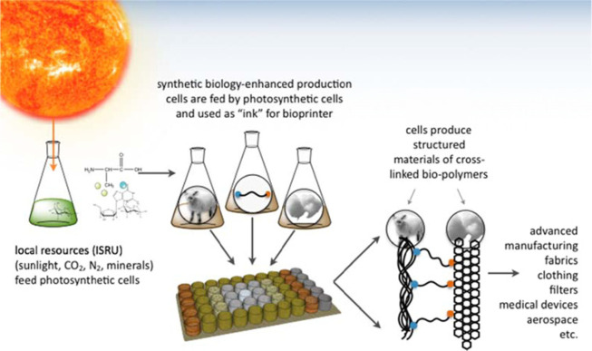
Bioprinting as an extension of 3D printing offers capabilities for printing tissues and organs for application in biomedical engineering. Conducting bioprinting in space, where the gravity is zero, can enable new frontiers in tissue engineering. Fabrication of soft tissues, which usually collapse under their own weight, can be accelerated in microgravity conditions as the external forces are eliminated. Furthermore, human colonization in space can be supported by providing critical needs of life and ecosystems by 3D bioprinting without relying on cargos from Earth, e.g., by development and long-term employment of living engineered filters (such as sea sponges-known as critical for initiating and maintaining an ecosystem). This review covers bioprinting methods in microgravity along with providing an analysis on the process of shipping bioprinters to space and presenting a perspective on the prospects of zero-gravity bioprinting.

Keywords: 3D bioprinting; microgravity; regenerative medicine; space exploration; tissue engineering.


Conflict of interest statement

The authors declare no competing financial interest.

Figures

Figure 1
Figure 1
Bioprinting in microgravity: Fabricating using bioprinting technology by shipping bioprinters into space where the gravity is zero can be beneficial in several ways: First, fabrication of soft tissues which usually collapse under their own weight can be accelerated in the microgravity conditions as the external forces are eliminated, resulting in advancing the tissue engineering applications. Second, human colonization in space can be supported by providing critical needs of life and ecosystems without relying on cargos from Earth, which is the potential to be the next step to start life on other planets. Adapted with permission, copyright 2018 OHB SE. The composition of the figure was done by OHB (ohb.de) with images from the European Space Agency (ESA) and the National Aeronautics and Space Administration Agency (NASA).
Figure 2
Figure 2
The 3D BioFabrication Facility (BFF) developed by Techshot. (A) BFF consists of the print volume on the left part and the power and data handling module in the lower right. The top right box is a separate device called the ADvanced Space Experiment Processor (ADSEP), where the cells printed with BFF are conditioned into tissue. Adapted from the Web site of Techshot (techshot.com) with permission, copyright 2019 Techshot. (B) BFF has already been shipped to the International Space Station (ISS) for studying bioprinting in microgravity. Adapted from the Web site of the ISS National Lab (issnationallab.org) with permission, copyright 2019 ISS National Laboratory. (C) Schematics view of tissue chips for recapitulating several tissue-level physiological functions, aiming for space medicine applications. Adapted from ref (81), copyright 2022 Elsevier, in accordance with Creative Commons CC-BY-NC-ND license.
Figure 3
Figure 3
The launched material into the destination planet must be light, stable, and reliable. Synthetic biology science can play an important role in this regard by enhancing production cells which are utilized to be fed into photosynthetic cells from local resources and be used as the required bioink for the bioprinting process. This process provides a vision for a biology-based Mars colony. Adapted from ref (65), copyright 2016 Portland Press, in accordance with the Creative Commons Attribution (CC BY) license.
Figure 4
Figure 4
The magnetic levitation setup used for biofabrication of in situ self-assembled 3D cell cultures inside a microcapillary channel system. (a) Mirrors are positioned at 45° on two sides to help visualize the cells with microscopes. Neodymium magnets are positioned on the sides with negative poles facing the inner microcapillary levitation and culturing channels. (b) Plot of cells’ levitation heights for different Gd concentrations. (c) Coefficient of variation (CV%) of levitation heights for different solutions based on Gd (Gd-BT-DO3A, Gd-DTPA, Gd-DTPA-BMA, Gd-DOTA, and Gd-BOPTA). Abbreviations: N: nonionic agents, I: ionic agents, Fd: fluidic drag force, Fi: inertial force, Fb: buoyancy force, and Fmag: magnetic force. Scale bar: 200 μm. Adapted from ref (73), copyright 2018 Springer Nature, in accordance with the Creative Commons Attribution (CC BY) license.
Figure 5
Figure 5
Magnetic levitational platform for soft living material fabrication. (a) Schematic view of fabricating units with photo-cross-linkable polymers using patterned mask photolithography. The glass sheets with a gel precursor solution were exposed to UV light (scale bar: 1 mm). (b) Fabrication of cell seeded microbead was performed by incubating laminin coated microbeads in cell suspension (scale bar: 500 μm). (c) Cell-encapsulating elements’ self-assembly in the magnetic levitational setup with two Neodymium magnets. (d) The particle moves from larger magnetic field strength (B) to lower magnetic field strength, if its magnetic susceptibility is lower than magnetic susceptibility of the suspending medium. (e) At equilibrium point, the two forces of magnetic (Fm) and corrected gravitational (Fg) (the difference between gravitational force and buoyancy force) act on the levitating particle. Adapted with permission from ref (74), copyright 2015 John Wiley & Sons.
Figure 6
Figure 6
Planaria, well-known flatworms for regeneration studies, were sent to space to investigate their behavior under microgravity conditions. (A) Immediately upon separation of their heads and tails, (B) they were sent to the ISS, for a 32-day period, with an identical control group kept on Earth. After 32 days, the group in the ISS was sent back to the Earth. (C) In an exceptionally rare occurrence, one of the worms from space was regenerated into a phenotype with two heads. Adapted from ref (78), copyright 2017 John Wiley & Sons, in accordance with the Creative Commons Attribution (CC BY) license.

References

    1. Gu Q.; et al. Three-dimensional bio-printing. Science China Life Sciences 2015, 58 (5), 411–419. 10.1007/s11427-015-4850-3. - DOI - PubMed
    1. Yigci D.; et al. 3D bioprinted glioma models. Progress in Biomedical Engineering 2022, 4 (4), 04200110.1088/2516-1091/ac7833. - DOI
    1. Rezapour Sarabi M.; Nakhjavani S. A.; Tasoglu S. 3D-Printed Microneedles for Point-of-Care Biosensing Applications. Micromachines 2022, 13 (7), 1099.10.3390/mi13071099. - DOI - PMC - PubMed
    1. Gungor-Ozkerim P. S.; et al. Bioinks for 3D bioprinting: an overview. Biomaterials science 2018, 6 (5), 915–946. 10.1039/C7BM00765E. - DOI - PMC - PubMed
    1. Seol Y.-J.; et al. Bioprinting technology and its applications. European Journal of Cardio-Thoracic Surgery 2014, 46 (3), 342–348. 10.1093/ejcts/ezu148. - DOI - PubMed

Publication types