. 2023 May 8;14(1):2639.
doi: 10.1038/s41467-023-38362-3.

Structural mechanisms of TRPM7 activation and inhibition

Affiliations

Structural mechanisms of TRPM7 activation and inhibition

Kirill D Nadezhdin et al. Nat Commun. .

Abstract

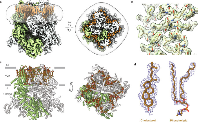
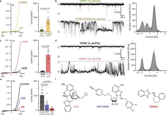
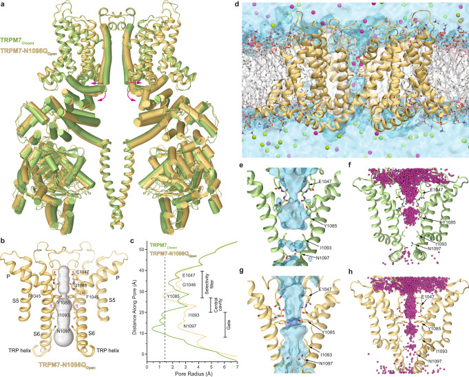
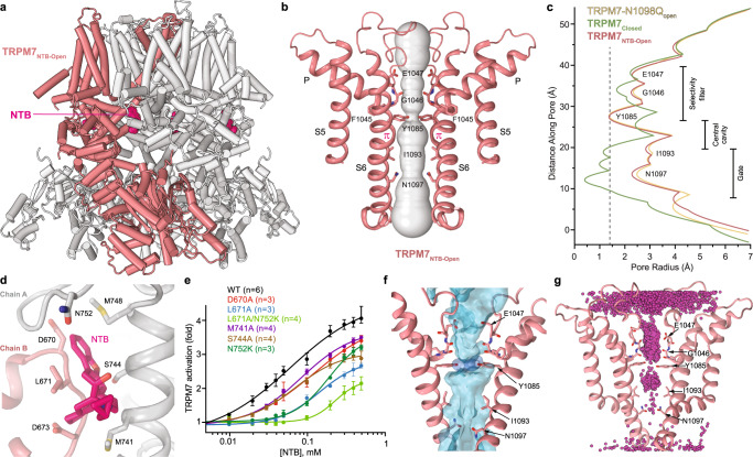
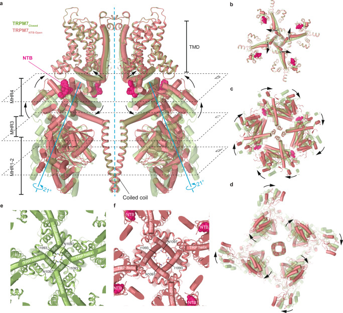
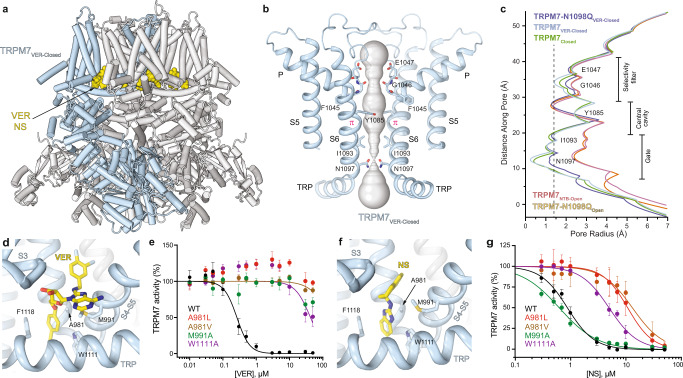
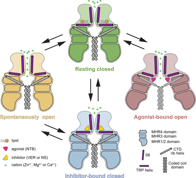
The transient receptor potential channel TRPM7 is a master regulator of the organismal balance of divalent cations that plays an essential role in embryonic development, immune responses, cell mobility, proliferation, and differentiation. TRPM7 is implicated in neuronal and cardiovascular disorders, tumor progression and has emerged as a new drug target. Here we use cryo-EM, functional analysis, and molecular dynamics simulations to uncover two distinct structural mechanisms of TRPM7 activation by a gain-of-function mutation and by the agonist naltriben, which show different conformational dynamics and domain involvement. We identify a binding site for highly potent and selective inhibitors and show that they act by stabilizing the TRPM7 closed state. The discovered structural mechanisms provide foundations for understanding the molecular basis of TRPM7 channelopathies and drug development.


Conflict of interest statement

The authors declare no competing interests.

Figures

Fig. 1
Fig. 1. Cryo-EM structure of TRPM7 in the apo state.
a Cryo-EM density map of TRPM7 in the closed, apo state at 2.19 Å resolution, viewed parallel to the membrane (left) and extracellularly (right). One of the four subunits is colored green, and the other three in grey. Lipid densities are highlighted in brown. The semi-transparent surface represents the lipid nanodisc density. b Close-up view of a map region in the TMD. c Structural model of TRPM7Closed reconstituted in lipid nanodiscs. One of the four subunits is colored in green, and the other three in grey. Lipids are shown as brown sticks. d Representative cryo-EM density (blue mesh) and models (brown sticks) for cholesterol and phospholipid.
Fig. 2
Fig. 2. Function of wild-type TRPM7 and the gain-of-function mutant N1098Q.
a, c, e Whole-cell patch-clamp recordings made from HEK 293T cells expressing wild-type (WT) (a, c, e) or mutant N1098Q (a) TRPM7 in response to a –100 to +100 mV voltage ramp. Left panels show representative current–voltage (I–V) relationships immediately after establishing the whole-cell configuration (a), without addition or after 170-s application of 500 μM NTB (c) and after 450-s exposure to the standard external solution in the absence (Control) or presence of 10 µM VER or 10 µM NS, following saturation of currents after 150 s of recordings (e). Right panels show current amplitudes measured at +80 mV (mean ± SEM). n is the number of cells. Statistical comparison was made using unpaired t test (a, c) or ordinary one-way ANOVA (e). Significance was accepted at P  ≤  0.05. b, d Representative single-channel currents recorded at the membrane potential of –100 mV from WT (b, d) or N1098Q mutant (b) TRPM7 reconstituted in lipid bilayers in the absence (b, d) or presence (d) of 2.5 μM NTB. The corresponding all-points amplitude histograms from three independent traces for TRPM7-N1098Q mutant (b) and for TRPM7 in the presence of 2.5 μM NTB (d) are shown on the right. The black curves are fits with three (b) or two (d) Gaussians. f Chemical structures of naltriben (NTB), VER155008 and NS8593.
Fig. 3
Fig. 3. Structure of TRPM7 with the gain-of-function mutation N1098Q.
a Superposition of TRPM7Closed (green) and TRPM7-N1098QOpen (yellow) structures. Only two of four subunits are shown, with the front and back subunits omitted for clarity. Pink arrows show domain movements. b Pore-forming domain of TRPM7-N1098QOpen with the residues contributing to the pore lining shown as sticks. Only two of four subunits are shown, with the front and back subunits omitted for clarity. The pore profile is shown as a space-filling model (grey). The π-bulge in the middle of S6 is labeled. c Pore radius for TRPM7-N1098QOpen (yellow) and TRPM7Closed (green) calculated using HOLE. The vertical dashed line denotes the radius of a water molecule, 1.4 Å. d A snapshot of the MD simulated system with TRPM7 shown as yellow ribbons, lipid bilayer acyl chains in grey and hydrophilic head groups as sticks, water as a light blue continuum, and Na+ and Cl ions as magenta and green spheres, respectively. e Water occupancy of the TRPM7Closed channel (blue surface) in MD simulations with no applied voltage. Residues lining the pore are shown in sticks. f Cumulative distribution of Na+ ions (magenta spheres) in the TRPM7Closed channel MD simulations with no applied voltage. g Water occupancy of the TRPM7-N1098Qopen channel (blue surface) in MD simulations with no applied voltage. The hydroxyl groups of Y1085 residues (shown as dark blue surface) contribute to the permeation pathway. Residues lining the pore are shown in sticks. h Cumulative distribution of K+ ions (magenta spheres) in the TRPM7-N1098Qopen channel MD simulations under applied voltage.
Fig. 4
Fig. 4. Structure of TRPM7 in complex with agonist naltriben.
a Open-state structure TRPM7NTB-open, with one subunit colored pink and the other three grey and molecules of NTB shown as space-filling models (bright pink). b Pore-forming domain of TRPM7NTB-Open with the residues contributing to pore lining shown as sticks. Only two of four subunits are shown, with the front and back subunits omitted for clarity. The pore profile is shown as a space-filling model (grey). The π-bulge in the middle of S6 is labeled. c Pore radius for TRPM7-N1098Qopen (yellow), TRPM7NTB-Open (pink), and TRPM7Closed (green) calculated using HOLE. The vertical dashed line denotes the radius of a water molecule, 1.4 Å. d Close-up view of the NTB binding site. The molecule of NTB (bright pink) and residues involved in its binding are shown in sticks. e Concentration-dependences for activation of wild-type (WT) and mutant TRPM7 channels by NTB, determined using the Ca2+ influx assay as outlined in Supplementary Fig. 7a. Curves through the points (mean ± SEM) are the logistic equation fits; n, the number of independent measurements. The corresponding values of IC50 and nHill are provided in Supplementary Table 4. Source data are provided. f Water occupancy of the TRPM7NTB-Open channel (blue surface) in MD simulations with no applied voltage. The hydroxyl groups of the Y1085 residues that contribute to the permeation pathway are shown as dark blue surfaces. Residues lining the pore are shown in sticks. g Cumulative distribution of K+ ions (magenta spheres) in TRPM7NTB-Open pore during MD simulations under applied voltage.
Fig. 5
Fig. 5. Mechanism of the agonist-induced opening of TRPM7.
a Superposition of TRPM7NTB-Open (red) and TRPM7Closed (green) structures viewed parallel to the membrane. NTB molecules are shown as space-filling models (bright pink). Only two of four subunits are shown, with the front and back subunits omitted for clarity. Rotation axes are shown in blue, while arrows depict domain movements during channel opening. bd Sections of TRPM7NTB-open (red) and TRPM7Closed (green) superposition indicated by dashed lines in (a), viewed perpendicular to the membrane, along the axis of fourfold rotational symmetry. Domain movements during NTB-induced channel opening are indicated by the black arrows. e, f Close-up view of the intracellular pore entrance in TRPM7Closed (e) and TRPM7NTB-Open (f) viewed along the axis of the 4-fold rotational symmetry. Residues forming the narrow constriction at the gate region of the pore are shown as sticks. NTB molecules are shown as space-filling models (bright pink).
Fig. 6
Fig. 6. Structures of TRPM7 in complex with inhibitors VER155008 and NS8593.
a Structure of TRPM7VER-Closed, with one subunit colored blue and the other three grey and molecules of VER155008 shown as space-filling models (yellow). b Pore-forming domain in TRPM7VER-Closed with the residues contributing to pore lining shown as sticks. Only two of four subunits are shown, with the front and back subunits omitted for clarity. The pore profile is shown as a space-filling model (grey). The π-bulge in the middle of S6 is labeled. c Pore radius for TRPM7Closed and TRPM7 in complex with inhibitors compared to the pore radius for the open-state structures, all calculated using HOLE. The vertical dashed line denotes the radius of a water molecule, 1.4 Å. d, f Close-up view of the vanilloid-like site in TRPM7VER-Closed (d) and TRPM7-N1098QNS-Closed (f), with the inhibitor molecule (yellow) and residues contributing to its binding shown in sticks . e, g Concentration-dependencies for inhibition of wild-type (WT) and mutant TRPM7 by VER155008 (e) or NS8593 (g) assessed using the Ca2+ influx assay. Curves through the points (mean ± SEM) are logistic equation fits. n, the number of independent measurements. The values of IC50 and nHill are provided in Supplementary Table 4. Source data are provided.
Fig. 7
Fig. 7. Mechanisms of TRPM7 activation and inhibition.
Schematic representation of conformational changes in TRPM7 during activation and inhibition. Upon spontaneous activation, the conformational changes are confined within the TMD. Conversely, binding of the agonist NTB induces substantial conformational rearrangements in both the N-terminal MHR1-4 domains and the TMD, leading to the channel opening. The inhibitor binding locks TRPM7 in the closed conformation resembling the resting apo state.

Similar articles

Cited by

References

    1. Clapham DE. TRP channels as cellular sensors. Nature. 2003;426:517–524. doi: 10.1038/nature02196. - DOI - PubMed
    1. Mederos y Schnitzler M, Waring J, Gudermann T, Chubanov V. Evolutionary determinants of divergent calcium selectivity of TRPM channels. FASEB J. 2008;22:1540–1551. doi: 10.1096/fj.07-9694com. - DOI - PubMed
    1. Hantute-Ghesquier, A., Haustrate, A., Prevarskaya, N. & Lehen’kyi, V. TRPM family channels in cancer. Pharmaceuticals11, 58 (2018). - PMC - PubMed
    1. Huang Y, Winkler PA, Sun W, Lu W, Du J. Architecture of the TRPM2 channel and its activation mechanism by ADP-ribose and calcium. Nature. 2018;562:145–149. doi: 10.1038/s41586-018-0558-4. - DOI - PubMed
    1. Ruan Z, et al. Structures of the TRPM5 channel elucidate mechanisms of activation and inhibition. Nat. Struct. Mol. Biol. 2021;28:604–613. doi: 10.1038/s41594-021-00607-4. - DOI - PMC - PubMed

Publication types

MeSH terms

Substances