. 2024 Mar;42(3):413-423.
doi: 10.1038/s41587-023-01784-x. Epub 2023 May 8.

Hypoimmune induced pluripotent stem cells survive long term in fully immunocompetent, allogeneic rhesus macaques

Affiliations

Hypoimmune induced pluripotent stem cells survive long term in fully immunocompetent, allogeneic rhesus macaques

Xiaomeng Hu et al. Nat Biotechnol. 2024 Mar.

Abstract

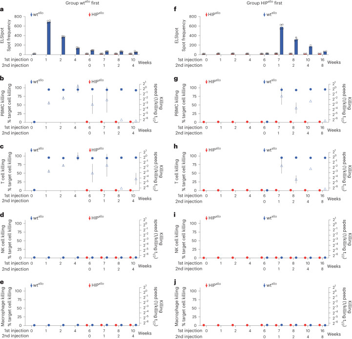
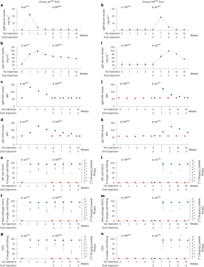
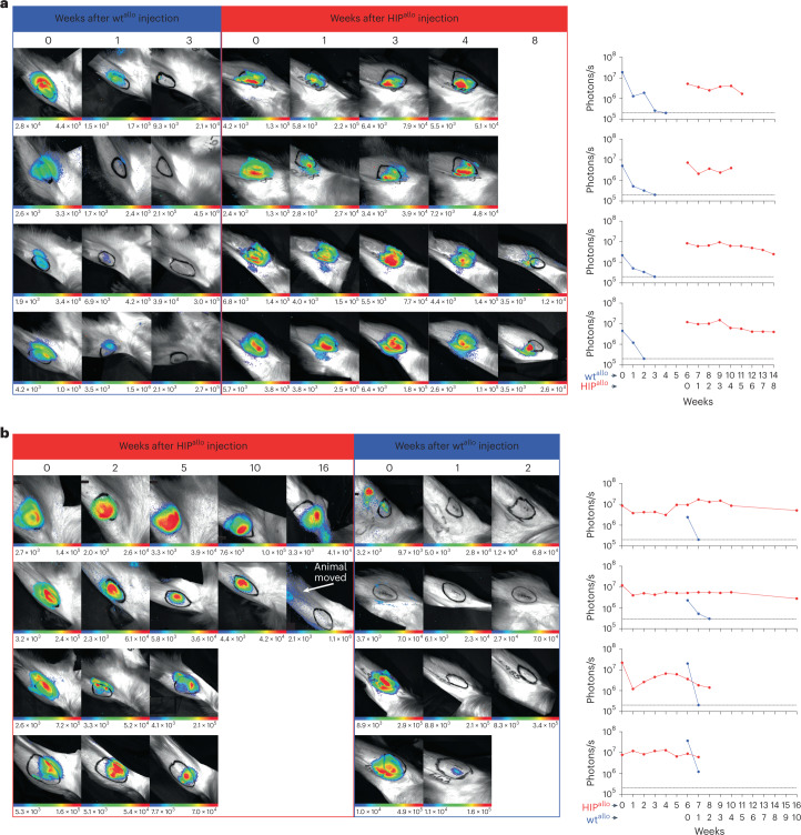
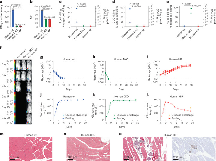
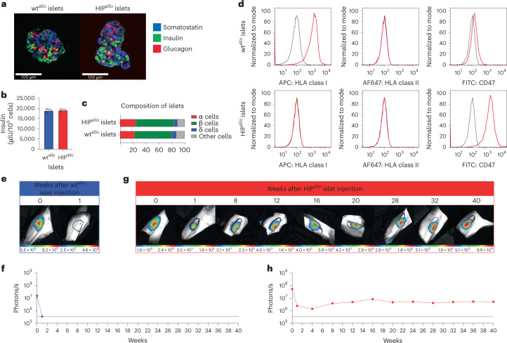
Genetic engineering of allogeneic cell therapeutics that fully prevents rejection by a recipient's immune system would abolish the requirement for immunosuppressive drugs or encapsulation and support large-scale manufacturing of off-the-shelf cell products. Previously, we generated mouse and human hypoimmune pluripotent (HIP) stem cells by depleting HLA class I and II molecules and overexpressing CD47 (B2M-/-CIITA-/-CD47+). To determine whether this strategy is successful in non-human primates, we engineered rhesus macaque HIP cells and transplanted them intramuscularly into four allogeneic rhesus macaques. The HIP cells survived unrestricted for 16 weeks in fully immunocompetent allogeneic recipients and differentiated into several lineages, whereas allogeneic wild-type cells were vigorously rejected. We also differentiated human HIP cells into endocrinologically active pancreatic islet cells and showed that they survived in immunocompetent, allogeneic diabetic humanized mice for 4 weeks and ameliorated diabetes. HIP-edited primary rhesus macaque islets survived for 40 weeks in an allogeneic rhesus macaque recipient without immunosuppression, whereas unedited islets were quickly rejected.


Conflict of interest statement

All experiments were conducted by or on behalf of Sana Biotechnology, Inc., and no data from Oregon Health & Science University, Washington University School of Medicine, University of California, San Francisco or Stanford University were used. L.L.L., A.J.C., T.D. and M.M.D. performed the work in this manuscript as consultants to Sana Biotechnology, Inc. T.D. and M.M.D. own stock and/or hold stock options in Sana Biotechnology, Inc. All other authors are employees of and own stock in Sana Biotechnology, Inc. Readers are welcome to comment on the online version of the paper. Correspondence and requests for materials should be addressed to S.S.

Figures

Fig. 1
Fig. 1. Cellular immune responses against allogeneic rhesus macaque wt and HIP grafts.
ae, Immune assays in the group receiving wtallo first. ELISpot assays with recipient monkey PBMCs drawn at scheduled timepoints (a, mean ± s.d., four monkeys). Killing assays with recipient monkey PBMCs (b), T cells (c), NK cells (d) and macrophages (e). Percent target cell killing is shown on the left y axis (mean ± s.d.), and killing speed is shown on the right y axis (killing t1/2−1, mean ± s.e.m., four monkeys). fj, Immune assays in the group receiving HIPallo first. ELISpot assays with recipient monkey PBMCs (f, mean ± s.d., four monkeys at weeks 0–7, three monkeys at week 8 and two monkeys at weeks 10–16). Killing assays with recipient monkey PBMCs (g), T cells (h), NK cells (i) and macrophages (j). Percent target cell killing is shown on the left y axis (mean ± s.d.), and killing speed is shown on the right y axis (killing t1/2−1, mean ± s.e.m., four monkeys at weeks 0–7, three monkeys at week 8 and two monkeys at weeks 10–16). All assays run against wtallo and HIPallo are shown in blue and red, respectively, at 6 weeks, and, later, all assays were run against both cell types.
Fig. 2
Fig. 2. Antibody-mediated responses against allogeneic rhesus macaque wt and HIP grafts.
ag, Antibody immune assays in the group receiving wtallo first. Total serum IgM (a) and IgG (b) levels and DSA IgM (c) and IgG (d) levels are shown (mean ± s.d., four monkeys). ADCC assays with de-complemented recipient monkey serum and NK cells (e) or macrophages (f) and CDC assays with complete recipient monkey serum (g) are shown. Percent target cell killing is shown on the left y axis (mean ± s.d.), and killing speed is shown on the right y axis (killing t1/2−1, mean ± s.e.m., four monkeys). hn, Antibody immune assays in the group receiving HIPallo first. Total serum IgM (h) and IgG (i) levels and DSA IgM (j) and IgG (k) levels are shown (mean ± s.d., four monkeys at weeks 0–7, three monkeys at week 8 and two monkeys at weeks 10–16). ADCC assays with de-complemented recipient monkey serum and NK cells (l) or macrophages (m) and CDC assays with complete recipient monkey serum (n) are shown. Percent target cell killing is shown on the left y axis (mean ± s.d.), and killing speed is shown on the right y axis (killing t1/2–1, mean ± s.e.m., four monkeys at weeks 0–7, three monkeys week at 8 and two monkeys at weeks 10–16). All assays run against wtallo and HIPallo are shown in blue and red, respectively, at 6 weeks, and, later, all assays were run against both cell types.
Fig. 3
Fig. 3. Graft survival assessed by quantitative BLI.
a,b, BLI images and BLI signals over time are shown for all four rhesus macaques receiving wtallo first (a) and all four monkeys receiving HIPallo first (b).
Fig. 4
Fig. 4. Histology of intramuscular allogeneic rhesus macaque wt and HIP grafts.
Representative images of at least two independent sections are shown from H&E staining and immunohistochemical staining for CD3 (T lymphocytes), CD20 (B lymphocytes) and CD68 (macrophages). a,b, wtallo injection site after 1 week at ×100 (a) and ×400 (b) magnification. c, wtallo injection site after 2 weeks at ×100 magnification. d, HIPallo cell injection site after 7 weeks at ×100 magnification. e, HIPallo cell injection site after 8 weeks at ×100 magnification.
Fig. 5
Fig. 5. Human HIP islets survive in immunocompetent allogeneic humanized mice and ameliorate diabetes.
ae, One million wt, DKO or HIP islet cells were injected into the thigh muscle of allogeneic humanized NSG-SGM3 mice. After 6 d, the immune response against the graft cells was assessed. IFN-γ ELISpot assays with mouse splenocytes were performed to assess the adaptive immune response against the islet grafts (a, mean ± s.d., three animals per group, one-way ANOVA with Bonferroni post hoc test). Serum DSAs of IgM type were quantified by flow cytometry (b, mean ± s.d., three animals per group, one-way ANOVA with Bonferroni post hoc test). Impedance killing assays of islet cells with recipient mouse T cells (c, mean ± s.d., three animals per group, one-way ANOVA with Bonferroni post hoc test) or serum (d, mean ± s.d., three animals per group, one-way ANOVA with Bonferroni post hoc test). Human NK cells were used for this impedance killing assay of islet cells (e, mean ± s.d., triplicates, one-way ANOVA with Bonferroni post hoc test). fi, Humanized NSG-SGM3 mice received streptozotocin and developed diabetes (f, one representative mouse is shown per group). Six days later, 1,000 FLuc+ allogeneic wt (g, five animals), DKO (h, five animals) or HIP islet clusters (i, eight animals) were injected intramuscularly, and their survival was monitored with BLI (all individual animals are shown). jl, Serial fasting blood glucose levels were quantified (mean ± s.d.) in the wt (j, five animals), DKO (k, five animals) or HIP (l, eight animals) groups. On day 30, animals underwent a glucose challenge with blood draw 30 min later (mean ± s.d.). mo, The injection sites of wt (m), DKO (n) or HIP islet clusters (o) were recovered and stained with H&E and for ISL-1 by immunohistochemistry (representative images).
Fig. 6
Fig. 6. Allogeneic HIP islets achieve long-term survival.
a, Immunofluorescence pictures of dissociated and reaggregated wtallo and HIPallo islets from rhesus monkeys (representative images). b, In vitro insulin secretion of wtallo and HIPallo islets (mean ± s.d., triplicates). c, The composition of wtallo and HIPallo islets of α, β, δ and other cells (mean, triplicates). d, Flow cytometry histograms for HLA class I and II and rhesus CD47 on wtallo and HIPallo islets (representative images). eh, The survival of FLuc+ wtallo and HIPallo islets in allogeneic rhesus monkeys was followed by BLI (one animal each).

References

    1. Yamanaka S. Pluripotent stem cell-based cell therapy—promise and challenges. Cell Stem Cell. 2020;27:523–531. doi: 10.1016/j.stem.2020.09.014. - DOI - PubMed
    1. Rezania A, et al. Reversal of diabetes with insulin-producing cells derived in vitro from human pluripotent stem cells. Nat. Biotechnol. 2014;32:1121–1133. doi: 10.1038/nbt.3033. - DOI - PubMed
    1. Pagliuca FW, et al. Generation of functional human pancreatic β cells in vitro. Cell. 2014;159:428–439. doi: 10.1016/j.cell.2014.09.040. - DOI - PMC - PubMed
    1. Kieffer TJ. Closing in on mass production of mature human beta cells. Cell Stem Cell. 2016;18:699–702. doi: 10.1016/j.stem.2016.05.014. - DOI - PubMed
    1. Maxwell KG, et al. Gene-edited human stem cell-derived β cells from a patient with monogenic diabetes reverse preexisting diabetes in mice. Sci. Transl. Med. 2020;12:eaax9106. doi: 10.1126/scitranslmed.aax9106. - DOI - PMC - PubMed