Review
. 2023 May;16(5):182-191.
doi: 10.1007/s12178-023-09831-5. Epub 2023 May 9.

Meniscus Radial Tears: Current Concepts on Management and Repair Techniques

Affiliations
Review

Meniscus Radial Tears: Current Concepts on Management and Repair Techniques

Enzo S Mameri et al. Curr Rev Musculoskelet Med. 2023 May.

Abstract

Purpose of review: This review provides a historical perspective on the approach to radial tears and collates the currently available evidence on repair techniques, rehabilitation, and outcomes following the treatment of meniscus radial tears.

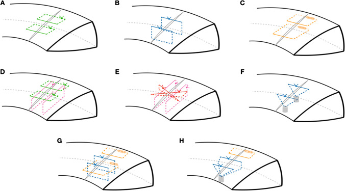
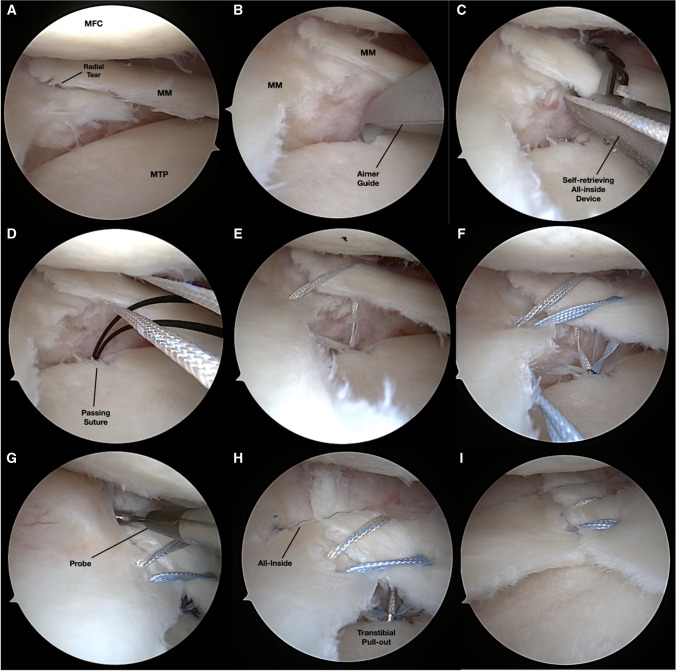
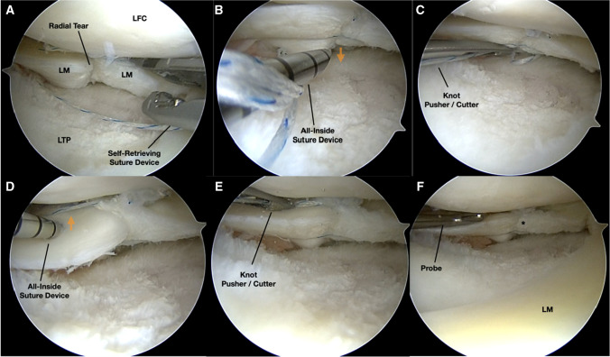
Recent findings: Recent literature shows that the repair of meniscus radial tears reports improved patient-reported outcome scores with high return to function and activity. However, no single technique nor construct was proven better than the other. Various methods of repairing radial tears can be employed, with biomechanical research supporting all-inside double vertical sutures, the addition of vertical "rip-stop" mattress sutures, and transtibial pullout augmentation. To ensure proper healing before undergoing physical therapy, it is crucial to abstain from weight-bearing and deep knee flexion for the first 6 weeks after surgery. Despite considerable heterogeneity in surgical techniques and rehabilitation protocols found in the current literature, studies reporting on radial repairs report positive results, with high healing rates and improved patient-reported outcomes.

Keywords: Meniscus radial tear; Meniscus repair; Postoperative outcomes.


Conflict of interest statement

The authors declare no competing interests.

Figures

Fig. 1
Fig. 1
Schematic representation of key radial repair techniques. A Conventional horizontal inside-out repair (green); B suture-based all-inside double vertical repair (blue); C anchor-based all-inside horizontal repair (orange); D “hash-tag” construct, with inside-out horizontal repair reinforced with vertical mattress “rip-stop” sutures (pink); E “cross-tag” construct, with suture-based all-inside figure-of-eight configuration (red) reinforced with vertical mattress “rip-stop” sutures; F two-tunnel transtibial pullout repair; G hybrid suture-based double vertical and anchor-based all-inside horizontal repair (see Fig. 3); H hybrid double-vertical transtibial pullout with all-inside horizontal repair (see Fig. 2)
Fig. 2
Fig. 2
Authors’ preference for medial meniscus radial tears, as represented in Fig. 1H and seen in a left knee arthroscopy following pie-crusting of the superficial medial collateral ligament (A). A single transtibial tunnel is created with the aid of a curved aimer guide and metallic cannula (FirstPass Mini, Smith & Nephew) (B). An all-inside self-retrieving device (C) is used to pass a suture tape in a vertical mattress configuration across the borders of the radial tear (DE); and pulled out of the tibial tunnel using a monofilament nitinol passing suture (D). These steps are then repeated with a second suture tape, producing a double vertical all-suture repair (F). The remaining gap identified with the use of a probe (G) is then closed using one anchor-based all-inside horizontal stitch (H). The all-inside suture in then tensioned and the knot cut, followed by manual tensioning of the transtibial pullout under arthroscopic visualization (I) and final fixation with an anchor (Footprint, Smith & Nephew) is carried out. MFC, medial femoral condyle; MM, medial meniscus; MTP, medial tibial plateau
Fig. 3
Fig. 3
Authors’ preference for lateral meniscus radial tears, as represented in Fig. 1G and seen in a right knee arthroscopy (A). A self-retrieving all-inside device (Novostitch, Smith & Nephew) is used to deploy suture-based double vertical side-to-side sutures (A). The construct is then completed with the addition of two anchor-based all-inside horizontal sutures, the first attaching the inferior surface of the meniscus to the capsule (B, C) and the second through the superior aspect of the meniscus into the capsule (D, E). The final hybrid construct is then visualized with satisfactory apposition of the borders (*) and probed for adequate stability. LFC, lateral femoral condyle; LM, lateral meniscus; LTP, lateral tibial plateau

Similar articles

Cited by

References

    1. Jackson GR, Meade J, Yu Z, Young B, Piasecki DP, Fleischli JE, et al. Outcomes and failure rates after revision meniscal repair: a systematic review and meta-analysis. Int Orthop. 2022;46(7):1557–1562. doi: 10.1007/s00264-022-05413-1. - DOI - PubMed
    1. Mameri ES, Dasari SP, Fortier LM, Verdejo FG, Gursoy S, Yanke AB, et al. Review of meniscus anatomy and biomechanics. Curr Rev Musculoskelet Med. 2022;15(5):323–335. doi: 10.1007/s12178-022-09768-1. - DOI - PMC - PubMed
    1. Adams BG, Houston MN, Cameron KL. The epidemiology of meniscus injury. Sports Med Arthrosc Rev. 2021;29(3):e24–e33. doi: 10.1097/JSA.0000000000000329. - DOI - PubMed
    1. Cullen KA, Hall MJ, Golosinskiy A. Ambulatory surgery in the United States, 2006. Natl Health Stat Report. 2009;11:1–25. - PubMed
    1. Jarraya M, Roemer FW, Englund M, Crema MD, Gale HI, Hayashi D, et al. Meniscus morphology: does tear type matter? A narrative review with focus on relevance for osteoarthritis research. Semin Arthritis Rheum. 2017;46(5):552–561. doi: 10.1016/j.semarthrit.2016.11.005. - DOI - PubMed