Review
. 2023 May 9;8(5):264-282.
doi: 10.1530/EOR-23-0047.

Biological aspects to enhance fracture healing

Affiliations
Review

Biological aspects to enhance fracture healing

Paul L Rodham et al. EFORT Open Rev. .

Abstract

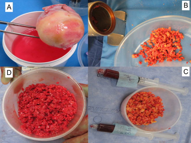
The ability to enhance fracture healing is paramount in modern orthopaedic trauma, particularly in the management of challenging cases including peri-prosthetic fractures, non-union and acute bone loss. Materials utilised in enhancing fracture healing should ideally be osteogenic, osteoinductive, osteoconductive, and facilitate vascular in-growth. Autologous bone graft remains the gold standard, providing all of these qualities. Limitations to this technique include low graft volume and donor site morbidity, with alternative techniques including the use of allograft or xenograft. Artificial scaffolds can provide an osteoconductive construct, however fail to provide an osteoinductive stimulus, and frequently have poor mechanical properties. Recombinant bone morphogenetic proteins can provide an osteoinductive stimulus; however, their licencing is limited and larger studies are required to clarify their role. For recalcitricant non-unions or high-risk cases, the use of composite graft combining the above techniques provides the highest chances of successfully achieving bony union.

Keywords: bone graft; bone substitute; fracture healing; non-union.


Conflict of interest statement

The authors declare that there is no conflict of interest that could be perceived as prejudicing the impartiality of the research reported.

Figures

Figure 1
Figure 1
Autologous cancellous bone graft. (A) Cancellous graft harvested from the posterior iliac crest of the pelvis. (B) Graft harvested from the femoral intramedullary cavity using the RIA device.
Figure 2
Figure 2
Demonstration of the use of a synthetic material Greenbone block for defect treatment of the pelvic iliac crest. (A) Comparison between the tricortical iliac crest harvested for a pubic symphysis fusion with the synthetic greenbone block. (B) Intra-operative picture showing the greenbone next to the iliac crest defect prior to implantation. (C) Cutting with an electric saw the green bone block scaffold to appropriate length. (D) Implantation of the green bone by press fitting application within the iliac crest defect. (E) AP pelvic radiograph showing the integration of the green bone scaffold at 12-month follow-up (red arrow). (F) Pelvic 3D model showing incorporation of the green bone scaffold within the iliac crest defect (red arrow).
Figure 3
Figure 3
Application of calcium sulphate (stimulant) for the treatment of a tibial partial bone defect following debridement for late presentation of infection after union that required also removal of the plate. (A) Intra-opertaive picture showing the footprint of the plate after removal and the defect created following debridement of the infected bone area. (B) Intra-operative picture showing the application of stimulant to the defect area. (C) Intra-operative picture with covering of the defect area with stimulant. (D) AP and lateral tibial radiographs at 8 weeks showing that the defect is healing. (E) AP and lateral tibial radiographs 16 weeks later showing complete healing of the defect area.
Figure 4
Figure 4
Graft materials used for composite grafting. (A) Autologous graft. (B) Allograft. (C) Xenograft (Orthos). (D) BMP-7. (5) Bone marrow aspirate.
Figure 5
Figure 5
Treatment of a tibial defect utilising the polytherapy approach. Implantation of (A) concentrated bone marrow aspirate (progenitor cells), (B) BMP-7, and (C) RIA graft.
Figure 6
Figure 6
Demonstration of enhancing the biological properties of allograft material. (A) Femoral head allograft. (B) Processing of femoral head into bone chips. (C) Loading of allograft chips with autologous PRP and concentrated bone marrow aspirate. (D) Difference in the macroscopic appearance of the allograft after enhancement appearing more viable (red) possessing inductivity and cellularity.
Figure 7
Figure 7
Overview of materials used in the clinical setting based on their properties and limitations of their use.

References

    1. Fesseha H & Fesseha Y. Bone grafting, its principle and application: a review. Osteology and Rheumatology 2020143–50. (10.17140/ORHOJ-1-113) - DOI
    1. Campana V Milano G Pagano E Barba M Cicione C Salonna G Lattanzi W & Logroscino G. Bone substitutes in orthopaedic surgery: from basic science to clinical practice. Journal of Materials Science. Materials in Medicine 2014252445–2461. (10.1007/s10856-014-5240-2) - DOI - PMC - PubMed
    1. Schlickewei W & Schlickewei C. The use of bone substitutes in the treatment of bone defects – the clinical view and history. Macromolecular Symposia 200725310–23. (10.1002/masy.200750702) - DOI
    1. Wang W & Yeung KWK. Bone grafts and biomaterials substitutes for bone defect repair: a review. Bioactive Materials 20172224–247. (10.1016/j.bioactmat.2017.05.007) - DOI - PMC - PubMed
    1. Azi ML Aprato A Santi I Kfuri MJ Masse A & Joeris A. Autologous bone graft in the treatment of post-traumatic bone defects: a systematic review and meta-analysis. BMC Musculoskeletal Disorders 201617 465. (10.1186/s12891-016-1312-4) - DOI - PMC - PubMed

LinkOut - more resources