Review
. 2023 May 9;8(5):351-360.
doi: 10.1530/EOR-23-0039.

Management of the stiff elbow: a literature review

Affiliations
Review

Management of the stiff elbow: a literature review

Mark F Siemensma et al. EFORT Open Rev. .

Abstract

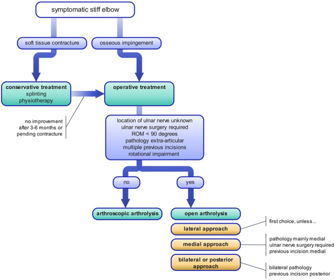
The elbow is prone to stiffness due to its unique anatomy and profound capsular reaction to inflammation. The resulting movement impairment may significantly interfere with a patient's activities of daily living. Trauma (including surgery for trauma), posttraumatic arthritis, and heterotopic ossification (HO) are the most common causes of elbow stiffness. In stiffness caused by soft tissue contractures, initial conservative treatment with physiotherapy (PT) and splinting is advised. In cases in which osseous deformities limit range of motion (e.g. malunion, osseous impingement, or HO), early surgical intervention is recommended. Open and arthroscopic arthrolysis are the primary surgical options. Arthroscopic arthrolysis has a lower complication and revision rate but has narrower indications. Early active mobilization using PT after surgery is recommended in postoperative rehabilitation and may be complemented by splinting or continuous passive motion therapy. Most results are gained within the first few months but can continue to improve until 12 months. This paper reviews the current literature and provides state-of-the-art guidance on the management regarding prevention, evaluation, and treatment of elbow stiffness.

Keywords: arthrofibrosis; osseous impingement; posttraumatic; soft tissue contracture; treatment.


Conflict of interest statement

The authors declare that there is no conflict of interest that could be perceived as prejudicing the impartiality of the research reported.

Figures

Figure 1
Figure 1
Plain elbow radiographs during maximum extension in a 23-year-old girl with posttraumatic stiffness of the right elbow. (A) Anteroposterior view showing degenerative joint changes along with the presence of osteophytes of the medial coronoid and deformation of the radial head. (B) Lateral view showing an additional osteophyte at the coronoid fossa, possibly causing impingement during elbow flexion.
Figure 2
Figure 2
Three-dimensional (3D) and four-dimensional (4D) bony reconstruction of the elbow of the patient shown in Figure 1. (A) Detailed images of the distal humerus (1), proximal ulna (2), and proximal radius (3). The mirrored non-affected side is overlain with the affected side (in transparent purple). Osteophytic bone spurs in the radial fossa and coronoid fossa and a deformed osteophytic border of the medial trochlea (1), together with profound osteophytes of the olecranon and coronoid process (2) and radial head deformation (3), can be seen. (B) Lateral view showing a maximum flexion of 110o (1) and an extension deficit of 55o (2).
Figure 3
Figure 3
A patient wearing two different types of static progressive splints for the conservative treatment of elbow stiffness. (A) A static progressive turnbuckle splint applying torque to the elbow in extension. Tuning of extension can be achieved by turning the red pin. Note: The red pin is shown for illustrative purposes but must be removed after the desired amount of flexion or extension torque is achieved. (B) A static progressive splint with a non-elastic strap and loop applying torque to the elbow in flexion.
Figure 4
Figure 4
Marked anatomical landmarks and portals for arthroscopic arthrolysis on the right arm in a patient lying in the lateral decubitus position. Note the free range of motion for extension and the space for the arthroscope on the axillary side. (A) Anatomical landmarks and portals shown for medial view. (B) Anatomical landmarks and portals shown for lateral view. U, ulnar nerve; M, medial epicondyle; T, triceps central band; O, olecranon; L, lateral epicondyle; R, radial head; S, soft spot; 1, straight posterior portal; 2, posterolateral portal; 3, anterolateral portal; 4, anteromedial portal; 5, soft spot portal.
Figure 5
Figure 5
Subsequent steps in the lateral approach during open arthrolysis. (A) Intraoperative view of the Kaplan approach following incision of the skin and subcutaneous tissue. (B) Further exposed extensor carpi radialis brevis (ECRB) and extensor digitorum communis (EDC) muscles. Note the yellow dotted line, highlighting the interval for the EDC split. (C) Opened joint capsule after EDC split with the exposed lateral epicondyle, capitellum, and radial head. Note the annular ligament is only incised if pathology of the proximal radioulnar joint exists and needs to be addressed.
Figure 6
Figure 6
Intraoperative view of the posterior approach during open arthrolysis of a posttraumatic stiff elbow. T, triceps, elevated (red); C, joint capsule (gray); S, ulnar nerve sulcus (blue); *, posterior band of medial collateral ligament (green) which forms the distal part of the floor of the sulcus; U, ulnar nerve (yellow), released and mobilized out of sulcus.
Figure 7
Figure 7
Treatment algorithm for the symptomatic stiff elbow.

References

    1. Morrey BF Askew LJ & Chao EY. A biomechanical study of normal functional elbow motion. Journal of Bone and Joint Surgery. American Volume 198163872–877. (10.2106/00004623-198163060-00002) - DOI - PubMed
    1. Sardelli M Tashjian RZ & MacWilliams BA. Functional elbow range of motion for contemporary tasks. Journal of Bone and Joint Surgery. American Volume 201193471–477. (10.2106/JBJS.I.01633) - DOI - PubMed
    1. Adolfsson L. Post-traumatic stiff elbow. EFORT Open Reviews 20183210–216. (10.1302/2058-5241.3.170062) - DOI - PMC - PubMed
    1. Myden C & Hildebrand K. Elbow joint contracture after traumatic injury. Journal of Shoulder and Elbow Surgery 20112039–44. (10.1016/j.jse.2010.07.013) - DOI - PubMed
    1. O'Driscoll SW & Giori NJ. Continuous passive motion (CPM): theory and principles of clinical application. Journal of Rehabilitation Research and Development 200037179–188. - PubMed

LinkOut - more resources