

Defining hippocampal area CA2 in the fox (Vulpes vulpes) brain
- PMID: 37159095
- PMCID: PMC10274530
- DOI: 10.1002/hipo.23546
Defining hippocampal area CA2 in the fox (Vulpes vulpes) brain
Abstract
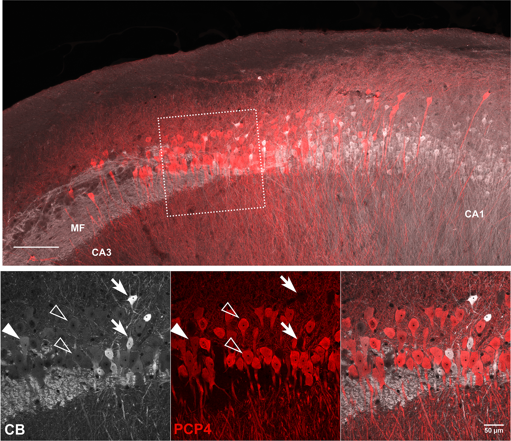
Since 1959, the Russian Farm-Fox study has bred foxes to be either tame or, more recently, aggressive, and scientists have used them to gain insight into the brain structures associated with these behavioral features. In mice, hippocampal area CA2 has emerged as one of the essential regulators of social aggression, and so to eventually determine whether we could identify differences in CA2 between tame and aggressive foxes, we first sought to identify CA2 in foxes (Vulpes vulpes). As no clearly defined area of CA2 has been described in species such as cats, dogs, or pigs, it was not at all clear whether CA2 could be identified in foxes. In this study, we cut sections of temporal lobes from male and female red foxes, perpendicular to the long axis of the hippocampus, and stained them with markers of CA2 pyramidal cells commonly used in tissue from rats and mice. We observed that antibodies against Purkinje cell protein 4 best stained the pyramidal cells in the area spanning the end of the mossy fibers and the beginning of the pyramidal cells lacking mossy fibers, resembling the pattern seen in rats and mice. Our findings indicate that foxes do have a "molecularly defined" CA2, and further, they suggest that other carnivores like dogs and cats might as well. With this being the case, these foxes could be useful in future studies looking at CA2 as it relates to aggression.
Keywords: PCP4; aggression; calbindin; mossy fibers; perineuronal nets; social behavior.
© 2023 The Authors. Hippocampus published by Wiley Periodicals LLC. This article has been contributed to by U.S. Government employees and their work is in the public domain in the USA.
Conflict of interest statement
The authors have no conflicts of interest to report.
Figures


Similar articles
-
Identification of hippocampal area CA2 in hamster and vole brain.bioRxiv [Preprint]. 2024 Feb 14:2024.02.12.579957. doi: 10.1101/2024.02.12.579957. bioRxiv. 2024. Update in: J Comp Neurol. 2024 Mar;532(3):e25603. doi: 10.1002/cne.25603. PMID: 38405991 Free PMC article. Updated. Preprint.
-
A marker set for construction of a genetic map of the silver fox (Vulpes vulpes).J Hered. 2004 May-Jun;95(3):185-94. doi: 10.1093/jhered/esh033. J Hered. 2004. PMID: 15220384
-
The brain of the silver fox (Vulpes vulpes): a neuroanatomical reference of cell-stained histological and MRI images.Brain Struct Funct. 2023 Jun;228(5):1177-1189. doi: 10.1007/s00429-023-02648-5. Epub 2023 May 9. Brain Struct Funct. 2023. PMID: 37160458 Free PMC article.
-
Genotyping-By-Sequencing (GBS) Detects Genetic Structure and Confirms Behavioral QTL in Tame and Aggressive Foxes (Vulpes vulpes).PLoS One. 2015 Jun 10;10(6):e0127013. doi: 10.1371/journal.pone.0127013. eCollection 2015. PLoS One. 2015. PMID: 26061395 Free PMC article.
-
Red fox genome assembly identifies genomic regions associated with tame and aggressive behaviours.Nat Ecol Evol. 2018 Sep;2(9):1479-1491. doi: 10.1038/s41559-018-0611-6. Epub 2018 Aug 6. Nat Ecol Evol. 2018. PMID: 30082739 Free PMC article.
Cited by
-
Identification of hippocampal area CA2 in hamster and vole brain.J Comp Neurol. 2024 Mar;532(3):e25603. doi: 10.1002/cne.25603. J Comp Neurol. 2024. PMID: 38497661 Free PMC article.
-
Identification of hippocampal area CA2 in hamster and vole brain.bioRxiv [Preprint]. 2024 Feb 14:2024.02.12.579957. doi: 10.1101/2024.02.12.579957. bioRxiv. 2024. Update in: J Comp Neurol. 2024 Mar;532(3):e25603. doi: 10.1002/cne.25603. PMID: 38405991 Free PMC article. Updated. Preprint.
References
-
- Amayasu H, Shoumura K, Ichinohe N, Yu S, & Yonekura H (1999). Cornu Ammonis of the dog: a rudimentary CA2 field is only present in a small part of the dorsal division, and is absent in the ventral division of the cornu Ammonis. J Hirnforsch, 39(3), 355–367. Retrieved from https://www.ncbi.nlm.nih.gov/pubmed/10536868 - PubMed
Publication types
MeSH terms
Grants and funding
LinkOut - more resources
Full Text Sources
Research Materials
Miscellaneous
