. 2023 Apr 5;3(4):100443.
doi: 10.1016/j.crmeth.2023.100443. eCollection 2023 Apr 24.

DNA ultra-sensitive quantification, a technology for studying HIV unintegrated linear DNA

Affiliations

DNA ultra-sensitive quantification, a technology for studying HIV unintegrated linear DNA

Hélène Marie Roux et al. Cell Rep Methods. .

Abstract

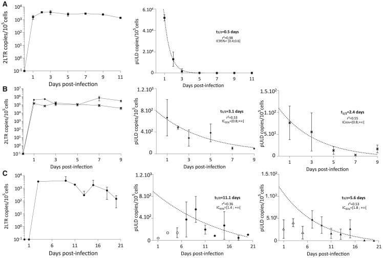
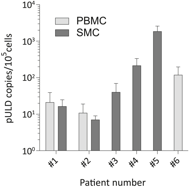
Unintegrated HIV DNA represents between 20% and 35% of the total viral DNA in infected patients. Only the linear forms (unintegrated linear DNAs [ULDs]) can be substrates for integration and for the completion of a full viral cycle. In quiescent cells, these ULDs may be responsible for pre-integrative latency. However, their detection remains difficult due to the lack of specificity and sensitivity of existing techniques. We developed an ultra-sensitive, specific, and high-throughput technology for ULD quantification called DUSQ (DNA ultra-sensitive quantification) combining linker-mediated PCR and next-generation sequencing (NGS) using molecular barcodes. Studying cells with different activity levels, we determined that the ULD half-life goes up to 11 days in resting CD4+ T cells. Finally, we were able to quantify ULDs in samples from patients infected with HIV-1, providing a proof of concept for the use of DUSQ in vivo to track pre-integrative latency. DUSQ can be adapted to the detection of other rare DNA molecules.


Conflict of interest statement

The authors declare no competing interests.

Figures

None
Graphical abstract
Figure 1
Figure 1
DUSQ library preparation workflow (A) Scheme of the DUSQ workflow. DNA is extracted by an organic phase method and ligated to a partially double-stranded adapter containing Illumina Read 1 sequence (blue), a unique molecular identifier (N)12 (red), and a linker with a 5′ GT cohesive end (green). A reparation is performed to obtain a full-length double-stranded DNA that is pre-amplified using a forward primer specific of the Illumina Read 1 sequence and a reverse primer specific to HIV-1 5′ LTR. A second round of amplification is performed using primers containing the other Illumina sequences. Sequencing is accomplished using the MiSeq technology. (B) Representative DNA size repartition of 96 samples pooled for sequencing at a concentration of 20 nM evaluated on a 2100 Bioanalyzer. Lower (35 bp) and upper markers (10,380 bp) are indicated. The library peak is around 520 bp, and genomic DNA can be seen at higher molecular sizes.
Figure 2
Figure 2
DUSQ analysis workflow Complete algorithm of the DUSQ bioinformatics analysis workflow. After sequencing, the FastQ files are exported from the MiSeq platform with trimmed adapters. The FastQ files are then preprocessed using the fastp tool, allowing quality filtering and UMI extraction. The output is then aligned on an HIV-1 reference sequence using the Bowtie2 tool, and the aligned sequences are filtered depending on their CIGAR string to store only the sequences that are aligned on the first 22 bases (only pULDs) using BAMTools. Aligned sequences are then extracted for UMI filtering and counting using the UMI-tools deduplicate (options that are not mentioned in the algorithm were disabled).
Figure 3
Figure 3
Sensitivity and reproducibility of DUSQ (A) Representative sequence logo of the probability of finding each nucleotide at each of the 12 positions of the UMIs. These data were generated by Sequence Logo Generator from the sequences issued from MDMs sampled 7 days post-infection after UMI filtering (156,136 reads). (B) Determination of the DUSQ quantification threshold on serial dilutions of HIV-1-infected MT4R5 cells in uninfected MT4R5 cells. Mean ± SD of n = 14 replicates is represented. The linear fitness is represented by the dotted line with its 95% confidence interval. The linear fitness coefficient R2 and its p value are indicated. (C) Reproducibility of pULD quantification by DUSQ. MT4R5 cells were treated with dolutegravir 2 h pre-infection and infected by NL-4.3 during 24 h. Three DUSQ quantifications were performed on each of n = 3 independent ligations. Each dot represents a DUSQ quantification, and the mean ± SD is represented. All the quantifications gave data within a 2-fold range, and no significant difference was observed between the 3 ligations (p values are indicated, unpaired two-tailed t test).
Figure 4
Figure 4
In vitro determination of pULDs half-life in MT4R5 cells, macrophages, and resting CD4+ T cells Quantification of 2LTR circles (left panel) and pULDs (middle and right panels) over time post-infection by (A) NL-4.3 in MT4R5 cell line (squares), (B) NL-AD8 in monocyte-derived macrophages from 2 different healthy donors (diamonds and crosses), and (C) NL-4.3 in resting CD4+ T cells from 2 different healthy donors (circles and triangles; empty symbols represent the pULD quantification before the addition of NVP; the 2LTR quantification could not be determined for the second experiment). Mean ± SD of n = 3 independent replicates is represented. pULD half-life has been determined using a one-phase decay non-linear regression; the goodness of the fit is indicated with the r2 coefficient and the half-life IC95% is indicated.
Figure 5
Figure 5
In vivo quantification of pULDs in splenocytes and blood of patients chronically infected with HIV-1 Quantification of pULDs in PBMCs or SMCs of 6 patients chronically infected with HIV-1. Mean ± SEM of n = 3 (patients #1, #2, 3, and #4) or n = 8 (patients #5 and #6) independent replicates is represented.

References

    1. Sloan R.D., Wainberg M.A. The role of unintegrated DNA in HIV infection. Retrovirology. 2011;8:52. doi: 10.1186/1742-4690-8-52. - DOI - PMC - PubMed
    1. Wu Y., Marsh J.W. Selective transcription and modulation of resting T cell activity by preintegrated HIV DNA. Science. 2001;293:1503–1506. doi: 10.1126/science.1061548. - DOI - PubMed
    1. Wu Y., Marsh J.W. Early transcription from nonintegrated DNA in human immunodeficiency virus infection. J. Virol. 2003;77:10376–10382. doi: 10.1128/jvi.77.19.10376-10382.2003. - DOI - PMC - PubMed
    1. Gelderblom H.C., Vatakis D.N., Burke S.A., Lawrie S.D., Bristol G.C., Levy D.N. Viral complementation allows HIV-1 replication without integration. Retrovirology. 2008;5:60. doi: 10.1186/1742-4690-5-60. - DOI - PMC - PubMed
    1. Esposito D., Craigie R. Sequence specificity of viral end DNA binding by HIV-1 integrase reveals critical regions for protein-DNA interaction. EMBO J. 1998;17:5832–5843. doi: 10.1093/emboj/17.19.5832. - DOI - PMC - PubMed

Publication types