. 2023 May 25;186(11):2410-2424.e18.
doi: 10.1016/j.cell.2023.04.015. Epub 2023 May 8.

Bacterial NLR-related proteins protect against phage

Affiliations

Bacterial NLR-related proteins protect against phage

Emily M Kibby et al. Cell. .

Abstract

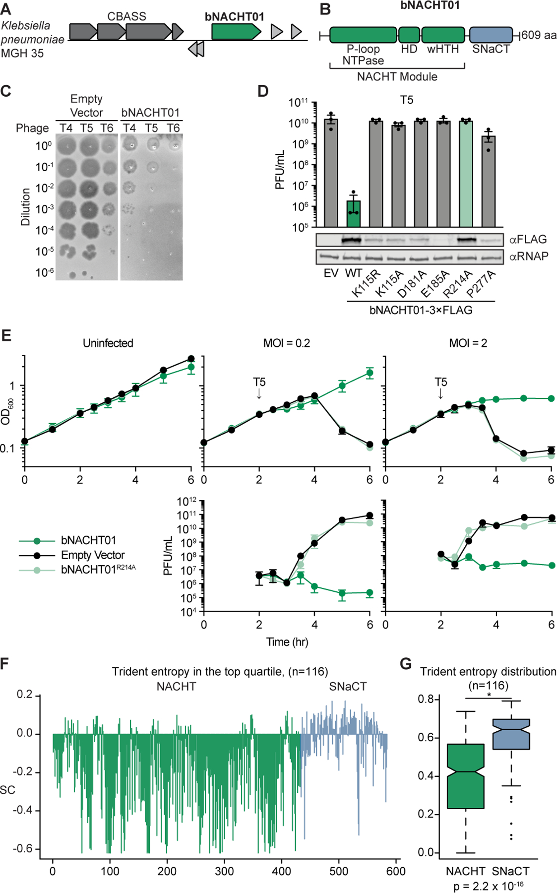
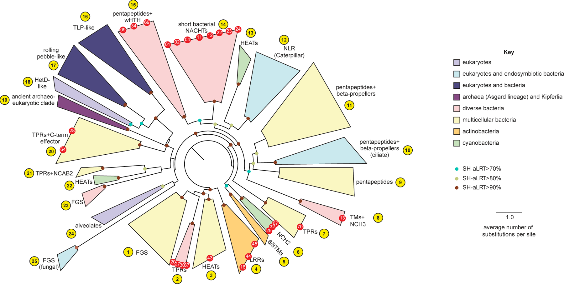
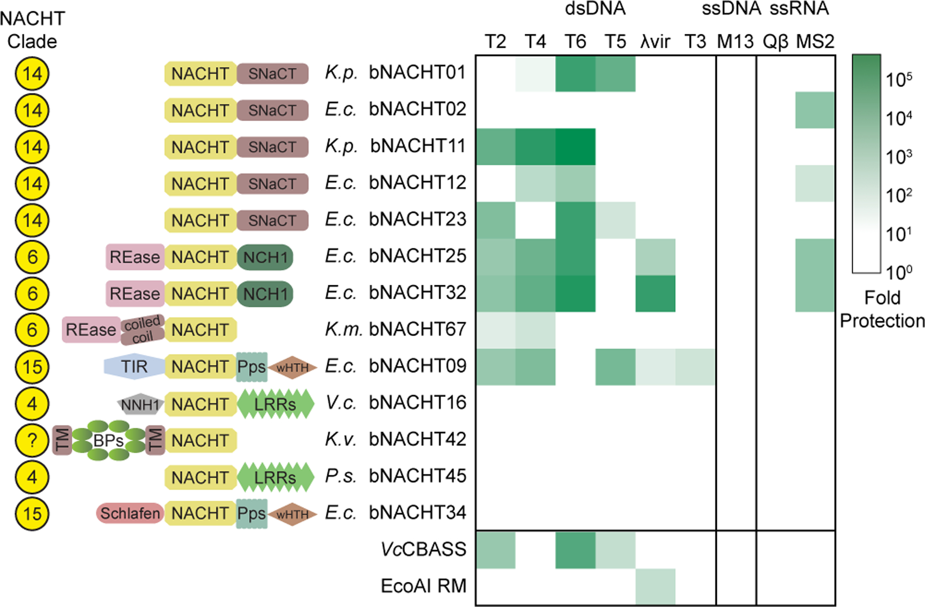
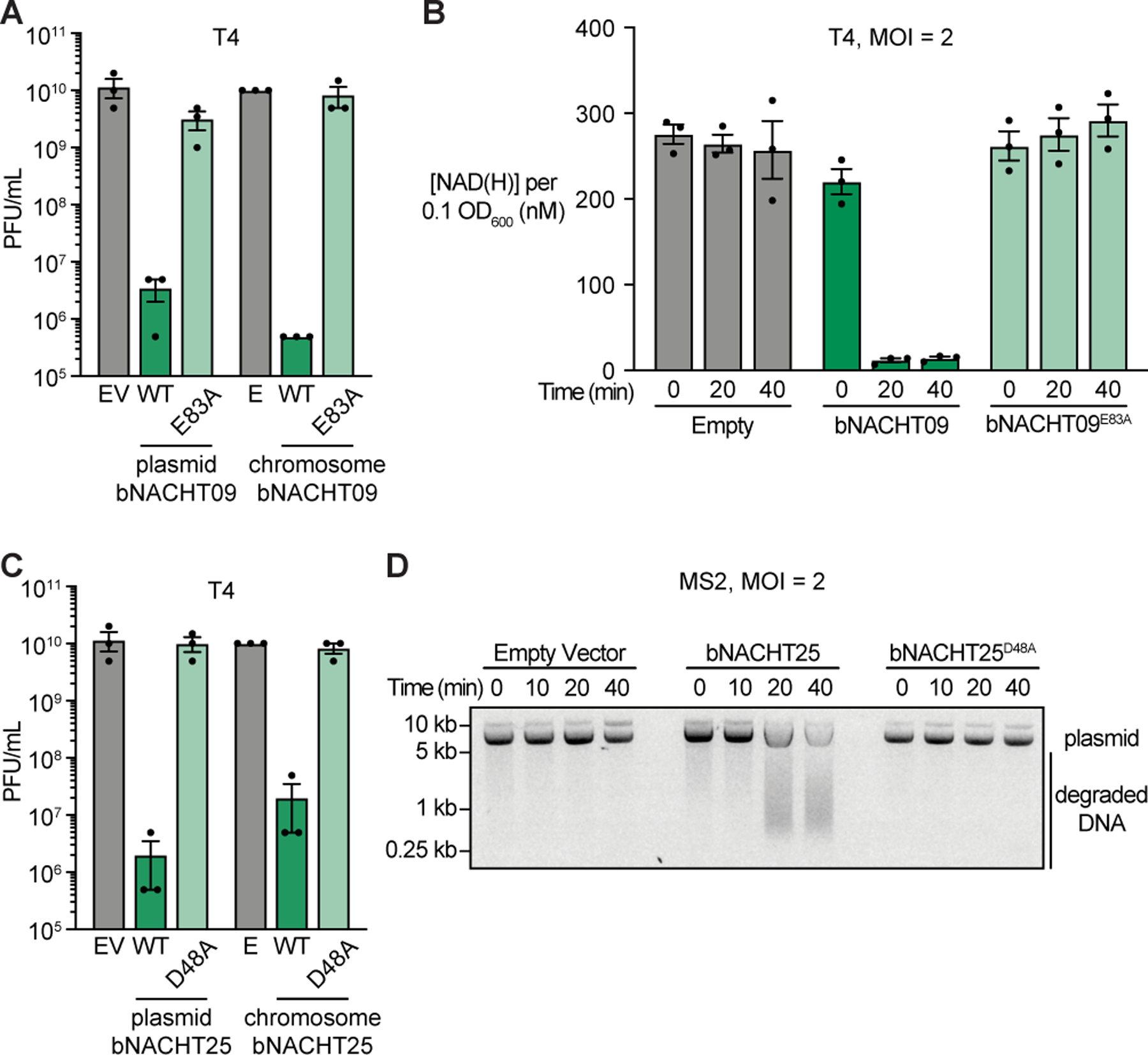
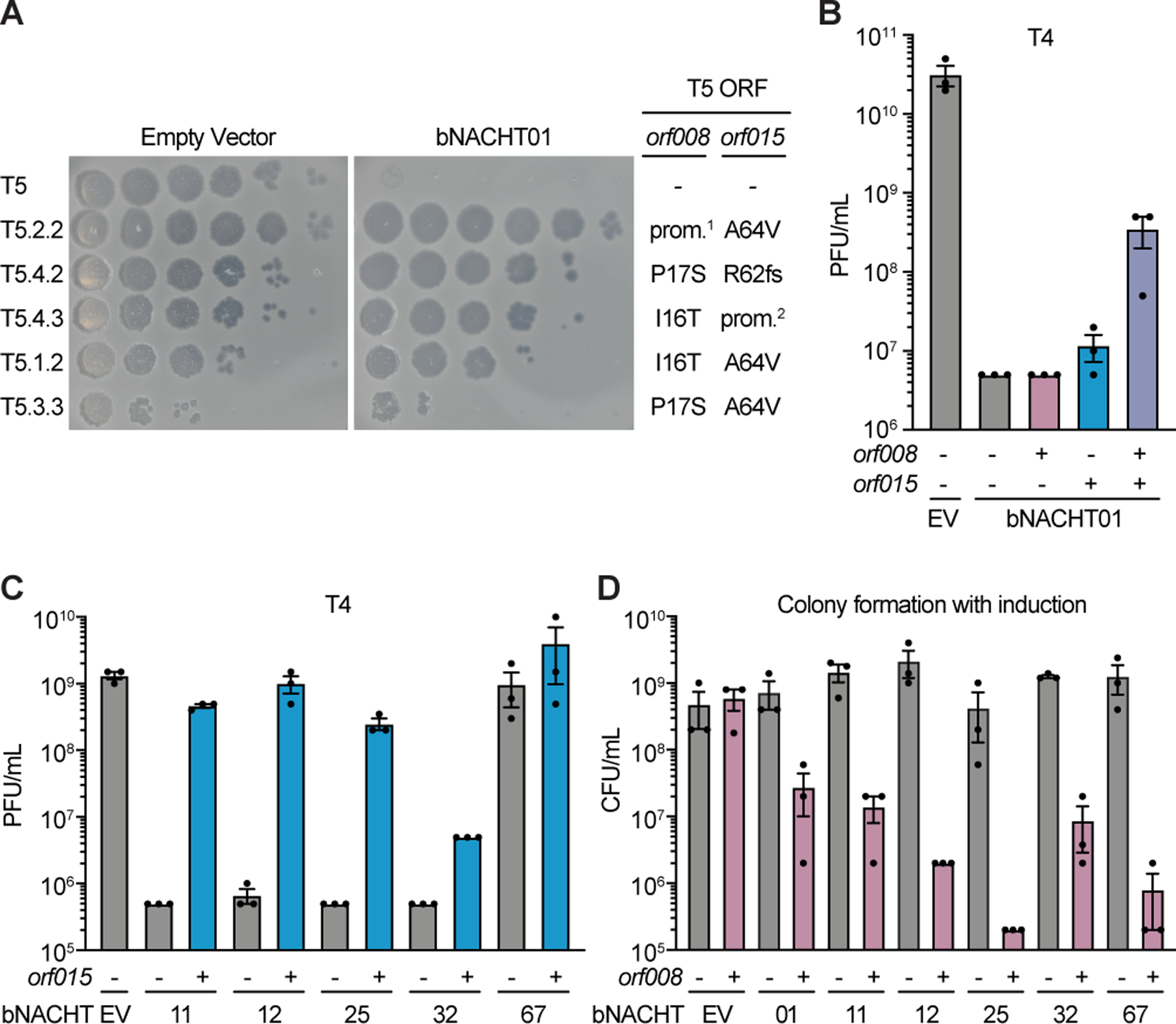
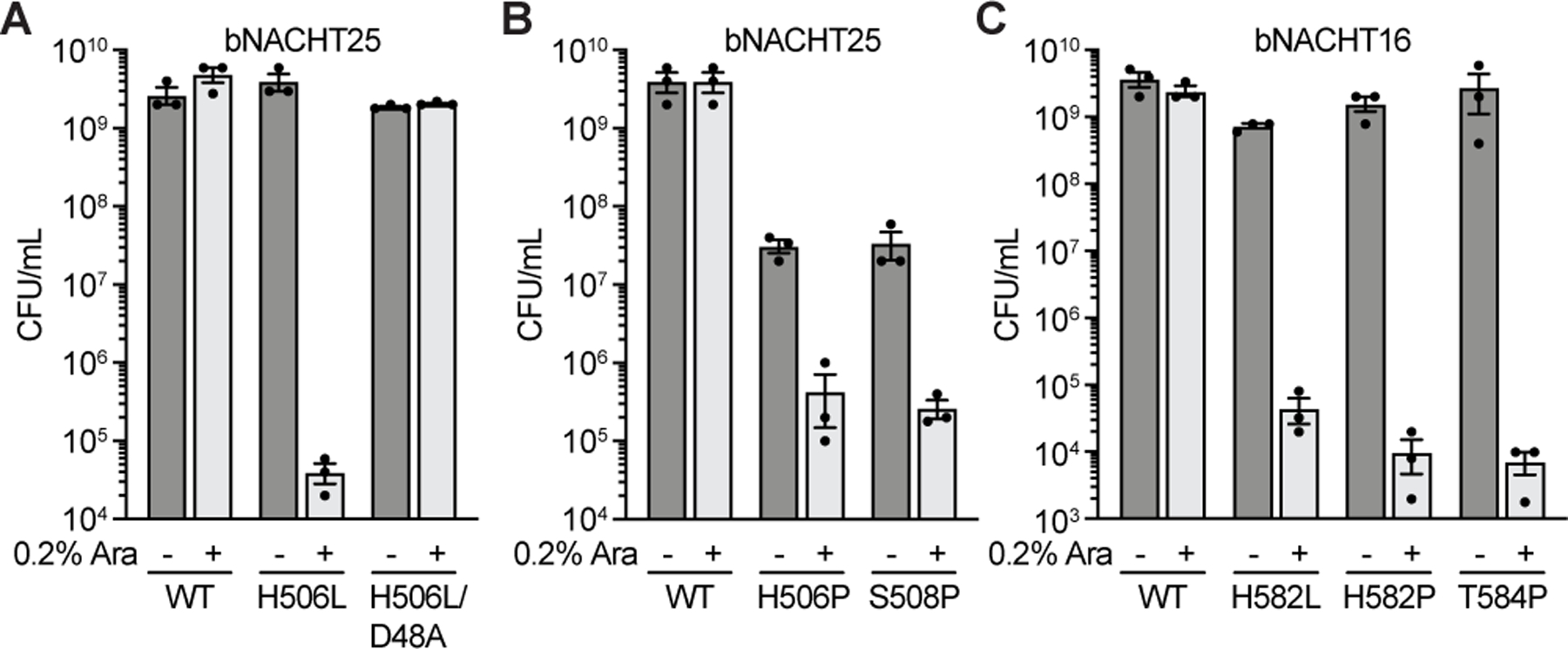
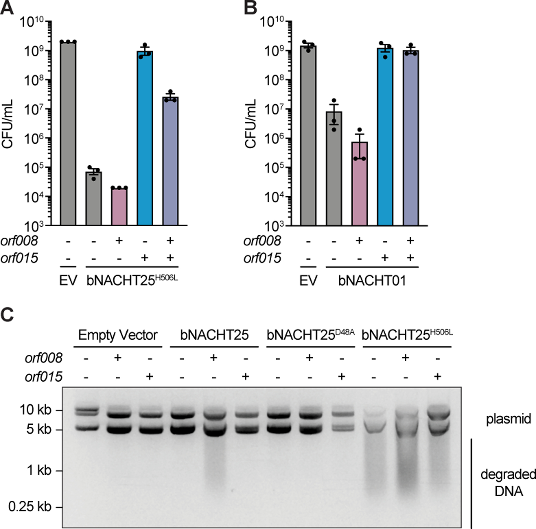
Bacteria use a wide range of immune pathways to counter phage infection. A subset of these genes shares homology with components of eukaryotic immune systems, suggesting that eukaryotes horizontally acquired certain innate immune genes from bacteria. Here, we show that proteins containing a NACHT module, the central feature of the animal nucleotide-binding domain and leucine-rich repeat containing gene family (NLRs), are found in bacteria and defend against phages. NACHT proteins are widespread in bacteria, provide immunity against both DNA and RNA phages, and display the characteristic C-terminal sensor, central NACHT, and N-terminal effector modules. Some bacterial NACHT proteins have domain architectures similar to the human NLRs that are critical components of inflammasomes. Human disease-associated NLR mutations that cause stimulus-independent activation of the inflammasome also activate bacterial NACHT proteins, supporting a shared signaling mechanism. This work establishes that NACHT module-containing proteins are ancient mediators of innate immunity across the tree of life.

Keywords: NACHT; NLR; STAND; bacteriophage; inflammasome; innate immunity; phage defense.


Conflict of interest statement

Declaration of interests The authors declare no competing interests.

Figures

Figure 1.
Figure 1.. A bacterial NACHT domain-containing protein is antiphage.
(A) Genome context of bNACHT01, which is located near a CBASS system in Klebsiella pneumoniae MGH 35. (B) Schematic of bNACHT01 (WP_015632533.1) protein domains, annotated by alignment to the NACHT module of NLRC4. The P-loop NTPase domain is also known as a nucleotide-binding domain (NBD), the helical domain (HD), and the winged helix-turn-helix (wHTH, also called WHD for winged helical domain) are indicated. See Figure S1 for a protein alignment of bNACHT01 with eukaryotic NACHT modules. (C) Efficiency of plating of indicated phages infecting E. coli expressing bNACHT01 or an empty vector (EV). Data are representative images of n = 3 biological replicates. (D) Above: Efficiency of plating of phage T5 infecting E. coli expressing the indicated genotype. Data represent the mean ± standard error of the mean (s.e.m.) of n = 3 biological replicates, shown as individual points. See Figure S1 for efficiency of plating of phage T4 and T6. Below: Western blot analysis of E. coli expressing empty vector or FLAG-tagged bNACHT01 of the indicated genotype. Representative image of n = 2 biological replicates. (E) Above: Growth curve of E. coli expressing the indicated plasmid. Arrows indicate the time each culture was infected with phage T5 at the indicated multiplicity of infection (MOI). Below: Efficiency of plating of the phage present in each sample at the indicated time points. Data represent the mean ± s.e.m. of n = 3 biological replicates. (F) Scaled Trident entropy (SC) values (see STAR Methods) for individual residues of bNACHT01-like proteins. The trident entropy for each column of the alignment, including both the NACHT module and SNACT domain, is scaled with respect to the top quartile. Positions with values greater than 0 are those with diversity in the top quartile. (G) Distribution of trident entropy (S) values across NACHT and SNaCT modules in bNACHT01-like proteins showing significantly different mean Trident entropy. Values were compared using a two-sample t-test.
Figure 2.
Figure 2.. NACHT module-containing proteins in bacteria are widespread and diverse.
A sequence-based phylogenetic tree of NACHT modules was generated using NACHT module-containing proteins from eukaryotes and prokaryotes. The NACHT module, not accessory domains, were used for tree building. Clades are color-coded based on the indicated key and numbered arbitrarily in yellow circles. Red dots indicate the bacterial NACHT proteins from each clade that were selected for analysis in this study. Bootstrap values are provided where applicable. See Figure S2 for bNACHT gene distribution, Figure S3 for representative domain architectures from each clade, and Table S4 for the most common domain architectures found in each clade. Additional details on genes used to construct the phylogenetic tree can be found in Table S2, and Table S1 contains a full list of all NACHT module-containing proteins identified. See Table S3 for tree topology tests used to validate proposed evolutionary relationships.
Figure 3.
Figure 3.. Bacterial NACHT proteins are antiphage.
Heat map of fold defense provided by the indicated bNACHT gene for a panel of diverse phages. E. coli expressing the indicated defense system was challenged with phages and fold defense was calculated for each defense system-phage pair by dividing the efficiency of plating (in PFU/mL) on empty vector by efficiency of plating on defense system-expressing bacteria. The NACHT clade, domain architecture, and species of origin for each bNACHT are shown. bNACHT genes displayed in this figure are a subset of the 27 candidates interrogated, selected based on their robust antiphage activity or the diversity of domain architectures sampled. Vibrio cholerae CBASS (VcCBASS) and E. coli UPEC-36 restriction modification system (EcoAI RM) were included as positive controls. Data represent the mean of n = 3 biological replicates. Escherichia coli (E.c.), Klebsiella michiganensis (K.m.), Klebsiella pneumoniae (K.p.), Klebsiella variicola (K.v.), Pseudomonas sp. LAIL14HWK12:I6 (P.s.), Vibrio campbellii (V.c.). Domain abbreviations as described in Figure S3. See Table S5 and Figures S4 and S5 for details on all 27 bNACHT genes analyzed. See Figure S6 for raw efficiency of plating data.
Figure 4.
Figure 4.. Bacterial NACHT effector modules are activated by phage.
(A) Efficiency of plating of phage T4 infecting E. coli expressing the indicated genotype from a low copy plasmid or from the chromosome. See Figure S5 for efficiency of plating data for phages T5 and T6. (B) Measurement of [NAD(H)] in each sample when normalized to an OD600 of 0.1 from E. coli expressing the indicated chromosomal genotype at the indicated time points after infection with phage T4. (C) Efficiency of plating of phage T4 infecting E. coli expressing the indicated genotype from a low copy plasmid or from the chromosome. See Figure S5 for efficiency of plating data for phages T5 and T6. For A–C, Empty (E) indicates E. coli with chromosomal expression of a Kanamycin resistance cassette and gfpmut3. Data represent the mean ± s.e.m. of n = 3 biological replicates, shown as individual points. (D) Visualization of plasmid integrity in E. coli expressing the indicated plasmid at the indicated time points after infection with MS2. Data are a representative image from n = 3 biological replicates.
Figure 5.
Figure 5.. Phage proteins modulate host immune responses.
(A) Efficiency of plating of wild-type or suppressor T5 phage when infecting E. coli expressing the indicated plasmid. Impact of orf008 and orf015 suppressor mutations are indicated. Data are representative images of n = 3 biological replicates. Wild-type alleles (−); mutations in the promoter region of orf008 (prom.); frame shift mutations at the indicated position (fs); and mutations deleting orf009–012 predicted to disrupt the promoter of orf015 (prom.) are indicated. See Table S6 for rates of suppressor phage isolation, suppressor mutations identified in orf008 and orf015, and a complete list of mutations identified. (B) Efficiency of plating of phage T4 when infecting E. coli expressing bNACHT01 or an empty vector (EV) on one plasmid and the indicated phage T5 gene(s) on a second plasmid. See Figure S5 for efficiency of plating of phage T6. (C) Efficiency of plating of phage T4 when infecting E. coli expressing the indicated bNACHT gene on one plasmid and phage T5 orf015 on a second plasmid. See Figure S7 for efficiency of plating of phages T2 and T6. (D) Quantification of colony formation of E. coli expressing the indicated bNACHT system on one plasmid and orf008 on a second plasmid. See Figure S7 for colony formation in the presence of orf015. For B–D, expression of orf008, orf015, and mCherry is IPTG-inducible. (−) symbols denote induction of an mCherry negative control. (+) symbols denote induction of the indicated phage gene. Data represent the mean ± s.e.m. of n = 3 biological replicates, shown as individual points.
Figure 6.
Figure 6.. Human disease-associated mutations hyperactivate bacterial NACHT proteins.
(A)–(B) Quantification of colony formation of E. coli expressing wild-type (WT) bNACHT25 or alleles with the indicated mutations. (C) Quantification of colony formation of E. coli expressing bNACHT16 with the indicated mutations. See Figure S7 for an alignment of NLRC4, bNACHT16, and bNACHT25. For A–C, gene expression was induced with arabinose. Symbols denote induction (+) or lack of induction (−). Data represent the mean ± s.e.m. of n = 3 biological replicates, shown as individual points.
Figure 7.
Figure 7.. Phage proteins alter the activity of hyperactive bacterial NACHT proteins.
(A)–(B) Quantification of colony formation of E. coli expressing a bNACHT gene with the indicated genotype on one plasmid and the indicated phage T5 gene(s) on a second plasmid. Data represent the mean ± s.e.m. of n = 3 biological replicates, shown as individual points. (C) Visualization of plasmid integrity in E. coli expressing a bNACHT gene with the indicated genotype on one plasmid and the indicated phage T5 gene on a second plasmid. For A–C, expression of orf008, orf015, and sfGFP was induced with IPTG. Symbols denote induction of an sfGFP negative control gene (−) or induction of the indicated phage gene (+). Expression of the indicated bNACHT gene or empty vector (EV) was arabinose-inducible. Data are an image representative of n = 3 biological replicates.

Comment in

References

    1. Bernheim A, and Sorek R (2020). The pan-immune system of bacteria: antiviral defence as a community resource. Nat. Rev. Microbiol 18, 113–119. 10.1038/s41579-019-0278-2. - DOI - PubMed
    1. Doron S, Melamed S, Ofir G, Leavitt A, Lopatina A, Keren M, Amitai G, and Sorek R (2018). Systematic discovery of antiphage defense systems in the microbial pangenome. Science 359, eaar4120. 10.1126/science.aar4120. - DOI - PMC - PubMed
    1. Gao L, Altae-Tran H, Böhning F, Makarova KS, Segel M, Schmid-Burgk JL, Koob J, Wolf YI, Koonin EV, and Zhang F (2020). Diverse enzymatic activities mediate antiviral immunity in prokaryotes. Science 369, 1077–1084. 10.1126/science.aba0372. - DOI - PMC - PubMed
    1. Rousset F, Depardieu F, Miele S, Dowding J, Laval A-L, Lieberman E, Garry D, Rocha EPC, Bernheim A, and Bikard D (2022). Phages and their satellites encode hotspots of antiviral systems. Cell Host Microbe 30, 740–753.e5. 10.1016/j.chom.2022.02.018. - DOI - PMC - PubMed
    1. Vassallo CN, Doering CR, Littlehale ML, Teodoro GIC, and Laub MT (2022). A functional selection reveals previously undetected anti-phage defence systems in the E. coli pangenome. Nat. Microbiol 7, 1568–1579. 10.1038/s41564-022-01219-4. - DOI - PMC - PubMed

Publication types

LinkOut - more resources