

Thermal inkjet bioprinting drastically alters cell phenotype
- PMID: 37160133
- PMCID: PMC10399642
- DOI: 10.1088/1758-5090/acd3b3
Thermal inkjet bioprinting drastically alters cell phenotype
Abstract
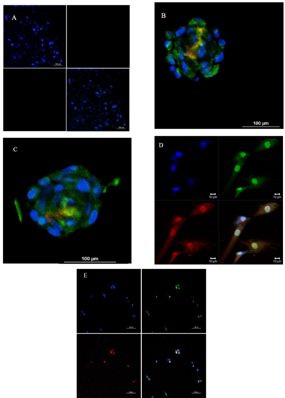
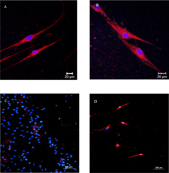
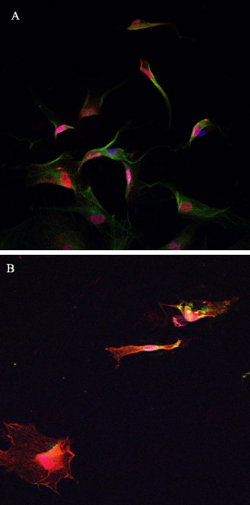
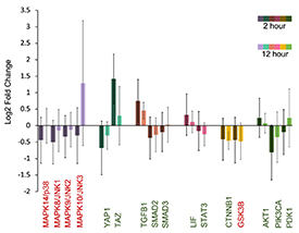
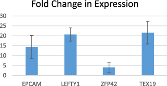
Since the first description of inkjet bioprinting of cells in 2003, quantifying the input and measuring the output of the printers has been the hallmark of the field of bioprinting, as it is virtually impossible to characterize cells that are inside the printing orifices or extrusion needles. We will describe here some recent discoveries of cell behavior due to inkjet bioprinting. Primary and immortalized adult dermal fibroblasts were expanded for 2-3 passages upon receiving. The cells were harvested, resuspended in PBS, and bioprinted into a 96-well plate with pluriSTEM media. Cells were then transferred either into precoated 96-well plates or 20µl drops were pipetted for hanging drop culture. IPC differentiation protocols were applied and the induction was begun approximately 45 min after printing. When differentiating aggregates, the initiation happened 45 min after the aggregates were transferred into the 96 wells. Standard immunostaining and RNA sequencing (RNA-Seq) were used to analyze the cell phenotypes. Preliminary results indicate that all cells expressed the three pluripotency markers oct-4, nanog, and sox-2. After applying a cardiomyocyte differentiation protocol, the cells stained positively for troponin-3. The cells also elongated and became more cardiomyocyte-like in their morphology. We analyzed bulk RNA seq data and our preliminary results show upregulation of some genes that have been implicated as stem cell markers: EPCAM, LEFTY1, ZFP42, and TEX19. In addition, differential expression of genes associated with pluripotency-relevant pathways shows some pathways are off like the MAPK/p38, MAPK/JNK1-3 which is expected for a pluripotent state. We also have data supporting the activation of the hippo pathway with transcriptional co-activator with PDZ binding motif (TAZ) highly upregulated and yes-associated protein staining the cell body. In addition, GSK3B is off and TGFB1, LIF/PIK3, and AKT1 are on as expected for pluripotency. Examining the gene network of upregulated genes, one can clearly distinguish the pivotal role of FOS, FOXO1, and PIK3 all related to pluripotency. Bioprinted fibroblasts will at least temporarily adopt a more primitive or dedifferentiated state, reminiscent of pluripotency. While immunochemistry shows the classic transcription factors required for pluripotency, gene expression shows a more nuanced picture of the transformations that occur upon printing. Understanding these transformations, even if temporary will be crucial when trying to build tissues using bioprinting technologies.
Keywords: differentiation; pluripotency; thermal inkjet bioprinting.
© 2023 IOP Publishing Ltd.
Figures

References
-
- Campbell A, Mohl J E, Gutierrez D A, Varela-Ramirez A, Boland T. Thermal bioprinting causes ample alterations of expression of LUCAT1, IL6, CCL26, and NRN1L genes and massive phosphorylation of critical oncogenic drug resistance pathways in breast cancer cells. Front. Bioeng. Biotechnol. 2020;8:82. doi: 10.3389/fbioe.2020.00082. - DOI - PMC - PubMed
Publication types
MeSH terms
Substances
Grants and funding
LinkOut - more resources
Full Text Sources
Other Literature Sources
Research Materials
Miscellaneous