. 2023 May 9;13(1):7491.
doi: 10.1038/s41598-023-34749-w.

Acute exercise as a modifier of neocortical plasticity and aperiodic activity in the visual cortex

Affiliations

Acute exercise as a modifier of neocortical plasticity and aperiodic activity in the visual cortex

Claire J Cadwallader et al. Sci Rep. .

Abstract

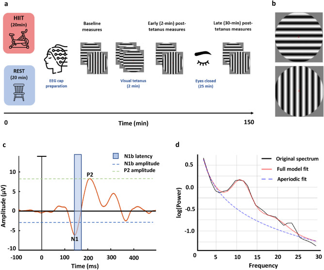
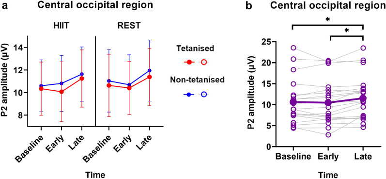
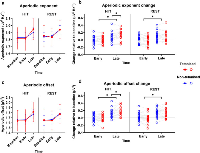
Long-term potentiation (LTP) is a form of neuroplasticity commonly implicated in mechanistic models of learning and memory. Acute exercise can boost LTP in the motor cortex, and is associated with a shift in excitation/inhibition (E:I) balance, but whether this extends to other regions such as the visual cortex is unknown. We investigated the effect of a preceding bout of exercise on LTP induction and the E:I balance in the visual cortex using electroencephalography (EEG). Young adults (N = 20, mean age = 24.20) engaged in 20 min of high-intensity interval training (HIIT) exercise and rest across two counterbalanced sessions. LTP was induced using a high frequency presentation of a visual stimulus; a "visual tetanus". Established EEG markers of visual LTP, the N1b and P2 component of the visual evoked potential, and an EEG-derived measure of the E:I balance, the aperiodic exponent, were measured before and after the visual tetanus. As expected, there was a potentiation of the N1b following the visual tetanus, with specificity to the tetanised stimulus, and a non-specific potentiation of the P2. These effects were not sensitive to a preceding bout of exercise. However, the E:I balance showed a late shift towards inhibition following the visual tetanus. A preceding bout of exercise resulted in specificity of this E:I balance shift to the tetanised stimulus, that was not seen following rest. This novel finding suggests a possible exercise-induced tuning of the visual cortex to stimulus details following LTP induction.


Conflict of interest statement

The authors declare no competing interests.

Figures

Figure 1
Figure 1
(a) Schematic of the experimental procedure. (b) The visual stimuli used in the paradigm consisted of a vertical and horizontal circular sine grating. Only one of these stimuli were presented during the visual tetanus, while the other served as the control stimulus. (c) An example VEP taken from an individual participant in the baseline measurement block, HIIT condition. The N1b was measured as the mean amplitude of the section of the VEP extending from the peak of the N1, to the midpoint between the N1 and the P2 peaks. The peak amplitude of the P2 was also recorded. (d) An example of the FOOOF model fit for an individual participant in the baseline measurement block.
Figure 2
Figure 2
(a) N1b amplitude in the left LO region. (b) N1b amplitude in the right LO region. (c) and (d) N1b amplitude change relative to baseline. Individual data points are plotted. Data is collapsed across levels of Session. *p < .05; error bars show 95% confidence interval.
Figure 3
Figure 3
Scalp maps display the VEP amplitude change relative to baseline, for the tetanised stimulus minus the non-tetanised stimulus. Note that in the early post-tetanus time there appears to be a larger increase in negativity in the HIIT session compared to the rest session. Cluster-based permutation testing did not reveal significant effects.
Figure 4
Figure 4
(a) P2 amplitude in the central occipital region. (b) P2 amplitude change across time. Individual data points are plotted. Data is collapsed across levels of Session and Stimulus. *p < .05; error bars show 95% confidence interval.
Figure 5
Figure 5
Aperiodic exponent and offset results in the central occipital region (electrode POz). (a) Aperiodic exponent. (b) Change in the aperiodic exponent relative to baseline. Individual data points are plotted. (c) Aperiodic offset. (d) Change in the aperiodic offset relative to baseline. Individual data points are plotted. *p < .05; error bars show 95% confidence interval.

References

    1. Bliss TV, Collingridge GL. A synaptic model of memory: Long-term potentiation in the hippocampus. Nature. 1993;361:31. doi: 10.1038/361031a0. - DOI - PubMed
    1. Takeuchi T, Duszkiewicz AJ, Morris RG. The synaptic plasticity and memory hypothesis: Encoding, storage and persistence. Philos. Trans. R. Soc. B. 2014;369:20130288. doi: 10.1098/rstb.2013.0288. - DOI - PMC - PubMed
    1. Kirkwood A, Rioult MG, Bear MF. Experience-dependent modification of synaptic plasticity in visual cortex. Nature. 1996;381:526–528. doi: 10.1038/381526a0. - DOI - PubMed
    1. Maren S. Long-term potentiation in the amygdala: a mechanism for emotional learning and memory. Trends Neurosci. 1999;22:561–567. doi: 10.1016/S0166-2236(99)01465-4. - DOI - PubMed
    1. Rioult-Pedotti M-S, Friedman D, Donoghue JP. Learning-induced LTP in neocortex. Science. 2000;290:533–536. doi: 10.1126/science.290.5491.533. - DOI - PubMed

Publication types