

Age-related changes in human skeletal muscle microstructure and architecture assessed by diffusion-tensor magnetic resonance imaging and their association with muscle strength
- PMID: 37162031
- PMCID: PMC10352548
- DOI: 10.1111/acel.13851
Age-related changes in human skeletal muscle microstructure and architecture assessed by diffusion-tensor magnetic resonance imaging and their association with muscle strength
Abstract
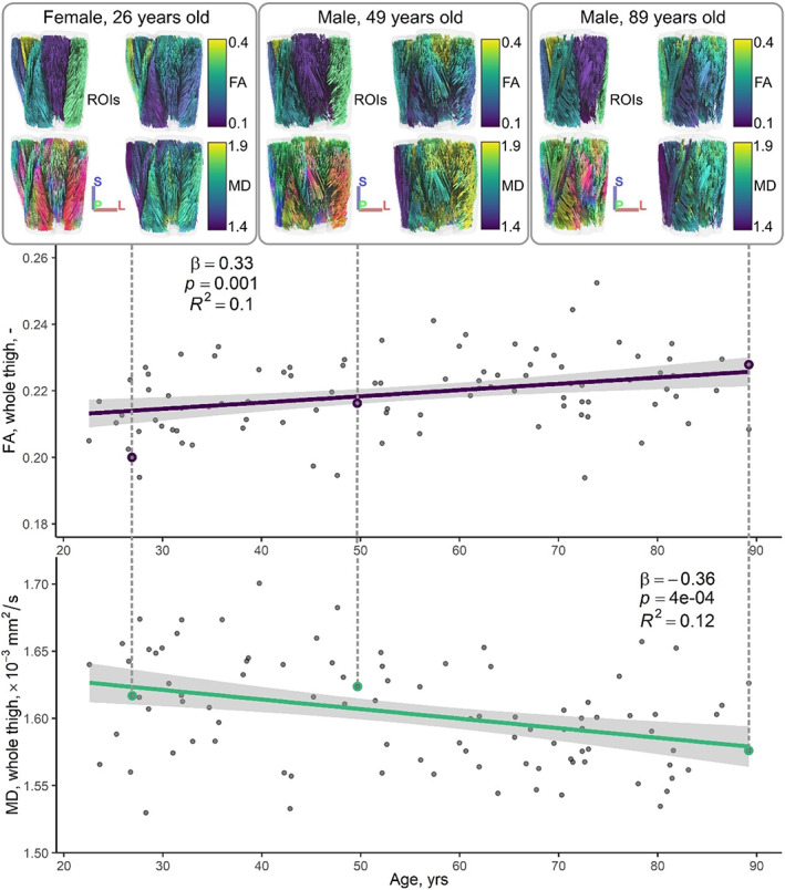
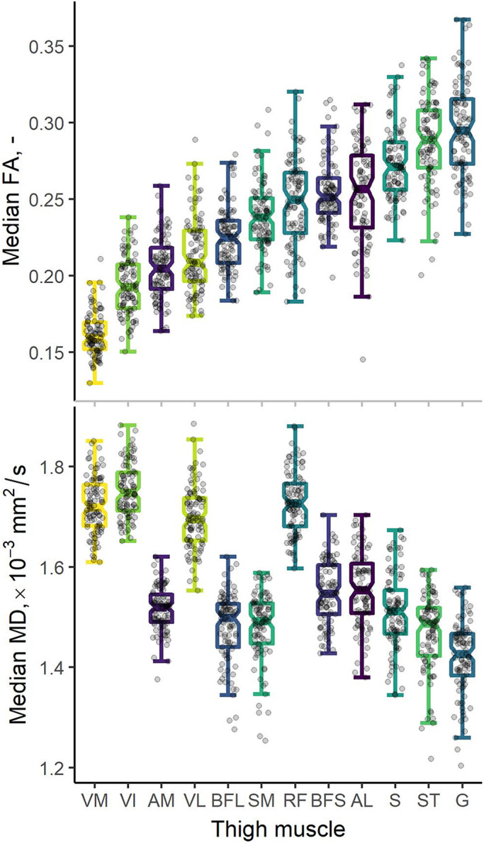
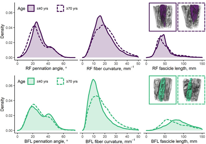
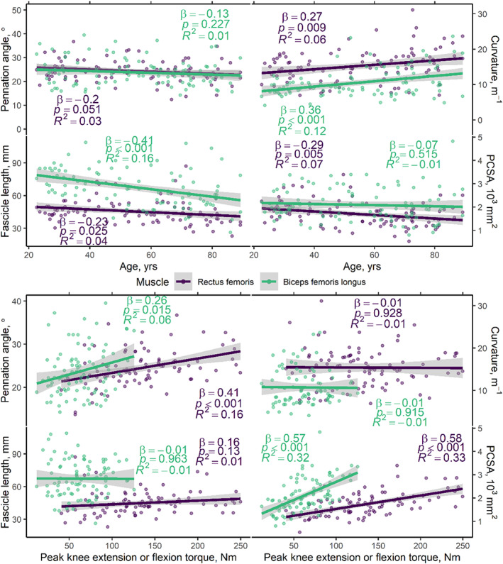
Diffusion-tensor magnetic resonance imaging (DT-MRI) offers objective measures of muscle characteristics, providing insights into age-related changes. We used DT-MRI to probe skeletal muscle microstructure and architecture in a large healthy-aging cohort, with the aim of characterizing age-related differences and comparing these to muscle strength. We recruited 94 participants (43 female; median age = 56, range = 22-89 years) and measured microstructure parameters-fractional anisotropy (FA) and mean diffusivity (MD)-in 12 thigh muscles, and architecture parameters-pennation angle, fascicle length, fiber curvature, and physiological cross-sectional area (PCSA)-in the rectus femoris (RF) and biceps femoris longus (BFL). Knee extension and flexion torques were also measured for comparison to architecture measures. FA and MD were associated with age (β = 0.33, p = 0.001, R2 = 0.10; and β = -0.36, p < 0.001, R2 = 0.12), and FA was negatively associated with Type I fiber proportions from the literature (β = -0.70, p = 0.024, and R2 = 0.43). Pennation angle, fiber curvature, fascicle length, and PCSA were associated with age in the RF (β = -0.22, 0.26, -0.23, and -0.31, respectively; p < 0.05), while in the BFL only curvature and fascicle length were associated with age (β = 0.36, and -0.40, respectively; p < 0.001). In the RF, pennation angle and PCSA were associated with strength (β = 0.29, and 0.46, respectively; p < 0.01); in the BFL, only PCSA was associated with strength (β = 0.43; p < 0.001). Our results show skeletal muscle architectural changes with aging and intermuscular differences in the microstructure. DT-MRI may prove useful for elucidating muscle changes in the early stages of sarcopenia and monitoring interventions aimed at preventing age-associated microstructural changes in muscle that lead to functional impairment.
Keywords: aging; diffusion tensor imaging; muscle strength; sarcopenia; skeletal muscle fibers; thigh.
© 2023 The Authors. Aging Cell published by Anatomical Society and John Wiley & Sons Ltd. This article has been contributed to by U.S. Government employees and their work is in the public domain in the USA.
Conflict of interest statement
The authors declare no competing financial interests.
Figures

References
-
- Adelnia, F. , Cameron, D. , Bergeron, C. M. , Fishbein, K. W. , Spencer, R. G. , Reiter, D. A. , & Ferrucci, L. (2019). The role of muscle perfusion in the age‐associated decline of mitochondrial function in healthy individuals. Frontiers in Physiology, 10, 427. 10.3389/fphys.2019.00427 - DOI - PMC - PubMed
-
- Bischoff‐Ferrari, H. A. , Orav, J. , Kanis, J. A. , Rizzoli, R. , Schlögl, M. , Staehelin, H. , Willett, W. C. , & Dawson‐Hughes, B. (2015). Comparative performance of current definitions of sarcopenia against the prospective incidence of falls among community‐dwelling seniors age 65 and older. Osteoporosis International, 26(12), 2793–2802. 10.1007/s00198-015-3194-y - DOI - PubMed
Publication types
MeSH terms
Substances
Grants and funding
LinkOut - more resources
Full Text Sources
Medical
