Review
. 2023 Jul;10(20):e2207334.
doi: 10.1002/advs.202207334. Epub 2023 May 10.

An Update on Adipose-Derived Stem Cells for Regenerative Medicine: Where Challenge Meets Opportunity

Affiliations
Review

An Update on Adipose-Derived Stem Cells for Regenerative Medicine: Where Challenge Meets Opportunity

Yi Qin et al. Adv Sci (Weinh). 2023 Jul.

Abstract

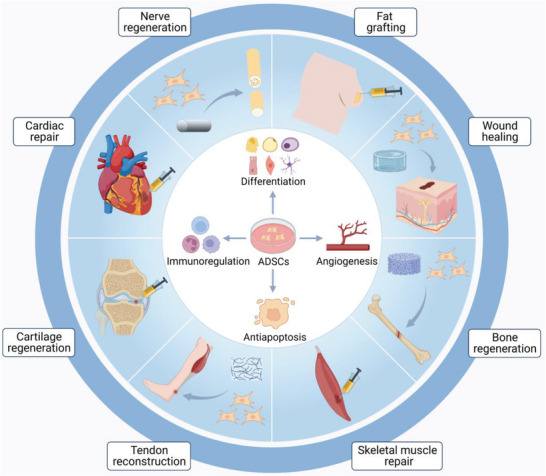
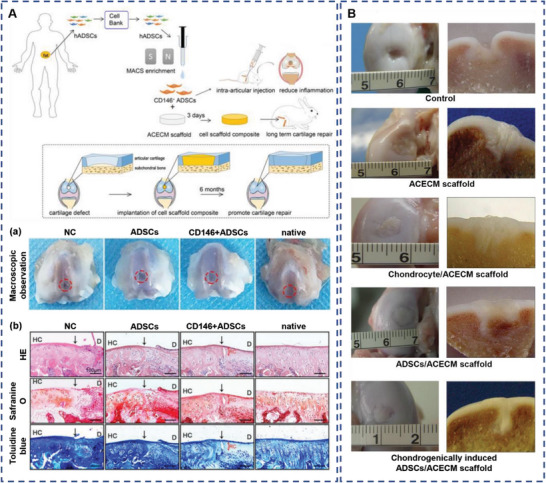
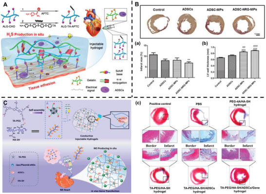
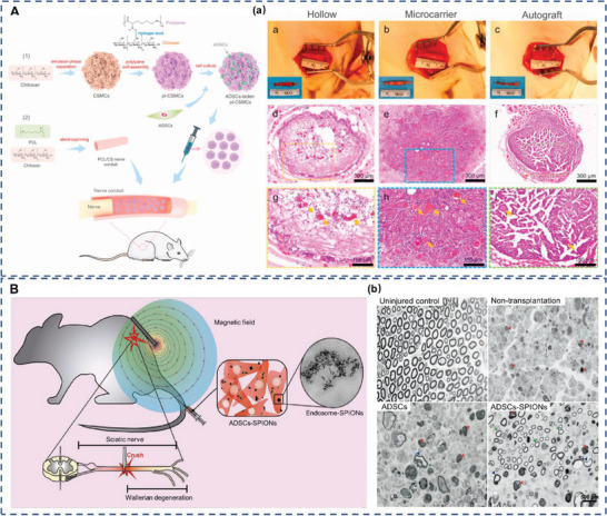
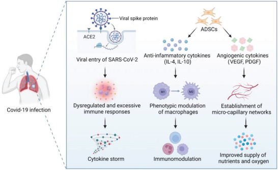
Over the last decade, adipose-derived stem cells (ADSCs) have attracted increasing attention in the field of regenerative medicine. ADSCs appear to be the most advantageous cell type for regenerative therapies owing to their easy accessibility, multipotency, and active paracrine activity. This review highlights current challenges in translating ADSC-based therapies into clinical settings and discusses novel strategies to overcome the limitations of ADSCs. To further establish ADSC-based therapies as an emerging platform for regenerative medicine, this review also provides an update on the advancements in this field, including fat grafting, wound healing, bone regeneration, skeletal muscle repair, tendon reconstruction, cartilage regeneration, cardiac repair, and nerve regeneration. ADSC-based therapies are expected to be more tissue-specific and increasingly important in regenerative medicine.

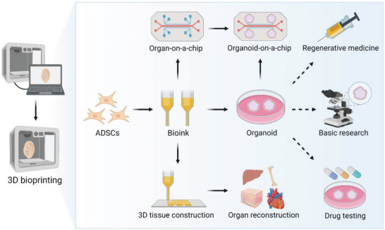
Keywords: 3D bioprinting; adipose-derived stem cells; cell-free therapy; clinical application; regenerative medicine.


Conflict of interest statement

The authors declare no conflict of interest.

Figures

Figure 1
Figure 1
Schematic representation of adipose‐derived stem cells (ADSCs) for regenerative therapies. ADSCs serve as an ideal candidate for regenerative medicine owing to their ability to differentiate into multilineages, facilitate angiogenesis, suppress apoptosis, and participate in immunoregulation. Numerous preclinical studies and clinical trials have demonstrated the therapeutic potential of ADSCs in fat grafting, wound healing, bone regeneration, skeletal muscle repair, tendon reconstruction, cartilage regeneration, cardiac repair, and nerve regeneration.
Figure 2
Figure 2
Schematic illustration of the biogenesis and therapeutic potential of adipose‐derived stem cell‐derived exosomes (ADSC‐Exos). The process begins with endocytosis and the formation of early endosomes, which can mature into multivesicular bodies (MVBs) with the accumulation of intraluminal vesicles (ILVs). When MVBs fuse with the plasma membrane, ILVs are released as exosomes. ADSC‐Exos contain tetraspanins (CD9, CD63, and CD81), biogenesis‐related proteins (TSG101 and ALIX), heat shock proteins (HSP70 and HSP90) and nucleic acids (DNA, mRNA, miRNA, and rRNA). ADSC‐Exos exhibit therapeutic potential by enhancing cell proliferation, cell migration, and angiogenesis, participating in immunoregulation, and suppressing apoptosis.
Figure 3
Figure 3
Application of 3D bioprinting for organoid production and organ reconstruction. Bioinks containing ADSCs are printed as organoids, organ‐on‐a‐chip systems, and organ‐level 3D tissues. Organoids have been widely used in basic research, drug testing, and regenerative medicine. The Organoids‐on‐a‐chip technology combines the best features of organoids and organ‐on‐a‐chip systems, becoming a promising approach for recapitulating the key aspects of human physiology and pathophysiology.
Figure 4
Figure 4
Procedures for applying adipose‐derived stem cells (ADSCs). Green arrows indicate the isolation of ADSCs. Adipose tissues are harvested from healthy donors or patients using liposuction and processed using enzymatical or mechanical procedures to separate stromal vascular fraction (SVF). Then ADSCs are obtained through seeding SVF in culture. Yellow arrows indicate the clinical use of ADSCs. According to the damaged organs of patients, ADSCs can be directly administered, preconditioned to enhance therapeutic effects, or combined with biomaterials for tissue engineering. Blue arrows indicate the storage of ADSCs. Cryopreserved ADSCs can be transported to medical institutions at a distance or ADSCs banks for long‐term storage until needed.
Figure 5
Figure 5
Adipose‐derived stem cells (ADSCs) increase the survival rate of fat grafts. A) Schematic illustration of the co‐transplantation of fat grafts with phosphate‐buffered saline (PBS), ADSCs transfected with Luciferase (ADSCmodLuc) and ADSCs transfected with VEGF modRNA (ADSCmodVEGF). (a) Representative gross morphological images of fat grafts at 15, 30 and 90 d following transplantation. (b) Representative gross morphological assessment and hematoxylin and eosin (H&E) staining of extracted fat grafts at 1‐week post‐implantation. Yellow arrows indicate new blood vessel formation within the fat grafts. Reproduced under terms of the CC‐BY license.[ 92 ] Copyright 2020, The Authors, Published by Springer Nature. B) Schematic illustration of isolation of CD34+CD146+, CD34+CD146−, and CD34+ unfractionated (UF) ADSCs by fluorescence‐activated cell sorting (FACS). (c) Micro‐computed tomography of fat grafts at 8 weeks post‐transplantation. Reproduced under terms of the CC‐BY license.[ 93 ] Copyright 2020, Oxford University Press. C) Magnetic resonance imaging (MRI) of the same areas of healthy participants before implantation and day 0 and 121 after implantation. Reproduced with permission.[ 94 ] Copyright 2013, Elsevier.
Figure 6
Figure 6
Adipose‐derived stem cells (ADSCs) promote wound healing. A) Representative images of the wounds with phosphate‐buffered saline (PBS), platelet‐rich plasma (PRP), ADSCs and ADSCs+PRP treatment. (a) Hematoxylin and eosin (H&E) staining on day 14 following surgery. Reproduced under terms of the CC‐BY license.[ 102 ] Copyright 2021, The Authors, Published by Springer Nature. B) Schematic illustration of an ovine burn wound model. (b) Representative images of the wounds on day 15. (c) Western blot staining for vascular endothelial growth factor (VEGF). Reproduced with permission.[ 105 ] Copyright 2020, Oxford University Press. C) Representative images of the wounds with PBS, monolayer ADSCs and ADSCs sheet treatment. (d) H&E staining of the wound tissue. Reproduced with permission.[ 106 ] Copyright 2018, Elsevier.
Figure 7
Figure 7
Clinical application of adipose‐derived stem cells (ADSCs) for cranio‐maxillofacial hard‐tissue regeneration and external anal sphincter injury treatment. A) Intraoperative photograph and computed tomography (CT) of frontal sinus regeneration (a), cranial repair (b), mandibular reconstruction (c), and nasal septal repair (d) using ADSCs. Reproduced with permission.[ 135 ] Copyright 2014, Oxford University Press. B) Images of endorectal sonography (e), the area occupied by the muscle (f), the median percentage of area occupied by the muscle (g) and sample electromyography (h) of the control group (left) and the ADSCs group (right) at 2 months after surgery, arrows indicate fibrous tissue. Reproduced under terms of the CC‐BY license.[ 147 ] Copyright 2017, The Authors, Published by Springer Nature.
Figure 8
Figure 8
Spatially control of adipose‐derived stem cells (ADSCs) differentiation mimicking tendon‐to‐bone attachment. A) The hierarchical structure of the tendon‐to‐bone attachment. (a) Schematic of transitional tissue. (b) Transmission electron microscopy (TEM) of the mineral gradient. (c) Raman microprobe results of mineral content. Color dots indicating mineral content (red, low; blue, high). (d) TEM‐electron energy loss spectroscopy image (red, mineral; green, tropocollagen). Reproduced with permission.[ 154 ] Copyright 2017, Springer Nature. B) Fabrication of a platelet‐derived growth factor (PDGF) gradient aligned nanofiber surface by controlling oxidative polymerization of dopamine. (e) The relative percentage of immobilized PDGF at different positions on a PDGF gradient nanofiber. Reproduced with permission.[ 155 ] Copyright 2018, Elsevier.
Figure 9
Figure 9
Adipose‐derived stem cells (ADSCs) combined with articular cartilage extracellular matrix (ACECM) promote cartilage regeneration. A) Overview of experimental design. (a) Representative images of defects treated with phosphate‐buffered saline (PBS) (negative control), ADSCs, CD146+ ADSCs, and the sham‐operated group at 2 weeks following surgery. (b) Histological analysis of the defected area using hematoxylin and eosin (H&E), safranin O, and toluidine blue. Black solid arrows denote the repair interface. Red solid arrows denote the depth of the repaired cartilage. HC, host cartilage; D, defect area; RC, repaired cartilage. Reproduced under terms of the CC‐BY license.[ 175 ] Copyright 2022, Ivyspring International Publisher. B) General joint observations (left) and longitudinal section (right) of the cartilage regeneration at 3 months after surgery. Reproduced with permission.[ 176 ] Copyright 2020, John Wiley and Sons.
Figure 10
Figure 10
Adipose‐derived stem cells (ADSCs) facilitate cardiac repair. A) Schematic illustration of the ADSCs‐loaded conductive hydrogen sulfide‐releasing hydrogel. Reproduced with permission.[ 185 ] Copyright 2019, American Chemical Society. B) Sirius red staining of the infarcted hearts at 3 months after different treatments. Quantification of infarct size (a) and the left ventricular (LV) wall thickness (b). Reproduced with permission.[ 186 ] Copyright 2017, Elsevier. C) Schematic illustration of the construction of an injectable conductive hydrogel encapsulating plasmid DNA‐eNOs and ADSCs. (c) Masson's trichrome staining of cardiac structures. Reproduced with permission.[ 187 ] Copyright 2018, Elsevier.
Figure 11
Figure 11
Adipose‐derived stem cells (ADSCs) promote peripheral nerve regeneration. A) Schematic of the preparation process of the ADSCs‐laden polylysine‐decorated macroporous chitosan microcarriers (pl‐CSMCs) and their application in nerve repair. (a) Representative images and hematoxylin and eosin (H&E) staining of the regenerated nerve tissue at the midportion of the nerve guide conduits (NGCs) or autograft 12 weeks after implantation. Yellow arrows indicate blood vessels. Reproduced under terms of the CC‐BY license.[ 202 ] Copyright 2021, The Authors, Published by Elsevier. B) Schematic illustration of magnetic targeted ADSCs therapy. (b) Analysis of semithin sections of axonal bundles in uninjured control sciatic nerve and the distal stump of non‐transplanted, transplanted with ADSCs and ADSCs‐superparamagnetic iron oxide nanoparticles (SPIONs) transplanted group at 7 days post‐injury. Arrows indicate intact axons; arrowheads indicate irregular axons; asterisks indicate myelin and axon debris. Reproduced with permission.[ 203 ] Copyright 2021, Elsevier.
Figure 12
Figure 12
Schematic illustration of adipose‐derived stem cells (ADSCs) for treating coronavirus disease 2019 (COVID‐19). Severe acute respiratory syndrome coronavirus 2 (SARS‐CoV‐2) can enter cells through angiotensin‐converting enzyme 2 (ACE2), thus lung tissues with high expression of ACE2 become the main targets for the novel coronavirus to invade. Dysregulated and excessive immune responses arising from virus infection cause a cytokine storm. ADSCs can secrete anti‐inflammatory cytokines that facilitate the phenotypic modulation of macrophages and immunomodulation. In addition, angiogenic cytokines secreted by ADSCs can enhance the establishment of micro‐capillary networks, which ensure the supply of nutrients and oxygen. These activities may synergistically promote fibrotic lung tissue remodeling.

Similar articles

Cited by

References

    1. Cossu G., Birchall M., Brown T., De Coppi P., Culme‐Seymour E., Gibbon S., Hitchcock J., Mason C., Montgomery J., Morris S., Muntoni F., Napier D., Owji N., Prasad A., Round J., Saprai P., Stilgoe J., Thrasher A., Wilson J., Lancet 2018, 391, 883. - PubMed
    1. Karagiannis P., Takahashi K., Saito M., Yoshida Y., Okita K., Watanabe A., Inoue H., Yamashita J. K., Todani M., Nakagawa M., Osawa M., Yashiro Y., Yamanaka S., Osafune K., Physiol. Rev. 2019, 9, 79. - PubMed
    1. Rowe R. G., Daley G. Q., Nat. Rev. Genet. 2019, 20, 377. - PMC - PubMed
    1. Erices A., Conget P., Minguell J. J., Br. J. Haematol. 2000, 109, 235. - PubMed
    1. Igura K., Zhang X., Takahashi K., Mitsuru A., Yamaguchi S., Takashi T. A., Cytotherapy 2004, 6, 543. - PubMed

Publication types

LinkOut - more resources