. 2023 Jun 15;91(6):e0003123.
doi: 10.1128/iai.00031-23. Epub 2023 May 10.

Mucins Shed from the Laminated Layer in Cystic Echinococcosis Are Captured by Kupffer Cells via the Lectin Receptor Clec4F

Affiliations

Mucins Shed from the Laminated Layer in Cystic Echinococcosis Are Captured by Kupffer Cells via the Lectin Receptor Clec4F

Anabella A Barrios et al. Infect Immun. .

Erratum in

Abstract

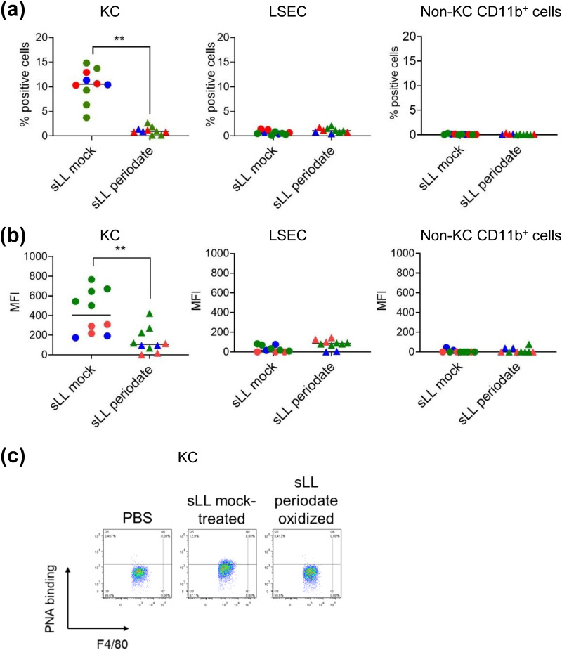
Cystic echinococcosis is caused by the larval stages (hydatids) of cestode parasites belonging to the species cluster Echinococcus granulosus sensu lato, with E. granulosus sensu stricto being the main infecting species. Hydatids are bladderlike structures that attain large sizes within various internal organs of livestock ungulates and humans. Hydatids are protected by the massive acellular laminated layer (LL), composed mainly of mucins. Parasite growth requires LL turnover, and abundant LL-derived particles are found at infection sites in infected humans, raising the question of how LL materials are dealt with by the hosts. In this article, we show that E. granulosus sensu stricto LL mucins injected into mice are taken up by Kupffer cells, the liver macrophages exposed to the vascular space. This uptake is largely dependent on the intact mucin glycans and on Clec4F, a C-type lectin receptor which, in rodents, is selectively expressed in Kupffer cells. This uptake mechanism operates on mucins injected both in soluble form intravenously (i.v.) and in particulate form intraperitoneally (i.p.). In mice harboring intraperitoneal infections by the same species, LL mucins were found essentially only at the infection site and in the liver, where they were taken up by Kupffer cells via Clec4F. Therefore, shed LL materials circulate in the host, and Kupffer cells can act as a sink for these materials, even when the parasite grows in sites other than the liver.

Keywords: Clec4F; Echinococcus; Kupffer cells; laminated layer; lectin; mucin.


Conflict of interest statement

The authors declare no conflict of interest.

Figures

FIG 1
FIG 1
Soluble LL mucins are taken up selectively by KCs, dependent on mucin glycans. C57BL/6 mice were injected i.v. with 100 μg dry mass of solubilized, biotin-derivatized LL mucin preparation (sLL), either treated with periodate to oxidize their terminal monosaccharide residues or mock treated. Twenty minutes later, mice were sacrificed, and sLL uptake was quantitated in permeabilized liver nonparenchymal cells using streptavidin-PE. (a and b) Data are shown for the cell types that are relevant in the context of the article, namely, KCs, LSECs, and non-KC CD11b+ cells in terms of percentages of positive cells (a) and geometric means of fluorescence intensity (b). (c) Representative dot plots for KCs are shown. The signal threshold for each cell type was established using cells from mice injected with vehicle only and similarly stained; the fluorescence intensity data had the median values corresponding to vehicle-injected mice subtracted for plotting. Results are pooled from 3 independent experiments (represented in different colors); individual mice and their overall median values are shown. The statistical analyses were carried out on the crude data by the Mack-Skillings test.
FIG 2
FIG 2
PNA and the E492 antibody are suitable probes for LL mucins captured by mouse KCs. (a and b) The binding of PNA, the E492 antibody, and soluble recombinant Clec4F to LL mucins coated onto ELISA plate wells (a) or present in LL particles in suspension (b) was studied. (c) The capacity of PNA and the E492 antibody to compete with the binding of recombinant Clec4F to LL mucins was analyzed in a competition ELISA format; results are expressed in relation to the response in the absence of competitor. In panels a and c, analytical duplicates are shown. (d) WT C57BL/6 mice were injected i.v. either with PBS only or with solubilized LL mucins (200 μg/mouse), and 22 h later, KCs and LSECs were analyzed by flow cytometry using either PNA or the E492 antibody as probes after cell permeabilization. Staining controls shown in panels b and d correspond to samples stained in the same way as the positive ones except that the primary probe for LL material was omitted, and the analogous controls in panel a gave no signal.
FIG 3
FIG 3
Generation of Clec4f gene-deficient mice. (a) Schematic structure of the mouse Clec4f gene. Coding exons appear as solid black bars and noncoding regions as white bars. Introns are represented as lines. (b) The target sequences for CRISPR/Cas9, 5′-TGGACGTGCCGCAGGGTCCT-3′ and 5′-GCTTCTCCCCTGCAGGACTG-3′, are located in the second exon and first intron-second exon junction of the gene, respectively. An 8-bp deletion corresponding to nucleotides 5′-CAGGACCC-3′ was identified by tail biopsy. The frameshift mutation produces a protein 480 amino acids (aa) shorter. (c) The liver nonparenchymal fraction was prepared from Clec4f−/− and WT mice and analyzed for Clec4F expression by flow cytometry without or with cellular permeabilization. KC were defined as shown in Fig. S2 in the supplemental material. The staining controls contain all the fluorophore-coupled antibodies but lack the primary anti-Clec4F antibody.
FIG 4
FIG 4
Fast uptake of soluble LL mucins by Kupffer cells depends largely on Clec4F. WT and Clec4F−/− mice were injected i.v. with 200 μg dry mass of sLL, either untreated or mock-treated as in Fig. 1 or vehicle only (PBS). Twenty minutes later, mice were sacrificed, and sLL uptake was quantitated in nonparenchymal liver cell populations by flow cytometry in permeabilized cells using PNA. (a and b) Results are shown for KCs, LSECs, and non-KC CD11b+ cells in terms of percentages of positive cells (a) and mean fluorescence intensities (b). (c) Representative flow cytometry plots are shown for KCs and LSECs. (d) An estimation of the fraction of sLL taken up by each of the cell populations under study, calculated as explained in Materials and Methods, is shown; the statistics shown compare the fractions taken up by KCs. The positive cell threshold was established using cells from the mice injected with PBS only. The data arise from two independent experiments, represented in blue and red in panels a to c and indicated as 1 and 2 in panel d. (a to c) Individual mice and their median values are shown. (d) Only medians are shown. The statistical analyses were carried out by the Mack-Skillings test.
FIG 5
FIG 5
Clec4F is important for the capture of soluble LL mucins by Kupffer cells even after allowing a long uptake time. WT and Clec4f−/− C57BL/6 mice were injected i.v. with 200 μg dry mass per mouse of sLL (mock treated as in Fig. 1) or PBS only as a control. Twenty-two hours later, mice were sacrificed, and sLL uptake was quantitated in liver nonparenchymal cells by flow cytometry using PNA after cell permeabilization. (a and b) Results are shown for KCs, LSECs, and non-KC CD11b+ cells in terms of percentages of positive cells (a) and mean fluorescence intensities (b). (c) Representative flow plots for KCs and LSECs are shown. (d) An estimation of the fraction of sLL taken up by each of the cell populations under study, calculated as explained in Materials and Methods, is shown; the statistics shown compare the fractions taken up by KCs. The positive cell threshold was established using staining controls (in which only biotinylated PNA was omitted). The PNA signal in nonpermeabilized cells was negligible. The data arise from two independent experiments, represented in blue and red in panels a to c and indicated as 1 and 2 in panel d. (a to c) Individual mice and their median values are shown. (d) Only medians are shown. The statistical analyses were carried out by the Mack-Skillings test (a to c) and the modified Wilcoxon-Mann-Whitney tests (d).
FIG 6
FIG 6
The uptake by Kupffer cells of LL mucins injected in particulate presentation depends largely on Clec4F. WT and Clec4f−/− C57BL/6 mice were injected i.p. with 450 μg dry mass of pLL or PBS only as a control. Twenty-two hours later, mice were sacrificed, and pLL uptake was quantitated in liver nonparenchymal cells by flow cytometry using PNA after cell permeabilization. (a and b) Results are shown for KCs, LSECs, and non-KC CD11b+ cells in terms of percentages of positive cells (a) and mean fluorescence intensities (b). (c) Representative dot plots for the three cell types are shown. (d) An estimation of the fraction of sLL taken up by each of the cell populations under study, calculated as explained in Materials and Methods, is shown; the statistics shown compare the fractions taken up by KC. The positive cell threshold was established using staining controls. The data arise from two independent experiments, represented in blue and red in panels a to c and indicated as 1 and 2 in panel d. (a to c) Individual mice and their median values are shown. (d) Only medians are shown. The statistical analyses were carried out by the Mack-Skillings test. The analogous flow cytometry results obtained using the E492 antibody for detection are shown in Fig. S6 in the supplemental material. (e) In addition, pLL was detected in liver sections, using the E492 antibody, by chromogenic histology. Black arrows indicate KC-like cells staining strongly for LL material, and empty arrowheads indicate hepatocytes staining weakly for LL material. (f) The localization of LL materials in KCs from infected mice was confirmed by fluorescence histology, using the E492 antibody as probe and F4/80 as KC marker (original magnification, ×630).
FIG 7
FIG 7
LL materials circulate systemically in infected mice and are captured selectively by Kupffer cells via Clec4F. (a) BALB/c mice (WT) were intraperitoneally infected with E. granulosus protoscoleces or injected with the corresponding volume of buffer only. Seven months later, the presence of LL-derived materials in various organs was analyzed by histology, using the E492 antibody as a probe. Images from representative mice are shown out of 15 infected and 5 control mice; the bars represent 50 μm. Black arrows indicate KC-like cells staining for LL material; empty arrowheads indicate occasional staining for LL material in spleen and lymph node. The staining observed in adipocytes (upper left-hand corner of the infected mouse mesenteric lymph node section) was also observed in adipocytes present in kidney and lung sections, irrespective of infection, and must therefore represent a cross-reaction of the antibody. No staining was observed in sections probed with the secondary reagent only. (b) The localization of LL materials in KC from infected mice was confirmed by fluorescence histology, using the E492 antibody as probe and either Clec4F or F4/80 as KC markers (original magnification, ×630). The uptake of LL-derived materials by nonparenchymal liver cells of infected BALB/c mice was also analyzed by flow cytometry, using PNA as probe. (c and d) Results are shown for KCs, LSECs, and non-KC CD11b+ cells in terms of percentages of positive cells (c) and mean fluorescence intensities (d). (e) Representative dot plots for the three cell types are shown. The positive cell threshold was established using staining controls. (f to h) In addition, WT and Clec4f−/− C57BL/6 mice were similarly infected, and the uptake of LL-derived materials was analyzed by flow cytometry and presented as for the BALB/c mice. Results in panels a to e are from a single experiment, and results in panels f to h are pooled from two independent experiments, shown in blue and red. In panels c, d, f, and g, data shown correspond to individual mice, and their median values are indicated. The statistical analyses were carried out by the modified Wilcoxon-Mann-Whitney (c and d) and Kruskal-Wallis (f and g) tests.

Similar articles

Cited by

References

    1. Thompson RCA. 2017. Biology and systematics of Echinococcus. Adv Parasitol 95:65–109. doi:10.1016/bs.apar.2016.07.001. - DOI - PubMed
    1. Díaz Á. 2017. Immunology of cystic echinococcosis (hydatid disease). British Medical Bull 124:121–133. doi:10.1093/bmb/ldx033. - DOI - PubMed
    1. Cucher MA, Macchiaroli N, Baldi G, Camicia F, Prada L, Maldonado L, Avila HG, Fox A, Gutiérrez A, Negro P, López R, Jensen O, Rosenzvit M, Kamenetzky L. 2016. Cystic echinococcosis in South America: systematic review of species and genotypes of Echinococcus granulosus sensu lato in humans and natural domestic hosts. Trop Med Int Health 21:166–175. doi:10.1111/tmi.12647. - DOI - PubMed
    1. Díaz A, Casaravilla C, Irigoín F, Lin G, Previato JO, Ferreira F. 2011. Understanding the laminated layer of larval Echinococcus I: structure. Trends Parasitol 27:204–213. doi:10.1016/j.pt.2010.12.012. - DOI - PubMed
    1. Díaz A, Casaravilla C, Allen JE, Sim RB, Ferreira AM. 2011. Understanding the laminated layer of larval Echinococcus II: immunology. Trends Parasitol 27:264–273. doi:10.1016/j.pt.2011.01.008. - DOI - PubMed

Publication types

LinkOut - more resources