

The Role of Cardiac PET in Diagnosis and Prognosis of Ischemic Heart Disease: Optimal Modality Across Different Patient Populations
- PMID: 37162723
- PMCID: PMC10170052
- DOI: 10.1007/s11883-023-01107-0
The Role of Cardiac PET in Diagnosis and Prognosis of Ischemic Heart Disease: Optimal Modality Across Different Patient Populations
Abstract
Purpose of review: Despite single-photon emission computerized tomography (SPECT) being the most used nuclear imaging technique for diagnosis of coronary artery disease (CAD), many now consider positron emission tomography (PET) as a superior modality. This review will focus on the advances of cardiac PET in recent years and its advantages compared to SPECT in diagnosis and prognosis of CAD.
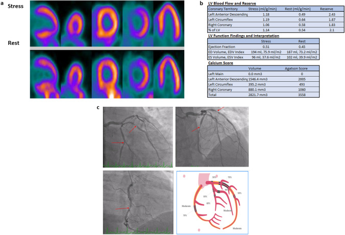
Recent findings: PET's higher resolution and enhanced diagnostic accuracy, as well as lower radiation exposure, all help explain the rationale for its wider spread and use. PET also allows for measurement of myocardial blood flow (MBF) and myocardial flow reserve (MFR), which aids in several different clinical scenarios, such as diagnosing multivessel disease or identifying non-responders. PET has also been shown to be useful in diagnosing CAD in various specific populations, such as patients with prior COVID-19 infection, cardiac transplant, and other comorbidities.
Keywords: Ischemia; Myocardial flow reserve; Myocardial perfusion; Positron emission tomography.
© 2023. The Author(s), under exclusive licence to Springer Science+Business Media, LLC, part of Springer Nature.
Conflict of interest statement
Dr. Mouaz Al-Mallah receives research support from Siemens unrelated to this study. All other authors certify that they have no affiliations with or involvement in any organization or entity with any financial interest or non-financial interest in the subject matter or materials discussed in this manuscript.
Figures
References
Publication types
MeSH terms
LinkOut - more resources
Full Text Sources
Medical
Research Materials
Miscellaneous
