This is a preprint.
HIV-1 infection of genetically engineered iPSC-derived central nervous system-engrafted microglia in a humanized mouse model
- PMID: 37162838
- PMCID: PMC10168358
- DOI: 10.1101/2023.04.26.538461
HIV-1 infection of genetically engineered iPSC-derived central nervous system-engrafted microglia in a humanized mouse model
Update in
-
HIV-1 infection of genetically engineered iPSC-derived central nervous system-engrafted microglia in a humanized mouse model.J Virol. 2023 Dec 21;97(12):e0159523. doi: 10.1128/jvi.01595-23. Epub 2023 Nov 30. J Virol. 2023. PMID: 38032195 Free PMC article.
Abstract
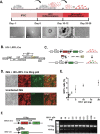
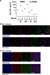
The central nervous system (CNS) is a major human immunodeficiency virus type 1 reservoir. Microglia are the primary target cell of HIV-1 infection in the CNS. Current models have not allowed the precise molecular pathways of acute and chronic CNS microglial infection to be tested with in vivo genetic methods. Here, we describe a novel humanized mouse model utilizing human-induced pluripotent stem cell-derived microglia to xenograft into murine hosts. These mice are additionally engrafted with human peripheral blood mononuclear cells that served as a medium to establish a peripheral infection that then spread to the CNS microglia xenograft, modeling a trans-blood-brain barrier route of acute CNS HIV-1 infection with human target cells. The approach is compatible with iPSC genetic engineering, including inserting targeted transgenic reporter cassettes to track the xenografted human cells, enabling the testing of novel treatment and viral tracking strategies in a comparatively simple and cost-effective way vivo model for neuroHIV.
Keywords: HIV encephalitis; HIV-1; HIV-associated neurocognitive disorder; induced pluripotent stem cell; latent reservoir; microglia.
Conflict of interest statement
Conflicts of Interest: The Authors do not declare a conflict. Data availability/accessibility: All protocols and materials will be provided by the Authors upon request.
Figures


References
-
- Organization WH. 2023. HIV/AIDS.
-
- Scaradavou A. 2023. Cord blood grafts for patients with HIV? Cell 186:1101–1102. - PubMed
-
- Siliciano JD, Kajdas J, Finzi D, Quinn TC, Chadwick K, Margolick JB, Kovacs C, Gange SJ, Siliciano RF. 2003. Long-term follow-up studies confirm the stability of the latent reservoir for HIV-1 in resting CD4+ T cells. Nat Med 9:727–8. - PubMed
-
- Lee E, von Stockenstrom S, Morcilla V, Odevall L, Hiener B, Shao W, Hartogensis W, Bacchetti P, Milush J, Liegler T, Sinclair E, Hatano H, Hoh R, Somsouk M, Hunt P, Boritz E, Douek D, Fromentin R, Chomont N, Deeks SG, Hecht FM, Palmer S. 2020. Impact of Antiretroviral Therapy Duration on HIV-1 Infection of T Cells within Anatomic Sites. J Virol 94. - PMC - PubMed
-
- Chun TW, Justement JS, Murray D, Hallahan CW, Maenza J, Collier AC, Sheth PM, Kaul R, Ostrowski M, Moir S, Kovacs C, Fauci AS. 2010. Rebound of plasma viremia following cessation of antiretroviral therapy despite profoundly low levels of HIV reservoir: implications for eradication. AIDS 24:2803–8. - PMC - PubMed
Publication types
Grants and funding
LinkOut - more resources
Full Text Sources
