[Preprint]. 2023 Apr 27:2023.04.26.538490.
doi: 10.1101/2023.04.26.538490.

Designer DNA NanoGripper

Affiliations

Designer DNA NanoGripper

Lifeng Zhou et al. bioRxiv. .

Update in

Abstract

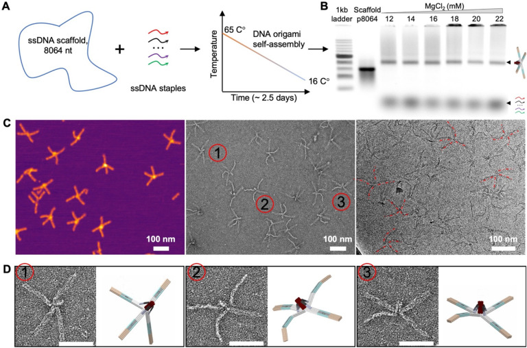
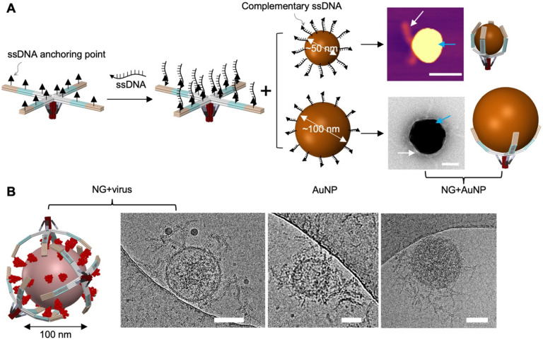
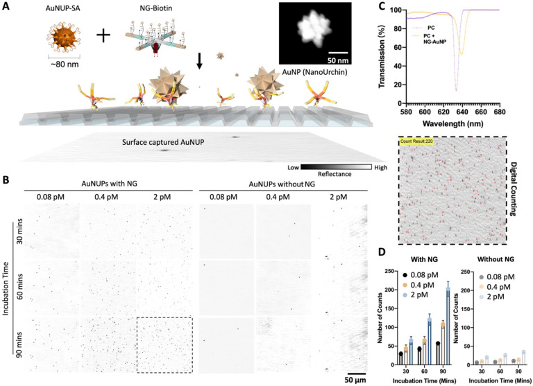
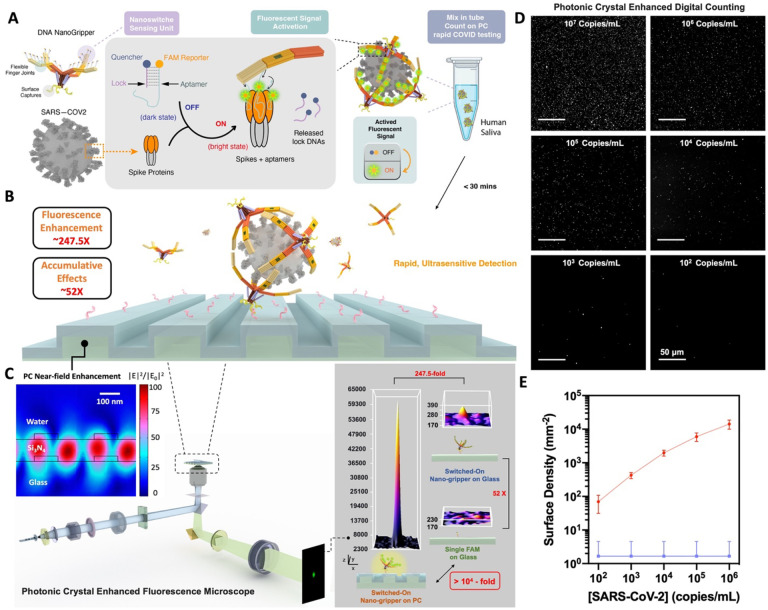
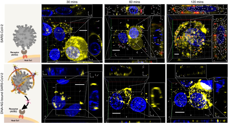
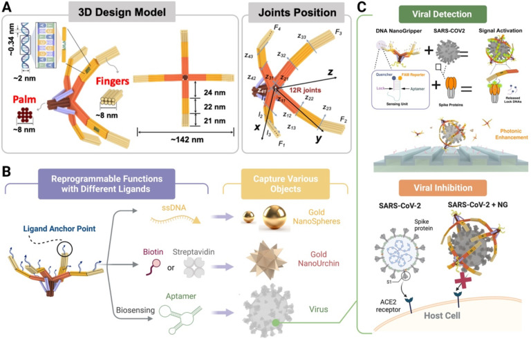
DNA has shown great biocompatibility, programmable mechanical properties, and structural addressability at the nanometer scale, making it a versatile material for building high precision nanorobotics for biomedical applications. Herein, we present design principle, synthesis, and characterization of a DNA nanorobotic hand, called the "NanoGripper", that contains a palm and four bendable fingers as inspired by human hands, bird claws, and bacteriophages evolved in nature. Each NanoGripper finger has three phalanges connected by two flexible and rotatable joints that are bendable in response to binding to other entities. Functions of the NanoGripper have been enabled and driven by the interactions between moieties attached to the fingers and their binding partners. We showcase that the NanoGripper can be engineered to interact with and capture various objects with different dimensions, including gold nanoparticles, gold NanoUrchins, and SARS-CoV-2 virions. When carrying multiple DNA aptamer nanoswitches programmed to generate fluorescent signal enhanced on a photonic crystal platform, the NanoGripper functions as a sensitive viral biosensor that detects intact SARS-CoV-2 virions in human saliva with a limit of detection of ~ 100 copies/mL, providing RT-PCR equivalent sensitivity. Additionally, we use confocal microscopy to visualize how the NanoGripper-aptamer complex can effectively block viral entry into the host cells, indicating the viral inhibition. In summary, we report the design, synthesis, and characterization of a complex nanomachine that can be readily tailored for specific applications. The study highlights a path toward novel, feasible, and efficient solutions for the diagnosis and therapy of other diseases such as HIV and influenza.


Figures

Fig 1.
Fig 1.. Design of DNA NanoGripper.
(A) Determination of optimized finger and palm cross-section sizes. (B) Schematic of the favorite finger-palm connection design that offers outward flip of the fingers to ensure the NG fingers stay in open configurations in free solution. (C) Schematic of scaffold DNA routing displayed using MagicDNA and oxView.
Fig. 2.
Fig. 2.. Synthesis and characterization of DNA NanoGripper.
(A) Schematic of DNA NG assembly using programmed thermal annealing. (B) Agarose gel electrophoresis (AGE) shows the NG formation (as a predominate band) in the buffer with different Mg2+ concentrations. NG aggregates are observed in higher Mg2+ concentration buffers. (C) Sample microscopy images of the DNA NG. Left: AFM image, middle: TEM image after negative staining, and right: cryoEM image. (D) Side-by-side comparison of DNA NG structure obtained from TEM imaging with the corresponding NG 3D model configurations. Scale bars indicate 100 nm.
Fig. 3.
Fig. 3.. Capture of gold nanoparticles and SARS-CoV-2 virus by DNA NG.
(A) AFM and TEM images show AuNPs’ interaction with DNA NG carrying ssDNA with a sequence complementary to the ssDNA coated on AuNPs (50 or 100 nm). Scale bars indicate 50 nm. (B) Cryo-EM images show the capture of SARS-CoV-2 by the DNA NG carrying multiple spike protein targeting aptamers. Scale bars indicate 50 nm.
Fig. 4.
Fig. 4.. Capture and counting of AuNUPs captured on a PC surface by DNA NGs.
(A) Schematic of the capture and counting of surface captured AuNUPs by DNA NG. NG is attached to the surface through the hybridization of the ssDNA on the palm (indicated as red arrows) and complementary ssDNA (pink arrows) on the PC surface. NG interacts with AuNUP via biotin-streptavidin (SA) binding. Size and shape of the AuNUP is characterized using SEM imaging. (B) PRAM microscopy images of surface captured AuNUPs (black dots) by DNA NGs tested at different particle concentrations and different incubation time points. (C) Transmission scan of the PC surface with or without surface immobilized NGs. (D) Plots for the counts of surface captured AuNUPs by DNA NGs tested at different concentrations and different incubation time points. Data are presented as mean ± s.d., n = 3 biologically independent samples.
Fig. 5.
Fig. 5.. Photonic crystal enhanced detection of SARS-CoV-2 by DNA NG sensor.
(A) Schematic of viral capture and fluorescent signal generation of functional NGs. (B) PC-NG hybrid system. (C) PC near-field enhancement and PCEF scanning microscope imaging system with nearly 104-fold signal enhancement compared to an individual FAM reporter on glass substrate. (D) PC enhanced digital counting and dose response of SARS-CoV-2 virus detection. Representative scanning images of PC surface captured SARS-CoV-2 at various viral concentrations after 10-min incubation with the NG sensor. (E) Quantification of captured SARS-CoV-2 virions on the PC surface at different viral concentrations (red curve). The assays were performed in triplicate with similar observations. Negative control (blue baseline) contains only buffer solution. Error bars represent the standard deviations of three independent measurements.
Fig. 6.
Fig. 6.. SARS-CoV-2 viral cell entry inhibition by DNA NG.
SARS-CoV-2 virions (red), cell membrane structures (yellow), and cell nuclei (blue) were labeled with FAM, Dil and Hoescht stains, respectively. Representative SARS-CoV-2 viral particles are indicated by white arrows. Top row: SARS-CoV-2 virions accumulate over time in the no-inhibitor treated condition. Bottom row: SARS-CoV-2 viral entry is inhibited over time by DNA NG treatment. Each panel is accompanied by three cross-sections of the main view (along the dotted lines in the main images). Confocal images show that DNA NG-unbound SARS-COV2 particles accumulate in the cell (top panels), while DNA NG-bound SARS-COV2 particles are prevented from cell entry (bottom panels). Scale bars indicate 10 μm. Confocal assays were performed in duplicate with similar observations.
Scheme 1.
Scheme 1.. Schematic of the DNA NanoGripper (NG) design, functionalization, and use in viral detection and inhibition.
(A) Configuration and dimension of the DNA NG that contains a central palm and four bendable fingers, where F, Z and R represent finger, rotation axis and rotation joint, respectively. (B) Functionalization of the DNA NG for interacting with different nanometer scale 3D objects. (C) Viral detection an inhibition using DNA NG.

References

    1. Hu C., Pané S., Nelson B. J., Soft Micro- and Nanorobotics. Annual Review of Control, Robotics, and Autonomous Systems 1, 53–75 (2018).
    1. Wang B., Kostarelos K., Nelson B. J., Zhang L., Trends in Micro-/Nanorobotics: Materials Development, Actuation, Localization, and System Integration for Biomedical Applications. Advanced Materials 33, (2020). - PubMed
    1. Lafontaine D. L. J., Tollervey D., The function and synthesis of ribosomes. Nature Reviews Molecular Cell Biology 2, 514–520 (2001). - PubMed
    1. Hirokawa N., Noda Y., Tanaka Y., Niwa S., Kinesin superfamily motor proteins and intracellular transport. Nature Reviews Molecular Cell Biology 10, 682–696 (2009). - PubMed
    1. Ali I., Yang W.-C., The functions of kinesin and kinesin-related proteins in eukaryotes. Cell Adhesion & Migration 14, 139–152 (2020). - PMC - PubMed

Publication types