[Preprint]. 2023 Apr 29:2023.04.28.538426.
doi: 10.1101/2023.04.28.538426.

Expansion of Disease Specific Cardiac Macrophages in Immune Checkpoint Inhibitor Myocarditis

Affiliations

Expansion of Disease Specific Cardiac Macrophages in Immune Checkpoint Inhibitor Myocarditis

Pan Ma et al. bioRxiv. .

Update in

  • Expansion of Pathogenic Cardiac Macrophages in Immune Checkpoint Inhibitor Myocarditis.
    Ma P, Liu J, Qin J, Lai L, Heo GS, Luehmann H, Sultan D, Bredemeyer A, Bajapa G, Feng G, Jimenez J, He R, Parks A, Amrute J, Villanueva A, Liu Y, Lin CY, Mack M, Amancherla K, Moslehi J, Lavine KJ. Ma P, et al. Circulation. 2024 Jan 2;149(1):48-66. doi: 10.1161/CIRCULATIONAHA.122.062551. Epub 2023 Sep 25. Circulation. 2024. PMID: 37746718 Free PMC article.

Abstract

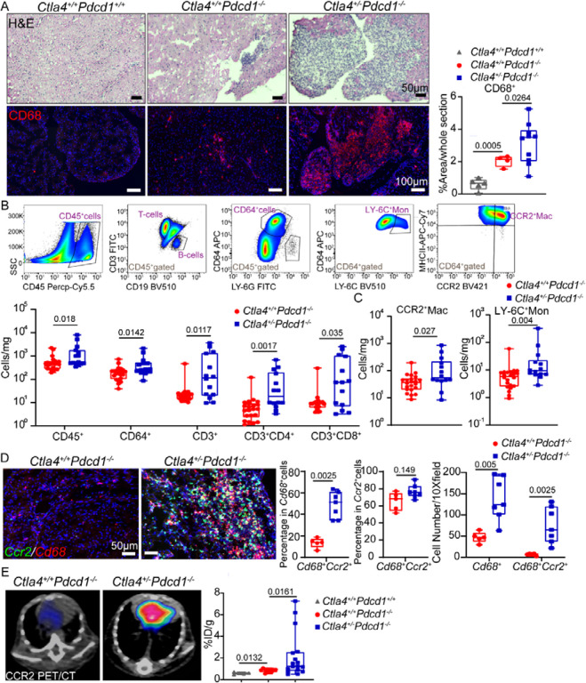
Background: Immune checkpoint inhibitors (ICIs), antibodies targeting PD-1/PD-L1 or CTLA4 have revolutionized cancer management but are associated with devastating immune-related adverse events (irAEs) including myocarditis. The main risk factor for ICI myocarditis is the use of combination PD-1 and CTLA4 inhibition. ICI-myocarditis is often fulminant and is pathologically characterized by myocardial infiltration of T lymphocytes and macrophages. While much has been learned regarding the role of T-cells in ICI-myocarditis, little is understood regarding the identity, transcriptional diversity, and functions of infiltrating macrophages.

Methods: We employed an established murine ICI myocarditis model ( Ctla4 +/- Pdcd1 -/- mice) to explore the cardiac immune landscape using single-cell RNA-sequencing, immunostaining, flow cytometry, in situ RNA hybridization and molecular imaging and antibody neutralization studies.

Results: We observed marked increases in CCR2 + monocyte-derived macrophages and CD8 + T-cells in this model. The macrophage compartment was heterogeneous and displayed marked enrichment in an inflammatory CCR2 + subpopulation highly expressing Cxcl9 , Cxcl10 , Gbp2b , and Fcgr4 that originated from CCR2 + monocytes. Importantly, a similar macrophage population expressing CXCL9 , CXCL10 , and CD16α (human homologue of mouse FcgR4) was found selectively expanded in patients with ICI myocarditis compared to other forms of heart failure and myocarditis. In silico prediction of cell-cell communication suggested interactions between T-cells and Cxcl9 + Cxcl10 + macrophages via IFN-γ and CXCR3 signaling pathways. Depleting CD8 + T-cells, macrophages, and blockade of IFN-γ signaling blunted the expansion of Cxcl9 + Cxcl10 + macrophages in the heart and attenuated myocarditis suggesting that this interaction was necessary for disease pathogenesis.

Conclusion: These data demonstrate that ICI-myocarditis is associated with the expansion of a specific population of IFN-γ induced inflammatory macrophages and suggest the possibility that IFN-γ blockade may be considered as a treatment option for this devastating condition.


Figures

Fig 1.
Fig 1.. Accumulation of CCR2+ monocytes and macrophages in Ctla4+/−Pdcd1−/ mouse hearts.
(A) Representative images of H&E and CD68 immunofluorescent staining (red) in wild type (Ctla4+/+Pdcd1+/+), Ctla4+/+Pdcd1−/−, and Ctla4+/−Pdcd1−/− hearts. Quantification of CD68+ cells. Data collected from two independent experiments. Ctla4+/+Pdcd1+/+ (n=5), Ctla4+/+Pdcd1−/− (n=4), Ctla4+/−Pdcd1−/− (n=10), Welch’s t test, two-tailed. Scale bar for H&E staining images, 50 μm. Scale bar for CD68 staining images, 100 μm (B) Quantification of CD45+, CD64+, CD3+, CD3+CD4+, CD3+CD8+ cells in the heart by flow cytometry. Data collected from four independent experiments. Ctla4+/+Pdcd1−/− (n=22), Ctla4+/−Pdcd1−/− (n=14), Mann-Whitney test, two-tailed. (C) Quantification of CCR2+ macrophages and LY-6Chigh monocytes by flow cytometry. Data collected from four independent experiments. Ctla4+/+Pdcd1−/− (n=22), Ctla4+/−Pdcd1−/− (n=14), Mann-Whitney test, two-tailed. (D) Ccr2 (green) and Cd68 (red) expression detected in Ctla4+/+Pdcd1−/− and Ctla4+/−Pdcd1−/− mouse hearts via RNA in situ hybridization. (Left) Representative images, scale bar, 50 μm. (Right) Quantification of the percentage of Cd68+Ccr2+cells in Cd68+cells and Ccr2+cells, respectively as well as the cell numbers per 10X field. Ctla4+/+Pdcd1−/− (n=5), Ctla4+/−Pdcd1−/− (n=7), Mann-Whitney test, two-tailed. (E) In vivo cardiac CCR2 signal was detected with a CCR2 specific radiotracer, 64Cu-DOTA-ECL1i using positron emission tomography (PET). Representative CCR2 PET/CT images (left) and quantification of CCR2 tracer uptake (right). Data collected from two independent experiments, Ctla4+/+Pdcd1+/+ (n=4), Ctla4+/+Pdcd1−/− (n=12), Ctla4+/−Pdcd1−/− (n=17), Mann-Whitney test, two-tailed.
Fig 2.
Fig 2.. Expansion of Cxcl9+Cxcl10+ macrophages in Ctla4+/−Pdcd1−/− mouse hearts.
(A) UMAP clustering of 23,606 cells from 14 mouse hearts (Ctla4+/+Pdcd1−/−, n=4; Ctla4+/−Pdcd1−/−, n=10), showing 8 major cell types. (B) UMAP clustering of 3,209 the myeloid cells spilt by experimental group highlighting 5 transcriptionally distinct subclusters. (C) The proportion of each myeloid subcluster in Ctla4+/+Pdcd1−/− and Ctla4+/−Pdcd1−/− mice. (D) Dot plots of differentially expressed genes in each myeloid subcluster. (E) Z-score feature plot of enriched genes in each myeloid subcluster and density plot of Ccr2 expression. Cell state marker genes (in black) were selected based on robust enrichment in their respective subclusters.
Fig 3.
Fig 3.. Cxcl9+Cxcl10+ macrophages exhibit an activated phenotype in ICI myocarditis.
(A) Volcano plot of differentially expressed genes amongst myeloid cells from Ctla4+/+ Pdcd1−/− and Ctla4+/−Pdcd1−/− hearts obtained by Wilcoxon Rank Sum test using R package Seurat (v4). (B) Z-score feature plot of the top 10 up-regulated genes in Ctla4+/+ Pdcd1−/− myeloid cells compared to Ctla4+/−Pdcd1−/− myeloid cells split by experimental group. Differentially expressed genes are selectively expressed in Cxcl9+Cxcl10+ macrophages (C) Increased Cxcl9, Cxcl10, Gbp2b, Ccl8, and Fcgr4 mRNA expression in Ctla4+/+ Pdcd1−/− compared to Ctla4+/−Pdcd1−/− heart tissue measured by RT-PCR. Data collected from two independent experiments, Ctla4+/+Pdcd1−/− (n=8), Ctla4+/−Pdcd1−/− (n=6), Mann-Whitney test, two-tailed. (D) Co-expression of Cxcl9 and Cxcl10 with Ccr2 in mouse hearts visualized by RNA in situ hybridization. (Left) Representative images in each condition, scale bar, 50 μm. (Right) Quantification of the cell number of Cxcl9+Ccr2+cells or Cxcl10+Ccr2+cells per 10X field in each condition as well as the percentage of Cxcl9+Ccr2+ or Cxcl10+Ccr2+cells in Cxcl9+ or Cxcl10+cells. Ctla4+/+Pdcd1+/+(n=4), Ctla4+/+Pdcd1−/− (n=4), Ctla4+/−Pdcd1−/− (n=9), Mann-Whitney test, two-tailed. (E) Quantification of FCGR4 protein expression on CD64+ macrophages by flow cytometry. Data collected from four independent experiments, Ctla4+/+Pdcd1−/− (n=20), Ctla4+/−Pdcd1−/− (n=14), Mann-Whitney test, two-tailed. (F) GO pathway enrichment analysis of up-regulated genes in Ctla4+/−Pdcd1−/− myeloid cells. The top five enriched pathways in Ctla4+/−Pdcd1−/− myeloid cells are displayed. Genes used in the analysis were selected from Seurat differential expression with P < 0.05 and log2FC > 0.5. P value calculated using hypergeometric distribution and corrected for multiple comparisons. (G) Z-score feature plots of enriched genes involving in response to Interferon-gamma; cytokine-mediated signaling pathway; myeloid leukocyte migration; antigen processing and presentation pathways in myeloid cells.
Fig 4.
Fig 4.. Cxcl9+Cxcl10+ macrophages originate from monocytes.
(A) tSNE force-directed layout plot of myeloid cells. Cells are colored by cell cluster annotations. (B) Pseudotime and entropy values of myeloid cells. Cxcl9+Cxcl10+ macrophages (Cxcl9 Cxcl10 Mac) have high pseudotime and low entropy values suggesting that they represent a differentiated cell state. (C) Terminal State Probability of cell states predicted as differentiated populations: Cxcl9 Cxcl10 Mac; Cd163 resident Mac; and DCs. (D) Box plots of entropy (upper) and Cxcl9 Cxcl10 Mac terminal state probability (lower) of myeloid subclusters split by experimental group. (E) Percentage of LY-6Chigh monocytes and CCR2+ macrophages of cardiac CD64+ cells from vehicle or MC-21 antibody treated mice quantified by flow cytometry. Displayed cells are CD45+LY-6GCD64+. Data collected from four independent experiments. vehicle group (n=12), MC-21 treated group (n=8), Mann-Whitney test, two-tailed. (F) Representative images (upper) and quantification (lower) of Cxcl9 and Cxcl10 positive cells in the heart 6 days after MC-21 antibody treatment. Data collected from four independent experiments. vehicle group (n=12), MC-21 treated group (n=8), Mann-Whitney test, two-tailed.
Fig 5.
Fig 5.. CXCL9+CXCL10+ macrophages in human ICI associated myocarditis.
(A) Expression of CXCL9 and CXCL10 via RNA in situ hybridization in human heart tissue from patients with ICI myocarditis (ICI, n=7), lymphocytic myocarditis (LM, n=5), ischemic cardiomyopathy (ICM, n=5), dilated cardiomyopathy (DCM, n=6), and donor control subject (n=6). Quantification of the number of CXCL9+ and CXCL10+ cells, Mann-Whitney test, two-tailed. Scale bar 50 μm. (B) Immunofluorescent staining of CD16α (green), CCR2 (red), CD68 (white) and DAPI (blue) in human heart tissue from patients with ICI (n=8), LM (n=5), ICM (n=5), DCM (n=6) and donor control subjects (n=6). Quantification of cell number and the percentage of CD68+CD16a+ cells in all CD68+cells, Mann-Whitney test, two-tailed. Scale bar, 50 μm.
Fig 6.
Fig 6.. T-cells are the primary source of IFN-γ in ICI myocarditis mouse hearts.
(A) Increased Ifng mRNA expression in Ctla-4+/−Pdcd1−/− mouse hearts measured by RT-PCR. Data collected from two independent experiments, Ctla4+/+Pdcd1−/− (n=8), Ctla4+/−Pdcd1−/− (n=6), Mann-Whitney test, two-tailed. (B) Feature plot of Ifng expression in all cell types recovered from the heart showing specific expression in the NK/T-cell cluster. (C) Feature plots of Ifng, Cd8a, and Cd4 expression in NK&T-cells showing CD8 T-cell expansion and enriched Ifng expression in CD8 T-cells from Ctla4+/−Pdcd1−/− hearts. (D) Percentages of IFNγ+CD4+, IFNγ+CD8+ T-cells, IFNγ+NK-cells, and IFNγ+CD64+ macrophages analyzed by flow cytometry. Data collected from two independent experiments, Ctla4+/+Pdcd1−/− (n=8), Ctla4+/−Pdcd1−/− (n=5), Mann-Whitney test, two-tailed. (E) The proportion of each NK&T subcluster per experimental group. (F) Z-score feature plots of top 10 up-regulated genes in Ctla4+/−Pdcd1−/− NK&T-cells compared to Ctla4+/+Pdcd1−/− NK&T cells split by group. (G) GO and KEGG enriched pathways using genes up-regulated in Ctla4+/−Pdcd1−/− NK&T cells compared to Ctla4+/+Pdcd1−/− NK&T cells. Genes used in the analysis were selected from Seurat differential expression with P < 0.05 and log2FC > 1. P-values calculated by hypergeometric distribution using R package ClusterProfiler.
Fig 7.
Fig 7.. T-cells are predicted to orchestrate the expansion and activation of Cxcl9+Cxcl10+ macrophages.
(A) Cell to cell communication analysis using CellChat predicted that T-cells signal to macrophages through IFN-γ. Violin plot showing the expression distribution of IFN-γ pathway ligand and receptors in T-cells and macrophages. (B) Heatmap showing the relative importance of each cell state based on the computed network of IFN-γ signaling. (C) Circle plot summarizing the inferred intercellular communication network between T-cells and macrophages for IFN-γ signaling. (D) Dotplot showing the strength of interaction between T-cell and macrophage cell states for IFN-γ signaling. P values were calculated using R package CellChat. (E) Violin plot showing the expression distribution of signaling genes involved in the inferred reciprocal CXCL signaling network (Cxcl9/ Cxcl10-Cxcr3) between macrophages and T-cells. (F) Heatmap displaying the relative importance of each cell state based on the computed network of CXCL signaling. (G) Circle plot depicting the inferred intercellular communication network between macrophage and T-cell states for CXCL signaling (Cxcl9/ Cxcl10-Cxcr3). (H) Quantification of cardiac Cxcl9+ and Cxcl10+ cells 6 days after anti-CD8 antibody treatment. Data collected from three independent experiments. vehicle (n=15); anti-CD8 (n=7), Mann-Whitney test, two-tailed.
Fig 8.
Fig 8.. IFN-γ blockade and macrophage depletion reduce cardiac Cxcl9+Cxcl10+ macrophages and prolong the survival of Ctla4+/−Pdcd1−/− mice.
(A) Survival of Ctla4+/− Pdcd1−/− mice treated with vehicle (isotype control) or anti-IFN-γ antibody (R46A2). Data collected from four independent experiments vehicle (n=25); anti-IFN-γ (n=26), Log-rank test. (B) Representative images (left) and quantification (right) of cardiac Cxcl9+ and Cxcl10+ cells as determined by RNA in situ hybridization 23 days after vehicle or anti-IFN-γ antibody treatment. Data collected from five independent experiments. vehicle (n=15); anti-IFN-γ (n=21), Mann-Whitney test, two-tailed. (C) Survival of Ctla4+/− Pdcd1−/− mice treated with vehicle (isotype control) or anti-CSF1R antibody (AFS98). vehicle (n=39); anti-CSF1R (n=32), Log-rank test. (D) Representative images (left) and quantification (right) of cardiac Cxcl9+ and Cxcl10+ cells as determined by RNA in situ hybridization 23 days after vehicle or anti-CSF1R antibody treatment. Data collected from four independent experiments. vehicle (n=14); anti-CSF1R (n=10), Mann-Whitney test, two-tailed. (E) Cardiac CD64+macrophages depletion was verified by flow cytometry. Representative images (left) and quantification (right) of CD64+ cells as determined by flow cytometry 60 days after first vehicle or anti-CSF1R antibody treatment. Displayed cells (left) are gated CD45+ cells. Data collected from three independent experiments. vehicle (n=7); anti-CSF1R (n=7), Mann-Whitney test, two-tailed.

References

    1. Wolchok JD, Kluger H, Callahan MK, Postow MA, Rizvi NA, Lesokhin AM, Segal NH, Ariyan CE, Gordon R-A and Reed K. Nivolumab plus ipilimumab in advanced melanoma. N Engl J Med. 2013;369:122–133. - PMC - PubMed
    1. Postow MA, Chesney J, Pavlick AC, Robert C, Grossmann K, McDermott D, Linette GP, Meyer N, Giguere JK and Agarwala SS. Nivolumab and ipilimumab versus ipilimumab in untreated melanoma. New England Journal of Medicine. 2015;372:2006–2017. - PMC - PubMed
    1. Motzer RJ, Tannir NM, McDermott DF, Frontera OA, Melichar B, Choueiri TK, Plimack ER, Barthélémy P, Porta C and George S. Nivolumab plus ipilimumab versus sunitinib in advanced renal-cell carcinoma. New England Journal of Medicine. 2018. - PMC - PubMed
    1. Heinzerling L, Ott PA, Hodi FS, Husain AN, Tajmir-Riahi A, Tawbi H, Pauschinger M, Gajewski TF, Lipson EJ and Luke JJ. Cardiotoxicity associated with CTLA4 and PD1 blocking immunotherapy. Journal for immunotherapy of cancer. 2016;4:1–11. - PMC - PubMed
    1. Varricchi G, Galdiero MR, Marone G, Criscuolo G, Triassi M, Bonaduce D, Marone G and Tocchetti CG. Cardiotoxicity of immune checkpoint inhibitors. ESMO open. 2017;2:e000247. - PMC - PubMed

Publication types