. 2023 Jul 1;34(8):ar80.
doi: 10.1091/mbc.E22-12-0572. Epub 2023 May 10.

Polo-like kinase 4 homodimerization and condensate formation regulate its own protein levels but are not required for centriole assembly

Affiliations

Polo-like kinase 4 homodimerization and condensate formation regulate its own protein levels but are not required for centriole assembly

John M Ryniawec et al. Mol Biol Cell. .

Abstract

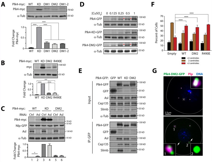
Polo-like kinase 4 (Plk4) is the master-regulator of centriole assembly, and cell cycle-dependent regulation of its activity maintains proper centrosome number. During most of the cell cycle, Plk4 levels are nearly undetectable due to its ability to autophosphorylate and trigger its own ubiquitin-mediated degradation. However, during mitotic exit, Plk4 forms a single aggregate on the centriole surface to stimulate centriole duplication. Whereas most Polo-like kinase family members are monomeric, Plk4 is unique because it forms homodimers. Notably, Plk4 trans-autophosphorylates a degron near its kinase domain, a critical step in autodestruction. While it is thought that the purpose of homodimerization is to promote trans-autophosphorylation, this has not been tested. Here, we generated separation-of-function Plk4 mutants that fail to dimerize and show that homodimerization creates a binding site for the Plk4 activator, Asterless. Surprisingly, however, Plk4 dimer mutants are catalytically active in cells, promote centriole assembly, and can trans-autophosphorylate through concentration-dependent condensate formation. Moreover, we mapped and then deleted the weak-interacting regions within Plk4 that mediate condensation and conclude that dimerization and condensation are not required for centriole assembly. Our findings suggest that Plk4 dimerization and condensation function simply to down-regulate Plk4 and suppress centriole overduplication.


Figures

FIGURE 1:
FIGURE 1:
Generating a Plk4 dimerization mutant. (A) Ribbon diagram showing the quaternary structure of the Drosophila Plk4 PB1-PB2, which forms a Z-shaped, end-to-end dimer. The dimerization interface (boxed) occurs between PB2 and PB2′ (yellow). (A′) Magnification of boxed region in A highlighting the interaction interface. Six hydrophobic residues (red and blue) interact at the PB2-PB2′ interface. Residues responsible for dimerization are (A′′) four residues (red) on each 2α1 helix and (A′′′) two residues (blue) on each 2β6 strand. The specific amino acid substitutions that comprise the dimerization mutants DM1 and DM2 are listed. (B) Anti-GFP immunoprecipitates were prepared from lysates of S2 cells transiently co-overexpressing the indicated inducible Plk4-GFP and myc-tagged Plk4 constructs. Blots of the input lysates and IPs were probed for α-tubulin, GFP, and myc. Note that all three dimer mutants block Plk4 dimerization.
FIGURE 2:
FIGURE 2:
Plk4 homodimerization does not stimulate kinase activation but instead functions to generate an Asl-A–binding site. (A) Asl-A stimulates WT Plk4 kinase activity but not the DM2 dimer mutant. Purified full-length WT or DM2 Plk4 (0.25 μM) was incubated with the indicated concentrations of purified Asl-A and γ32-ATP. The Plk4 in each sample (resolved by SDS–PAGE) was scintillation counted (cpm) and then normalized to the count of the WT/no AslA sample in that experiment. There were no significant differences in activity between any of the DM2 samples, regardless of Asl-A concentration. Asterisks mark significant differences as determined by one-way analysis of variance (ANOVA) test with Tukey’s multiple comparisons posttest. n = 3 experiments. Error bars, standard error of the mean (SEM). For all figures, *, 0.05 > P ≥ 0.01; **, 0.01 > P ≥ 0.001; ***, 0.001 > P ≥ 0.0001; ****, 0.0001 > P; ns, not significant. (B) Depletion of Plk4 activator, Asl, but not Ana2, increases Plk4 levels. Top panels, S2 cells were RNAi treated for 7 d. On day 4, cells were cotransfected with Nlp-GFP (transfection loading control) and inducible Plk4-myc. Starting on day 5, cells were induced to express Plk4-myc. Cell lysates were prepared on day 7 and probed on immunoblots for GFP, myc, Asl, Ana2, and α-tubulin (loading control). Graph shows relative amounts of Plk4-myc as determined by densitometry, normalized to Nlp-GFP, and plotted relative to control RNAi. Asterisks mark significant differences as determined by one-way ANOVA test with Tukey’s multiple comparisons posttest. Note that the Slimb RNAi treatment was not included in the ANOVA test because the differences between it and the other treatments were so large that it made statistical comparison between the other treatments invalid. n = 3 experiments per treatment. Error bars, SEM. (C) Schematic showing how dimeric Asl-A (red) could facilitate trans-autophosphorylation of Plk4 (green) by promoting homodimerization of the kinase as well as positioning two dimers in close proximity. For simplicity, PB3 of Plk4 is not shown. (D) Asl-A does not promote the dimerization of a Plk4 dimer mutant. S2 cells were Asl-depleted by RNAi targeting an exon in the gene region encoding Asl-C for 7 d. On day 4, cells were transfected with the indicated constructs and starting on day 5 induced to express for 48 h. Anti-GFP immunoprecipitates (IPs) were prepared from lysates, and Western blots of the IPs were probed for α-tubulin, GFP, V5, and myc. Note that DM2 Plk4-myc fails to immunoprecipitate with WT Plk4-GFP in cells coexpressing Asl-A-V5 (lane 4). Graph shows relative amounts of Asl-A-V5 in the immunoprecipitate as determined by densitometry and normalized to WT-Plk4-GFP. Asterisks mark significant differences as determined by one-way ANOVA test with Tukey’s multiple comparisons posttest. n = 3 experiments per treatment. Error bars, SEM. (E) Plk4 dimerization is required for Asl-A binding. Cells were prepared as described in D. Anti-GFP immunoprecipitates were prepared from lysates of Asl-depleted S2 cells transiently co-overexpressing the indicated inducible Plk4-EGFP and Asl-A-V5 constructs. Immunoblots of the input lysates and immunoprecipitates were probed for α-tubulin, GFP, and V5. Note that Asl-A binding to DM2 is negligible (lane 4) as well as the control Asl-binding mutant, R490E Plk4 (lane 5). (F) The Asl-binding mutation R490E does not prevent Plk4 dimerization. Anti-GFP immunoprecipitates were prepared from lysates of S2 cells transiently co-overexpressing the indicated inducible Plk4-EGFP and myc-tagged constructs. Blots of the input lysates and IPs were probed for α-tubulin, GFP, and myc.
FIGURE 3:
FIGURE 3:
Dimer mutant Plk4 is catalytically active in cells and induces centriole amplification. (A) Top panels, Plk4 dimerization mutants efficiently autodestruct. The stabilities of different Plk4 proteins were analyzed by immunoblotting lysates of S2 cells transiently expressing the indicated inducible Plk4-myc construct. Anti-myc and α-tubulin (loading control) immunoblots are shown. Graph, Plk4-myc intensities were measured by densitometry of the anti-myc immunoblots and normalized to their respective loading controls. Plotted values are the normalized Plk4 intensities relative to the KD sample. For all graphs in this figure, asterisks mark significant differences determined by one-way ANOVA with Tukey’s multiple comparisons posttest. n = 3 experiments. Error bars, SEM. (B, C) Asl is not required for Plk4 autodestruction. Top panels, The stabilities of different Plk4 proteins were analyzed by immunoblotting lysates of S2 cells transiently expressing the indicated inducible Plk4-myc constructs. Anti-myc, GFP, Asl, and α-tubulin (loading control) immunoblots are shown. Graphs, Plk4-myc intensities were measured by densitometry of the anti-myc immunoblots and normalized to their respective loading controls. Plotted values are fold changes of the normalized Plk4 intensities relative to the KD sample. Nlp-GFP, loading control for transfected cells in C. For Asl RNAi, cells were treated as described in Figure 2D. n = 3 experiments. Error bars, SEM. (D) Dimerization mutant Plk4 can autophosphorylate. The electrophoretic mobilities of different Plk4 proteins were examined by immunoblotting lysates of S2 cells transiently expressing the indicated inducible Plk4-GFP constructs, using increasing concentrations (mM) of CuSO4 to drive increasing expression levels. Anti-GFP and α-tubulin (loading control) immunoblots are shown. Arrowheads mark slower-migrating species of phospho-Plk4. (E) Dimerization mutant Plk4 binds Slimb. S2 cells were transfected with the indicated inducible Plk4-GFP or GFP constructs and their expression induced with 1 mM CuSO4 for 24 h. Anti-GFP IPs were prepared from cell lysates, and Western blots of the IPs were probed for GFP, Slimb, Asl, Cep135, and α-tubulin. (F) Plk4 homodimerization or Asl-A binding is not required to induce centriole amplification (>2 centrioles per cell). Transfected S2 cells were induced to express different Plk4-GFP constructs at low (0.25 mM CuSO4) levels for 3 d and then cells were fixed and immunostained for Plp (centriole marker) and their centrioles were manually counted. Centriole amplification occurs in cells expressing either WT, DM2, or R490E Plk4 compared with cells transfected with control empty vector. n = 3 experiments. Significance was determined by two-way ANOVA with Tukey’s multiple comparisons test. ****, P < 0.0001. Error bars, SEM. (G) Plk4 homodimerization is not required for its localization to centrioles. S2 cells expressing Plk4-DM2-GFP (green) were immunostained for Plp (magenta) to mark centrioles. DNA, blue. Dashed lines show cell borders. Insets show higher magnifications of the boxed regions. Top panel, Example of DM2-expressing cell containing centriole amplification. Bottom panel, Overexpressed DM2 not only localizes to centrioles but can also aggregate into round punctae (arrows) that do not colocalize with the Plp centriole marker.
FIGURE 4:
FIGURE 4:
Plk4 can phosphorylate in trans to promote its degradation without having to homodimerize. (A) Plk4 trans-autophosphorylation–mediated degradation does not require Asl-A to stimulate kinase activity. Top panels, The stabilities of different Plk4 proteins were analyzed by immunoblotting lysates of S2 cells transiently expressing the indicated inducible GFP (control) or Plk4-GFP constructs along with Plk4-KD-myc. Anti-myc, GFP, and α-tubulin (loading control) immunoblots are shown. Graph, Plk4-KD-myc intensities were measured by densitometry of the anti-myc immunoblots and normalized to their respective loading controls. Plotted values are the normalized Plk4-KD-myc intensities relative to the control GFP cotransfection treatment. Asterisks mark significant differences determined by one-way ANOVA with Tukey’s multiple comparisons test. Error bars, SEM. (B) WT Plk4 does not appreciably trans-activate the Plk4 dimerization mutant. The stabilities of different Plk4 proteins were analyzed by immunoblotting lysates of S2 cells transiently expressing the indicated inducible GFP (control) or Plk4-GFP and -myc constructs. Anti-myc, GFP, and α-tubulin (loading control) immunoblots are shown. (C–E) Coexpression of dimer mutant or Asl-A–binding mutant Plk4 alters the pattern of Plk4 KD aggregates in S2 cells. (C) Images show S2 cells expressing Plk4-KD-myc (green) whose localization fits three distinct patterns. After 3 d of expression, cells were stained for Plp-labeled centrioles (magenta) and DNA (blue). (D) DM2 and KD Plk4 coaggregate. S2 cells expressed inducible Plk4 constructs for 3 d and were stained for Plk4-DM2-GFP (green), Plk4-KD-myc (red), centriole marker Plp (white), and DNA (blue). Dashed yellow lines mark cell borders in all images. Dashed boxes are shown at higher magnification (insets). Arrowhead (pink) marks a centrosome. (E) Graph shows percentage of cells with the indicated Plk4-KD-GFP localization patterns. Note that all Plk4 constructs prevent KD Plk4 from forming large aggregates. Significant differences were determined using one-way ANOVA with Tukey’s multiple comparison test. n = 3 experiments per treatment. Error bars, SEM.
FIGURE 5:
FIGURE 5:
Drosophila Plk4’s linker regions are required to form condensates in cells, but condensate formation is not necessary for centriole assembly. (A) Plk4-GFP constructs used to map regions required to form condensates. Plk4-GFP–tagged constructs were transiently transfected into S2 cells, allowed 24 h recovery, and then induced to express by adding 0.7 mM CuSO4 for 2 d. Live cells were scored on whether they contained round GFP condensates (Y, yes; N, no; A, irregular-shaped aggregates). X denotes constructs containing the KD D156N mutation. DRE, downstream regulatory element. AA denotes the S293A/T297A Slimb-binding mutation (SBM) rendering the protein nondegradable. DM2 denotes dimer mutation in PB2. CT, carboxy-terminus; min, condensate-mutant Plk4. (B) Kinase activity and autophosphorylation status do not influence Plk4’s ability to form condensates in cells. Images of S2 cell overexpressing nondegradable, kinase-active SBM-Plk4-GFP (green). Example of KD-Plk4-GFP condensates is shown in Figure 4C. After 2 d of expression, cells were stained for DNA (blue). Dashed yellow lines mark cell borders. Inset shows higher magnification of the boxed region. (C) Plk4 protein within condensates can exchange with the cytoplasmic pool but its turnover requires kinase activity. FRAP analysis of cells expressing the indicated Plk4-GFP construct. Cells were prepared as described in B. n = >12 cells per construct. Error bars, SEM. (D, E) Plk4 condensation is not required for centriole targeting or centriole assembly. (C) Image shows that Plk4-min-GFP (green) colocalizes with Plp-immunostained centrioles (magenta). DNA, blue. Dashed yellow lines mark cell border. Inset of boxed region shows colocalization of Plk4-min and Plp at higher magnification. (D) Transfected S2 cells were induced to express GFP or different Plk4-GFP constructs (0.7 mM CuSO4) for 4 d and then cells were fixed, immunostained for Plp, and their centrioles manually counted. Centriole amplification occurs in cells expressing either SBM, min, or min-DM2 Plk4 as compared with control GFP transfected cells. n = 3 experiments. Significance was determined by two-way ANOVA with Tukey’s multiple comparisons test. **, P < 0.01; n.s., not significant. Error bars, SEM. (F) Dimerization or condensation is sufficient for Plk4 trans-autophosphorylation–mediated degradation. The stabilities of different Plk4 proteins were analyzed by immunoblotting lysates of S2 cells transiently expressing the indicated inducible GFP (control) or Plk4-GFP constructs along with Plk4-KD-myc. Anti-myc, GFP, and α-tubulin (loading control) immunoblots are shown. Note that Plk4-min is highly stable because it lacks the DRE containing the Slimb-binding phosphodegron (A). (G) Graph shows percentage of cells with the indicated Plk4-KD-GFP localization patterns. Note that Plk4-min-DM2-GFP, which fails to dimerize or form condensates, has a near-identical effect as KD Plk4-GFP on Plk4-KD-myc’s ability to form large spherical condensates. Significant differences were determined using two-way ANOVA with Tukey’s multiple comparison test. ****, P < 0.0001. n = 3 experiments per treatment. Error bars, SEM.
FIGURE 6:
FIGURE 6:
Model describing concentration-dependent multistep pathway in down-regulating Plk4 protein levels. (A) Plk4 is globally maintained at a low level due to a dimerization and Asl-dependent pathway of Plk4 activation and autodestruction. Newly synthesized Plk4 initially exists in an autoinhibited, monomeric form. Autoinhibition is exerted by L1, which binds to the activation loop of the kinase domain and prevents its autophosphorylation. Plk4 homodimerizes through hydrophobic interactions between two PB2 modules. The resultant Z-shaped, end-to-end dimer creates a unique Asl-binding site that extends across the dimer and makes multiple electrostatic contacts across both PB1 and PB2. The short acidic region within the N-terminus of Asl binds along one Plk4 Z-dimer surface and relieves autoinhibition by repositioning L1. Plk4 catalytic activity is dramatically activated by Asl binding (Boese et al., 2018; Figure 2). Plk4 then trans-autophosphorylates its activation loop as well as the Slimb-binding degron that promotes its ubiquitin-mediated proteolysis, consequently suppressing centriole duplication. (B) If global Plk4 protein levels rise above a critical threshold, then Asl becomes a rate-limiting component and Plk4 accumulates. However, in this scenario, centriole amplification is avoided due to an efficient compensatory mechanism that down-regulates Plk4 protein levels; Plk4 forms condensates (likely due to LLPS) through its linker domains 1 and 2. Naturally, condensate formation requires interactions between Plk4 molecules, and although homodimerization likely facilitates this, homodimerization is not required. Instead, weak interactions exist between linker domains and PBs to mediate condensate formation. Plk4 has an intrinsically low level of catalytic activity (Klebba et al., 2015a; Lopes et al., 2015), and within this condensate (likely a mixture of both monomers and dimers), the density of Plk4 is high enough to promote trans-autophosphorylation. Normally, this condensation pathway is not observed in interphase cells because Plk4 levels are too low (due to the Asl-mediated pathway) but the existence of this pathway comes into view in interphase cells forced to overexpress Plk4. Movement of autophosphorylated Plk4 subunits from the condensate pool to the cytoplasm exposes the protein to ubiquitin-mediated degradation.

References

    1. Adams PD, Afonine PV, Bunkóczi G, Chen VB, Davis IW, Echols N, Headd JL, Hung L-W, Kapral GJ, Grosse-Kunstleve RW, et al. (2010). PHENIX: a comprehensive Python-based system for macromolecular structure solution. Acta Crystallogr D Biol Crystallogr 66, 213–221. - PMC - PubMed
    1. Arquint C, Gabryjonczyk AM, Imseng S, Böhm R, Sauer E, Hiller S, Nigg EA, Maier T (2015). STIL binding to Polo-box 3 of PLK4 regulates centriole duplication. eLife 4, e07888. - PMC - PubMed
    1. Bettencourt-Dias M, Rodrigues-Martins A, Carpenter L, Riparbelli M, Lehmann L, Gatt MK, Carmo N, Balloux F, Callaini G, Glover DM (2005). SAK/PLK4 is required for centriole duplication and flagella development. Curr Biol 15, 2199–2207. - PubMed
    1. Boese CJ, Nye J, Buster DW, McLamarrah TA, Byrnes AE, Slep KC, Rusan NM, Rogers GC (2018). Asterless is a Polo-like kinase 4 substrate that both activates and inhibits kinase activity depending on its phosphorylation state. Mol Biol Cell 29, 2874–2886. - PMC - PubMed
    1. Brownlee CW, Klebba JE, Buster DW, Rogers GC (2011). The protein phosphatase 2A regulatory subunit Twins stabilizes Plk4 to induce centriole amplification. J Cell Biol 195, 231–243. - PMC - PubMed

Publication types

Substances

LinkOut - more resources