. 2023 May 10;18(5):e0274065.
doi: 10.1371/journal.pone.0274065. eCollection 2023.

A validated protocol to UV-inactivate SARS-CoV-2 and herpesvirus-infected cells

Affiliations

A validated protocol to UV-inactivate SARS-CoV-2 and herpesvirus-infected cells

Timothy K Soh et al. PLoS One. .

Abstract

Downstream analysis of virus-infected cell samples, such as reverse transcription polymerase chain reaction (RT PCR) or mass spectrometry, often needs to be performed at lower biosafety levels than their actual cultivation, and thus the samples require inactivation before they can be transferred. Common inactivation methods involve chemical crosslinking with formaldehyde or denaturing samples with strong detergents, such as sodium dodecyl sulfate. However, these protocols destroy the protein quaternary structure and prevent the analysis of protein complexes, albeit through different chemical mechanisms. This often leads to studies being performed in over-expression or surrogate model systems. To address this problem, we generated a protocol that achieves the inactivation of infected cells through ultraviolet (UV) irradiation. UV irradiation damages viral genomes and crosslinks nucleic acids to proteins but leaves the overall structure of protein complexes mostly intact. Protein analysis can then be performed from intact cells without biosafety containment. While UV treatment protocols have been established to inactivate viral solutions, a protocol was missing to inactivate crude infected cell lysates, which heavily absorb light. In this work, we develop and validate a UV inactivation protocol for SARS-CoV-2, HSV-1, and HCMV-infected cells. A fluence of 10,000 mJ/cm2 with intermittent mixing was sufficient to completely inactivate infected cells, as demonstrated by the absence of viral replication even after three sequential passages of cells inoculated with the treated material. The herein described protocol should serve as a reference for inactivating cells infected with these or similar viruses and allow for the analysis of protein quaternary structure from bona fide infected cells.


Conflict of interest statement

The authors have declared that no competing interests exist.

Figures
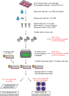
Fig 1
Fig 1. Schematic of validation procedure of UV inactivation protocol for SARS-CoV-2.
Samples were generated with 3 biological replicates under the conditions that lead to no detectable virus. Fresh cells were treated with the UV irradiated samples, and these treated cells were passaged for three weeks to test for viral replication. The inactivation protocol is the same for HSV-1 and HCMV, except for differences in the number of cells and passaging times, which are described in the Materials and Methods section.
Fig 2
Fig 2. Mixing cells during irradiation increases inactivation efficiency.
Vero B4 cells were infected with HSV-1 and irradiated in tissue vials. Cells were treated with the indicated irradiation dose in a single interval (No Mixing, black line) or were treated repeatedly with a fluence of 2,500 mJ/cm2 followed by mixing for the total dose stated (With Mixing, green line). Sample inactivation was evaluated by plaque assay with a limit of detection (LoD) of 5x10² PFU/mL. Each dose curve was performed with n = 1.
Fig 3
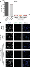
Fig 3. Inactivation of HSV-1 infected cells.
Vero B4 cells infected with HSV-1 were UV-inactivated. (A) The virus titres of samples taken during inactivation validation were determined with a limit of detection (LoD) of 5 PFU/mL. The before inactivation and UV inactivated samples were performed with biological replicates, n = 3, and the standard deviation is shown. The untreated control sample was performed with n = 1. (B) Immunofluorescence imaging of cells treated with the UV-inactivated infected cells was performed with biological replicates, n = 3. Cells were stained for the HSV-1 protein ICP0 and with DAPI. The scale bar represents 100 μm.
Fig 4
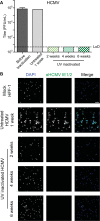
Fig 4. Inactivation of HCMV infected cells.
HFF-1 cells infected with HCMV were UV-inactivated. (A) The virus titres of samples taken during inactivation validation were determined with a limit of detection (LoD) of 5 PFU/mL. The before inactivation and UV inactivated samples were performed with biological replicates, n = 3, and the standard deviation is shown. The untreated control sample was performed with n = 1. (B) Immunofluorescence imaging of cells treated with the UV-inactivated infected cells was performed with biological replicates, n = 3. Cells were stained for the HCMV proteins IE1/2 and with DAPI. The scale bar represents 100 μm.
Fig 5
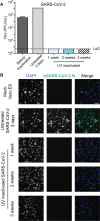
Fig 5. Inactivation of SARS-CoV-2 infected cells.
Vero E6 cells infected with SARS-CoV-2 were UV-inactivated. (A) The virus titres of samples taken during inactivation validation were determined with a limit of detection (LoD) of 5 PFU/mL. The before inactivation and UV inactivated samples were performed with biological replicates, n = 3, and the standard deviation is shown. The untreated control sample was performed with n = 1. (B) Immunofluorescence imaging of cells treated with the UV-inactivated infected cells was performed with biological replicates, n = 3. Cells were stained for the SARS-CoV-2 protein N and with DAPI. The scale bar represents 100 μm.

References

    1. Olschewski S, Thielebein A, Hoffmann C, Blake O, Muller J, Bockholt S, et al.. Validation of Inactivation Methods for Arenaviruses. Viruses. 2021;13(6). doi: 10.3390/v13060968 ; PubMed Central PMCID: PMC8225210. - DOI - PMC - PubMed
    1. Ravanat JL, Douki T, Cadet J. Direct and indirect effects of UV radiation on DNA and its components. J Photochem Photobiol B. 2001;63(1–3):88–102. doi: 10.1016/s1011-1344(01)00206-8 . - DOI - PubMed
    1. Nishisaka-Nonaka R, Mawatari K, Yamamoto T, Kojima M, Shimohata T, Uebanso T, et al.. Irradiation by ultraviolet light-emitting diodes inactivates influenza a viruses by inhibiting replication and transcription of viral RNA in host cells. J Photochem Photobiol B. 2018;189:193–200. doi: 10.1016/j.jphotobiol.2018.10.017 . - DOI - PubMed
    1. Tsunetsugu-Yokota Y. Large-scale preparation of UV-inactivated SARS coronavirus virions for vaccine antigen. Methods Mol Biol. 2008;454:119–26. doi: 10.1007/978-1-59745-181-9_11 ; PubMed Central PMCID: PMC7122600. - DOI - PMC - PubMed
    1. Heilingloh CS, Aufderhorst UW, Schipper L, Dittmer U, Witzke O, Yang D, et al.. Susceptibility of SARS-CoV-2 to UV irradiation. Am J Infect Control. 2020;48(10):1273–5. doi: 10.1016/j.ajic.2020.07.031 ; PubMed Central PMCID: PMC7402275. - DOI - PMC - PubMed

Publication types