. 2023 May 10;18(5):e0285253.
doi: 10.1371/journal.pone.0285253. eCollection 2023.

Heterozygosity for ADP-ribosylation factor 6 suppresses the burden and severity of atherosclerosis

Affiliations

Heterozygosity for ADP-ribosylation factor 6 suppresses the burden and severity of atherosclerosis

Venkateswara R Gogulamudi et al. PLoS One. .

Abstract

Atherosclerosis is the root cause of major cardiovascular diseases (CVD) such as myocardial infarction and stroke. ADP-ribosylation factor 6 (Arf6) is a ubiquitously expressed GTPase known to be involved in inflammation, vascular permeability and is sensitive to changes in shear stress. Here, using atheroprone, ApoE-/- mice, with a single allele deletion of Arf6 (HET) or wildtype Arf6 (WT), we demonstrate that reduction in Arf6 attenuates atherosclerotic plaque burden and severity. We found that plaque burden in the descending aorta was lower in HET compared to WT mice (p˂0.001) after the consumption of an atherogenic Paigen diet for 5 weeks. Likewise, luminal occlusion, necrotic core size, plaque grade, elastic lamina breaks, and matrix deposition were lower in the aortic root atheromas of HET compared to WT mice (all p≤0.05). We also induced advanced human-like complex atherosclerotic plaque in the left carotid artery using partial carotid ligation surgery and found that atheroma area, plaque grade, intimal necrosis, intraplaque hemorrhage, thrombosis, and calcification were lower in HET compared to WT mice (all p≤0.04). Our findings suggest that the atheroprotection afforded by Arf6 heterozygosity may result from reduced immune cell migration (all p≤0.005) as well as endothelial and vascular smooth muscle cell proliferation (both p≤0.001) but independent of changes in circulating lipids (all p≥0.40). These findings demonstrate a critical role for Arf6 in the development and severity of atherosclerosis and suggest that Arf6 inhibition can be explored as a novel therapeutic strategy for the treatment of atherosclerotic CVD.


Conflict of interest statement

The authors have declared that no competing interests exist.

Figures
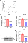
Fig 1
Fig 1. Low oscillatory shar stress Arf6 heterozygosity decreases Arf6 expression.
(A) Gene expression of Arf6 in unligated (right) and partially ligated (left) carotid arteries, (B,C) Gene expression fo Arf from endothelial cell (EC) enriched and vascular smooth muscle cell (VSMC) enriched carotid artery lysates from wildtype (WT) and Arf6 heterozygote (HET) mice (N = 3-4/group), (D) primary lung EC protein expression fo Arf6 and β-actin from WT and Arf6 HET mice, (E) densitometric quantification of western blotting data. (N = 3/replicate, ECs for each replicate were pooled from lungs of 3–5 mice). Data are presented as individual pairs of unligated and partially ligated datapoints (A) or mean ± SEM with individual datapoints (B-E). Group differences were assessed using paired (A) or unpaired (B-E) student’s t test.
Fig 2
Fig 2. Arf6 heterozygosity attenuates burden and severity of spontaneous atherosclerotic plaque.
(A, B) representative images and quantification of descending aortic plaque from wildtype (WT) and Arf6 heterozygote (HET) mice (N = 15/group), (C) representative images of Movat’s pentachrome stained aortic root sections for atheroma and necrotic core areas, luminal occlusion and plaque grades. Note: The fibrous caps overlying necrotic cores in the WT sample (blue arrowheads), (D) aortic root atheroma area (N = 11/group), (E) percent luminal occlusion of aortic root (n = 11-12/group), (F) area of necrotic core (1 = 11/group) and (G) plaque grade (N = 11/group). (H) Representative images of Movat’s pentachrome stained aortic root sections for non-muscular matrix (teal or yellow color) and the elastic laminae (black lines). (I) Matrix deposition score (N = = 9/group) and (J) counts of elastin lamina breaks in WT and Arf6 HET mice(N = 9/group). Data are presented as mean ± SEM with individual datapoints. Scale bars indicate 100 μm. Group differences were assessed using unpaired student’s t test for continuous variables and Mann-Whitney tests for scaled variables.
Fig 3
Fig 3. Arf6 heterozygosity attenuates the size and severity of oscillatory shear stress induced advanced human-like carotid artery atherosclerotic plaque.
Quantification and scoring of left carotid artery sections after five weeks of partial carotid ligation surgery and atherogenic diet consumption (A) Representative images of hematoxylin and eosin as well as Masson’s trichrome stained left carotid artery sections. The arterial lumen is indicated by *. In the WT sample, there is loss of the endothelial lining, and a luminal thrombus is present. Hemorrhage within the plaque is also present (black arrowhead), (B) total atheroma area (N = 8-10/group), (C) plaque grade (N = 7-10/group) (D) intimal necrosis (N = 8-10/group), (E) intraplaque hemorrhage (N = 8-10/group), (F) thrombosis (N = 8-10/group), and (G) calcification (N = 8-10/group), of WT and Arf6 HET mice. Data are presented as mean ± SEM with individual datapoints. Scale bars indicate 100 μm. Group differences were assessed using unpaired student’s t test for continuous variables and Mann-Whitney tests for scaled variables.
Fig 4
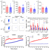
Fig 4. Arf6 heterozygosity does not alter plasma lipids but reduces immune cell migration as well as endothelial and smooth muscle cell proliferation.
(A) Plasma total cholesterols (N = 7-8/group), (B) plasma low-density and very low-density lipoprotein (LDL/VLDL) (N = 7-8/group), (C) plasma high-density lipoprotein (HDL) (N = 7-8/group), (D) plasma triglycerides (N = 7-8/group). (E) Representative images of characterization and counting of total and migrated T cells using flow cytometry, (F) baseline and C-C motif ligand 2 (CCL2)-induced migration of lymphocytes, total T cells, helper T cells and cytotoxic T cells (N = 6/replicate) (G, H) in vitro resistance, a measure of proliferation, of primary vascular smooth muscle cell (VSMC) and lung endothelial cell (EC). N = 6/replicate for SMC proliferation and 3/replicate for EC proliferation. SMC and EC for each replicate were pooled from thoracic aorta and lungs of 3–5 mice, respectively. Data are presented as mean ± SEM. Group differences were assessed using unpaired student’s t test (lipid profile), two-way ANOVA (T cell migration) or repeated measure ANOVA (EC and VSMC proliferation).

References

    1. Islam T. Impact of statins on vascular smooth muscle cells and relevance to atherosclerosis. J Physiol. 2020;598(12):2295–6. Epub 2020/04/25. doi: 10.1113/JP279774 . - DOI - PubMed
    1. Bentzon JF, Otsuka F, Virmani R, Falk E. Mechanisms of plaque formation and rupture. Circ Res. 2014;114(12):1852–66. Epub 2014/06/07. doi: 10.1161/CIRCRESAHA.114.302721 . - DOI - PubMed
    1. Bloom SI, Islam MT, Lesniewski LA, Donato AJ. Mechanisms and consequences of endothelial cell senescence. Nat Rev Cardiol. 2022. Epub 20220719. doi: 10.1038/s41569-022-00739-0 . - DOI - PMC - PubMed
    1. Gimbrone MA Jr., García-Cardeña G. Endothelial Cell Dysfunction and the Pathobiology of Atherosclerosis. Circ Res. 2016;118(4):620–36. Epub 2016/02/20. doi: 10.1161/CIRCRESAHA.115.306301 . - DOI - PMC - PubMed
    1. Islam MT, Hall SA, Lesniewski LA. Chronic aerobic exercise: targeting two birds with one stone. J Physiol. 2021;599(4):1015–6. Epub 20210104. doi: 10.1113/JP280929 . - DOI - PMC - PubMed

Publication types

Substances