. 2023 Aug 7;222(8):e202211074.
doi: 10.1083/jcb.202211074. Epub 2023 May 10.

Centralspindlin proteins Pavarotti and Tumbleweed along with WASH regulate nuclear envelope budding

Affiliations

Centralspindlin proteins Pavarotti and Tumbleweed along with WASH regulate nuclear envelope budding

Kerri A Davidson et al. J Cell Biol. .

Abstract

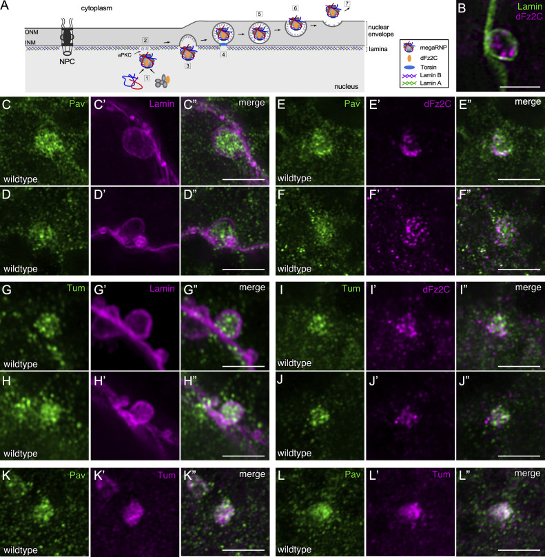
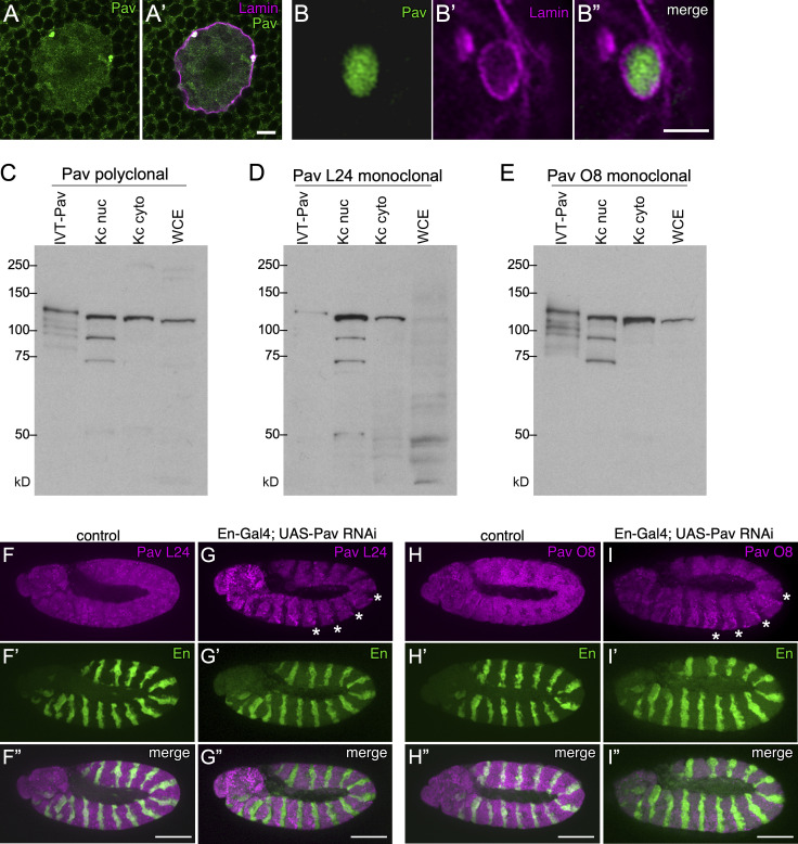
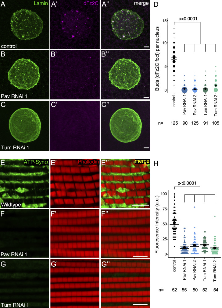
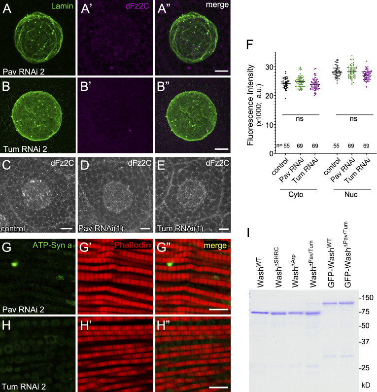
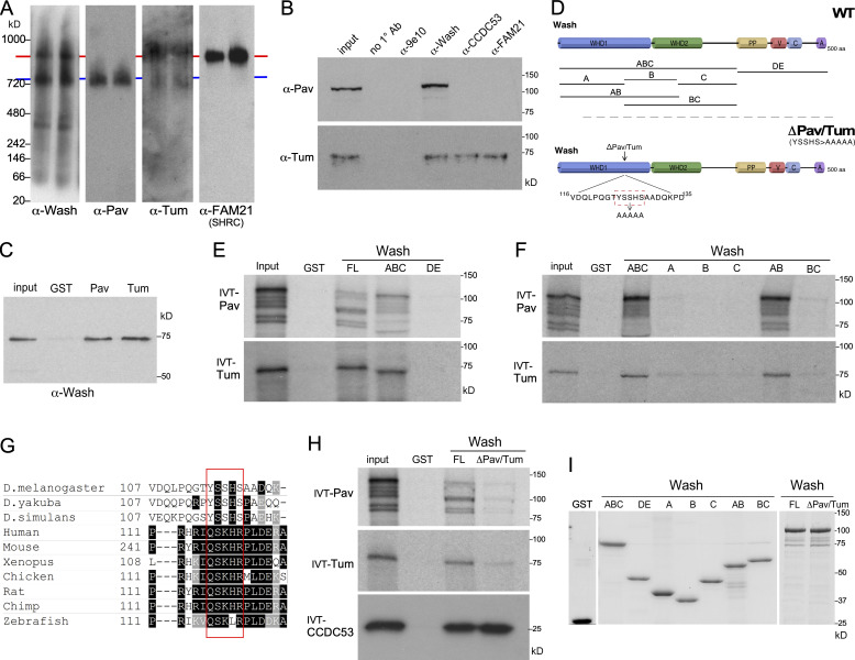
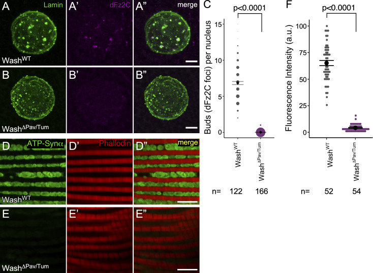
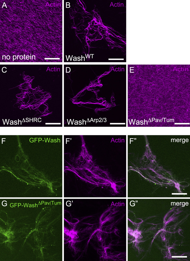
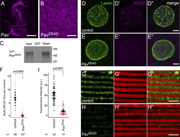
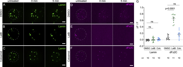
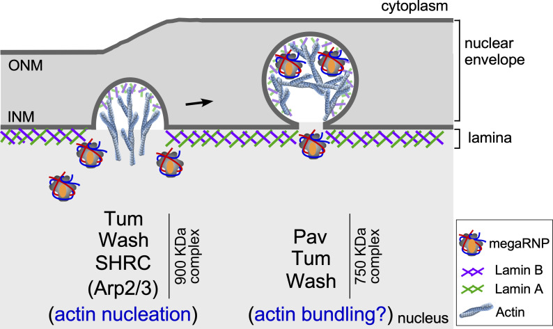
Nuclear envelope (NE) budding is a nuclear pore-independent nuclear export pathway, analogous to the egress of herpesviruses, and required for protein quality control, synapse development, and mitochondrial integrity. The physical formation of NE buds is dependent on the Wiskott-Aldrich Syndrome protein, Wash, its regulatory complex (SHRC), and Arp2/3, and requires Wash's actin nucleation activity. However, the machinery governing cargo recruitment and organization within the NE bud remains unknown. Here, we identify Pavarotti (Pav) and Tumbleweed (Tum) as new molecular components of NE budding. Pav and Tum interact directly with Wash and define a second nuclear Wash-containing complex required for NE budding. Interestingly, we find that the actin-bundling activity of Pav is required, suggesting a structural role in the physical and/or organizational aspects of NE buds. Thus, Pav and Tum are providing exciting new entry points into the physical machineries of this alternative nuclear export pathway for large cargos during cell differentiation and development.


Conflict of interest statement

Disclosures: The authors declare no competing interests exist.

Figures

Figure 1.
Figure 1.
Pav and Tum are expressed in the nucleus and accumulate at NE buds. (A) Schematic of the proposed steps in the NE budding process: megaRNPs visualized by dFz2C are assembled [1], the nuclear lamina is modified by aPKC [2], the INM and lamina begins to bend and megaRNPs enter this membrane deformation [3] and subsequently are encapsulated by the INM [4], scission of the INM occurs [5], followed by INM fusion with the outer nuclear membrane (ONM) [6], and megaRNP exit into cytoplasm [7]. (B) Super-resolution (Airyscan) micrograph projection of wildtype larval salivary gland nucleus stained with antibodies to Lamin B and dFz2C. Large Lamin B and dFz2C positive puncta indicate NE buds. (C–L″) Super-resolution micrograph projections of NE buds from wildtype larval salivary gland nuclei stained with antibodies to Lamin B and Pav (C–D″), Pav and dFz2C (E–F″), Lamin B and Tum (G–H″), Tum and dFz2C (I–J″), and Pav and Tum (K–L″). Scale bars: 0.5 μm.
Figure S1.
Figure S1.
Pav antibody specificity. (A–B″) Super-resolution (Airyscan) micrograph projection of wildtype larval salivary gland nucleus (A and A′) or higher magnification NE bud (B–B″) stained with antibodies to Lamin and Pav. This rabbit polyclonal Pav antibody is from the Glover lab and described in Adams et al. (1998). (C–E) Specificity of Pav mouse polyclonal and monoclonal lines L24 and O8. Western blot of in vitro translated (IVT) full-length Pav protein, Kc cell nuclear extract, Kc cell cytoplasmic extract, and whole-cell 0–2 h Drosophila embryo extract (WCE) probed with Pav polyclonal (C) or Pav monoclonal lines L24 (D) or O8 (E). (F–I″) Staining of control (F–F″ and H–H″) or En-Gal4; UAS-Pav RNAi (G–G″ and I–I″) embryos with antibodies to the Pav L24 (F–G″) or Pav O8 (H–I″) monoclonal lines to show antibody specificity. Note that these monoclonal antibodies do not stain the En regions where Pav is knocked down (asterisks). Scale bars: A and A′: 5 μm, B–B″: 0.5 μm, F–I″: 100 μm. Source data are available for this figure: SourceData FS1.
Figure 2.
Figure 2.
Pav and Tum knockdown nuclei lack NE buds and display NE bud–associated phenotypes. (A–C″) Confocal micrograph projections of control (A–A″), Pav RNAi (B–B″), and Tum RNAi (C–C″) larval salivary gland nuclei stained with Lamin B and dFz2C. (D) Quantification of NE buds per nucleus from two independent Pav and Tum RNAi lines. The number of nuclei assayed (n) is indicated. (E–G″) Confocal micrograph projection of adult IFM from control (+/Sgs-Gal4; E–E″), Pav RNAi (F–F″), Tum RNAi (G–G″) aged 21 d then stained with the activity-dependent mitochondrial marker (ATP-Synthetase α) and with Phalloidin. (H) Quantification of mitochondrial fluorescence intensity in IFM. The number of IFM muscles assayed (n) is indicated. Error bars represent ± SEM. Kruskal–Wallis test (D and H); all P values indicated. Scale bars: 5 μm.
Figure S2.
Figure S2.
pav/tum mutant characterization. (A–B″) Confocal micrograph projections of Pav RNAi 2 (A–A″) and Tum RNAi 2 (B–B″) larval salivary gland nuclei stained with Lamin B and dFz2C. (C–E) Confocal micrograph of a larval salivary gland cell from control (C), Pav RNAi 1 (D), and Tum RNAi 1 (E) stained with dFz2C. (F) Quantification of dFz2C levels in the cytoplasm (Cyto) or nucleus (Nuc) for control, Pav RNAi 1, and Tum RNAi 1. The number of salivary gland cells assayed (n) is indicated. Error bars represent ± SEM. Kruskal–Wallis test was used. (G–H″) Confocal micrograph projections of adult IFM from Pav RNAi 2 (G–G″) and Tum RNAi 2 (H–H″) flies aged 21 d then stained for the activity-dependent mitochondrial marker ATP-Synthetase α and phalloidin. (I) Coomassie-stained gel of bacterially purified proteins used for the F-actin bundling assays. Scale bars: A–B″, G–H″: 5 μm; C–E: 20 μm. Source data are available for this figure: SourceData FS2.
Figure 3.
Figure 3.
Pav and Tum interact with Wash in a conserved hydrophobic region within Wash’s WHD1 domain. (A) Western blots from blue native PAGE of Drosophila Kc cell nuclear extracts probed with antibodies to Wash, Pav, Tum, and FAM21 (SHRC subunit). Putative ∼900 kD complex with Wash, Tum, and the SHRC (red line) and ∼720 kD complex with Wash, Pav, and Tum (blue line) are indicated. (B) Western blots of immunoprecipitations from Kc cell nuclear extracts with no primary antibody included (no 1° Ab), a non-specific antibody (9e10), Wash, CCDC53, and FAM21. Blots were probed with antibodies to Pav and Tum as indicated. (C) GST pulldown experiments with bacterially purified proteins demonstrating Pav and Tum bind directly to full-length Wash. (D) Schematic diagram of the Wash wildtype rescue and substitution mutation constructs indicating the position and specific substitution mutations for the WashΔPav/Tum construct and the Wash fragments used for mapping (not drawn to scale). PP: polyproline domain, V: Verprolin homology domain, C: central/connecting/cofilin-like domain, A: acidic domain. (E and F) GST pulldown assays with 35S-labeled in vitro translated (IVT) Pav and Tum and bacterially purified GST-Wash full length and fragments indicated. 10% input is shown. (G) Sequence alignment of the Wash region required for binding to Pav and Tum (red box). (H) GST pulldown assays with 35S-labeled in vitro translated Pav and Tum and bacterially purified GST-WashWT and GST-WashΔPav/Tum. 10% input shown. (I) Coomassie-stained gel of purified GST fusion proteins used in this study to examine protein interactions among Wash, Pav, and Tum. Source data are available for this figure: SourceData F3.
Figure 4.
Figure 4.
Wash binding to Pav and Tum is required for NE budding. (A–B″) Confocal micrograph projections of larval salivary gland nuclei from washWT (A–A″) and washΔPav/Tum (B–B″) stained with Lamin B and dFz2C. (C) Quantification of NE buds per nucleus in larval salivary gland nuclei. The number of nuclei assayed (n) is indicated. (D–E″) Confocal micrograph projections of adult IFM from washWT (D–D″) and washΔPav/Tum (E–E″) flies aged 21 d then stained for the activity dependent mitochondrial marker ATP-Synthetase α and with phalloidin. (F) Quantification of ATP-Synthetase α fluorescence intensity from adult IFMs. The number of IFM muscles assayed (n) is indicated. Error bars represent ± SEM. Kruskal–Wallis test (C and F); all P values indicated. Scale bars: 5 μm.
Figure 5.
Figure 5.
The washΔPav/Tum mutation binds, but can no longer bundle, actin. (A–E) Phalloidin-stabilized actin filaments incubated with no protein (A), WashWT (B), WashΔSHRC (C), WashΔArp2/3 (D), or WashΔPav/Tum (E) proteins. Note the lack of actin bundling in the presence of WashΔPav/Tum protein. (F–G″) Binding of sfGFP-Wash (F–F″) and sfGFP-WashΔPav/Tum (G–G″) to phalloidin-stabilized actin filaments that were bundled with Capu (formin) protein. Final protein concentrations for bundling assays: WashWT, 500 nM; WashΔSHRC, 500 nM; WashΔArp2/3, 500 nM; WashΔPav/Tum, 500 nM; CapuFH2, 500 nM; sfGFP-Wash, 50 nM; and sfGFP-WashΔPav/Tum, 50 nM. Scale bar: 30 µm.
Figure 6.
Figure 6.
Bundled actin is required for NE bud formation. (A and B) Phalloidin-stabilized actin filaments were incubated with PavWT (A) and PavDEAD (B) protein. Final protein concentrations for bundling assays: PavWT, 200 nM; PavDEAD, 200 nM. (C) GST pulldown assays with 35S-labeled in vitro translated PavDEAD and bacterially purified GST alone or GST-Wash full-length protein as indicated. 10% input is shown. (D–E″) Confocal micrograph projections of larval salivary gland nuclei from control (D–D″) and PavDEAD (E–E″) stained with Lamin B and dFz2C. (F) Quantification of NE buds per nucleus in larval salivary gland nuclei. The number of nuclei assayed (n) is indicated. (G–H″) Confocal micrograph projections of adult IFM from control (G–G″) and PavDEAD (H–H″) flies aged 21 d then stained for the activity dependent mitochondrial marker ATP-Synthetase α and phalloidin. (I) Quantification of ATP-Synthetase α fluorescence intensity from adult IFMs. The number of IFM muscles assayed (n) is indicated. Error bars represent ± SEM. Kruskal–Wallis test (F and I); all P values indicated. Scale bars: 30 μm in A and B; 5 μm in D–E″ and G–H″. Source data are available for this figure: SourceData F6.
Figure 7.
Figure 7.
Retention of dFz2C within NE buds requires actin. (A–F) Time-lapse confocal xy projection images from Drosophila salivary gland nuclei expressing GFP-Lamin (A–C) or dFz2-Scarlet (D–F) treated with DMSO (A and D), LatB (B and E), or colcemid (C and F) at the times indicated. (G) Quantification of the GFP-Lamin and dFz2-Scarlet in the conditions indicated. The number of nuclei assayed (n) is indicated. Error bars represent ± SEM. Two-tailed Student’s t test; all P values indicated. Scale bars: 10 μm.
Figure 8.
Figure 8.
Roles of Wash, Pav, and Tum in NE bud formation and/or organization. Schematic diagram of NE budding highlighting the proposed roles of the two Wash-containing nuclear complexes. The ∼900 kD Wash/SHRC/Tum complex is proposed to mediate branched actin polymerization that aids in physical NE bud formation. The ∼750 kD Wash/Pav/Tum complex is proposed to use actin bundling to aid in the internal infrastructure/organization of NE buds.

Similar articles

Cited by

References

    1. Adams, R.R., Tavares A.A., Salzberg A., Bellen H.J., and Glover D.M.. 1998. Pavarotti encodes a kinesin-like protein required to organize the central spindle and contractile ring for cytokinesis. Genes Dev. 12:1483–1494. 10.1101/gad.12.10.1483 - DOI - PMC - PubMed
    1. Bigalke, J.M., and Heldwein E.E.. 2015. The great (nuclear) escape: New insights into the role of the nuclear egress complex of herpesviruses. J. Virol. 89:9150–9153. 10.1128/JVI.02530-14 - DOI - PMC - PubMed
    1. Bigalke, J.M., and Heldwein E.E.. 2016. Nuclear exodus: Herpesviruses lead the way. Annu. Rev. Virol. 3:387–409. 10.1146/annurev-virology-110615-042215 - DOI - PMC - PubMed
    1. Blanchoin, L., Boujemaa-Paterski R., Sykes C., and Plastino J.. 2014. Actin dynamics, architecture, and mechanics in cell motility. Physiol. Rev. 94:235–263. 10.1152/physrev.00018.2013 - DOI - PubMed
    1. Buracco, S., Claydon S., and Insall R.. 2019. Control of actin dynamics during cell motility. F1000 Res. 8:F1000 Faculty Rev-1977. 10.12688/f1000research.18669.1 - DOI - PMC - PubMed

Publication types

MeSH terms

LinkOut - more resources