. 2023 May 10;9(19):eadf5336.
doi: 10.1126/sciadv.adf5336. Epub 2023 May 10.

Dodecamer assembly of a metazoan AAA+ chaperone couples substrate extraction to refolding

Affiliations

Dodecamer assembly of a metazoan AAA+ chaperone couples substrate extraction to refolding

Arpit Gupta et al. Sci Adv. .

Abstract

Ring-forming AAA+ chaperones solubilize protein aggregates and protect organisms from proteostatic stress. In metazoans, the AAA+ chaperone Skd3 in the mitochondrial intermembrane space (IMS) is critical for human health and efficiently refolds aggregated proteins, but its underlying mechanism is poorly understood. Here, we show that Skd3 harbors both disaggregase and protein refolding activities enabled by distinct assembly states. High-resolution structures of Skd3 hexamers in distinct conformations capture ratchet-like motions that mediate substrate extraction. Unlike previously described disaggregases, Skd3 hexamers further assemble into dodecameric cages in which solubilized substrate proteins can attain near-native states. Skd3 mutants defective in dodecamer assembly retain disaggregase activity but are impaired in client refolding, linking the disaggregase and refolding activities to the hexameric and dodecameric states of Skd3, respectively. We suggest that Skd3 is a combined disaggregase and foldase, and this property is particularly suited to meet the complex proteostatic demands in the mitochondrial IMS.


Figures
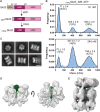
Fig. 1.
Fig. 1.. PARLSkd3 assembles into both hexamer and dodecamer.
(A) Scheme depicting the domain composition of Skd3 and its sequential proteolytic processing in mitochondria. (B) Mass photometry measurements of the oligomeric state of PARLSkd3_WB at different concentrations in the presence of 2 mM ATP. Gaussian fits of the data (black lines) yield the indicated population of particles in each assembly state. The measured masses of the three populations are indicated and consistent with the predicted molecular weight of PARLSkd3 monomer (66 kDa), hexamer (396 kDa), and dodecamer (792 kDa). (C) Representative top and side views of cryo-EM 2D class averages of ATPγS-bound PARLSkd3_WB. Scale bar, 100 Å. (D) Low pass–filtered cryo-EM maps of the Skd3 hexamer. Green highlights the EM density for the bound substrate. (E) Cryo-EM map of the Skd3 dodecamer.
Fig. 2.
Fig. 2.. Cryo-EM structures of substrate-bound PARLSkd3 hexamers.
(A and B) Top (left) and side (right) of the high-resolution sharpened EM density map (A) and molecular model (B) for the Skd3 NBD ring. The bound substrate is in green. (C) Left, overview of the arrangement of the pore loops surrounding the bound substrate in the open spiral conformation of the Skd3 NBD. Right, zoom in of the contacts of the pore 1 and pore 2 loops with substrate and with one another. (D) Active site interaction of the bound ATP in the P4 protomer. (E) Comparison of the 3DVA-derived EM density maps for the closed and open spiral conformations of the Skd3 NBD. A lower threshold for the EM density was used to visualize the flexible P6 protomer in the closed conformation. Color coding and the position of the individual protomers are indicated in the scheme below. (F) The Skd3 ARD and the interdomain linker were fit as a rigid body to the non-NBD EM density in the low-pass filtered density map of the PARLSkd3 hexamer.
Fig. 3.
Fig. 3.. PARLSkd3 dodecamer forms a fenestrated cylindrical cage with the ARD mediating the interface between hexamers.
(A) Side and face views of the EM density maps of the upper (red) and lower (blue) NBD rings of the Skd3 dodecamer after focused refinement. The masks used for focused refinement are shown. (B) Cut-through view of the PARLSkd3 dodecamer. Folded luciferase in surface representation [Protein Data Bank (PDB) #1LCI] is shown for comparison. (C) Left, overlay of the dodecamer map with model. The molecular model of the open Skd3 NBD (Fig. 2B) was fit as a rigid body to the NBD density. The AlphaFold-predicted model of the ARD-interdomain linker region was fit as a second rigid body to the non-NBD density. Inset, close-up of the ARDs highlighting conserved turn loop residues bridging the dodecamer interface. (D) CONSURF analysis of the ARD of human Skd3 isoform 2. Residues are colored by CONSURF scores (60) across 791 homologous Skd3 sequences. Mutated turn loop residues are labeled in red. Consensus ARM residues are shown above (black). (E) Aggregated luciferase (U4L0.2 protocol) was presented to the indicated Skd3 variants in the presence of 5 mM ATP and ARS, and the generation of enzymatically active luciferase was measured using chemiluminescence (see Materials and Methods). Data are represented as mean ± SD, with n = 3 technical replicates. (F) Mass photometry measurements of the assembly state of PARLSkd3_L1,L2GH in the presence of 2 mM ATP. The WB mutation was introduced to facilitate hexamer formation. Gaussian fits of the data (black lines) gave the indicated populations in the monomer, hexamer, and dodecamer states and are shown as mean ± SE.
Fig. 4.
Fig. 4.. PARLSkd3 releases luciferase in a conformation committed to folding.
(A) Alternative models for luciferase disaggregation and refolding mediated by PARLSkd3 and the use of GroEL-D87K to distinguish between the models. (B) Refolding of aggregated luciferase (U4L0.2 protocol) by PARLSkd3 was measured as in Fig. 3E in the presence and absence of the indicated concentrations of GroEL-D87K. (C) Luciferase denatured in 5 M GdmHCl was diluted to 50 nM in refolding buffer. Where indicated, the reaction also contained 1 μM PARLSkd3 and/or 320 nM GroEL-D87K. Refolded luciferase was detected by chemiluminescence. (D) Luciferase denatured in 5 M GdmHCl was diluted to 50 nM in refolding buffer containing 1 μM Skd3; where indicated, 320 nM GroEL-D87K was added 5 min later, and refolding was measured by chemiluminescence. Spontaneous luciferase refolding with and without GroEL-D87K was measured in parallel and subtracted. (E) Aggregated luciferase (U8L0.5 protocol) was mixed with 1 μM PARLSkd3 in the presence of 5 mM ATP and ARS to initiate disaggregation and refolding. Fifteen minutes later, soluble proteins were chromatographed on a Superdex 200 column, and 0.5 ml fractions was collected and analyzed. Enzymatically active luciferase in individual fractions was detected using chemiluminescence (blue). Individual fractions were also probed with anti-His (for luciferase, green) and anti-ClpB (for Skd3, magenta) antibodies. See fig. S9 for raw data. Data are shown as mean ± SEM in (B) to (D) or mean ± SD in (E), with n = 3 technical replicates. Error bars are plotted but may not be visible.
Fig. 5.
Fig. 5.. Dodecamer assembly–deficient mutants uncouple client refolding from disaggregation.
(A) Refolding of aggregated luciferase mediated by the indicated Skd3 variants, measured as in Fig. 3E. (B) Mass photometry measurement of MPPΔ3Skd3_WB in the presence of 2 mM ATP. Gaussian fits of the data (black lines) gave the indicated population in the monomer, hexamer, and dodecamer states. (C) Summary of the dodecamer/hexamer population ratio for PARLSkd3 and the dodecamer assembly–defective Skd3 mutants. (D and E) Disaggregation of luciferase by PARLSkd3, Skd3_L1,L2GH (D), and MPPΔ3Skd3 (E) was measured as described in Materials and Methods. Top, representative Western blot analysis of the soluble and total luciferase; bottom, quantification of the data. cpSRP43, a plant-derived protein, was used as a loading control. (F and G) Refolding of aggregated luciferase (U4L0.2 protocol) by Skd3_L1,L2GH (F) and MPPD3Skd3 (G) was measured as in Fig. 3D in the absence and presence of the indicated concentrations of GroEL-D87K. (H) The effect of GroEL-D87K on luciferase refolding is compared between wild-type PARLSkd3 (red), Skd3_L1,L2GH (green), and MPPΔ3Skd3 (dark green). The amount of refolded luciferase at 150 min for reactions containing GroEL-D87K was normalized to that without the trap for each Skd3 variant. Data are shown as mean ± SEM in (A) and (F) to (H) or mean ± SD in (D) and (E), with n = 3 technical replicates. Error bars are plotted but may not be visible.
Fig. 6.
Fig. 6.. Skd3 ARD can participate in substrate interaction.
(A) Side view of the ARD showing the surface that faces the internal cavity in the Skd3 dodecamer. Color coding for CONSURF scores is the same as in Fig. 3. In the inset, conserved hydrophobic residues (CONSURF ≥ 6) on the surface of the ARD (gray) and the insertion domain (aqua) are highlighted. Residues tested by mutagenesis in (B) and (C) are color-indicated. (B and C) Refolding of aggregated luciferase mediated by the indicated Skd3 variants, measured as in Fig. 3E. (D) Equilibrium titrations to measure the binding of FITC-casein to PARLSkd3 and ARD in the indicated nucleotide states. Lines are fits of the data to Eq. 2, and the obtained Kd values are summarized. (E and F) PARLSkd3 and ARD delay Aβ42 fibrillation under ATP-depleted conditions. Monomeric Aβ42 (5 μM) was incubated with and without the indicated concentrations of PARLSkd3 (E) or ARD (F) under quiescent conditions without added ATP and ARS. Fibril formation was monitored by ThT fluorescence every 5 min. The dark lines show the mean of three replicates, and the shaded area shows the range of data. The data were analyzed by the AmyloFit algorithm, from which fibrillation reaction halftimes were extracted and summarized in (G). (G) Summary of the effect of PARLSkd3 variants on the kinetics of Aβ42 fibrillation. “t1/2” denotes the half-time of Aβ42 fibrillation so that 1/t1/2 represents the apparent rate constant of the reaction. Data are shown as mean ± SEM, with n = 3 technical replicates. See fig. S12 for raw data. (H) Aβ42 fibrillation reactions were carried out with and without 2 μM of indicated chaperone. Reactions were imaged under TEM after 6 hours. Scale bar, 100 nm.
Fig. 7.
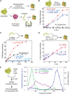
Fig. 7.. Working model for the coupled disaggregation and refolding reaction mediated by PARLSkd3.
Step 1, an Skd3 hexamer recognizes aggregated proteins and initiates substrate extraction. Step 2, an Skd3-bound substrate protein is dislodged from the aggregate. Step 3, Skd3 dodecamer assembles, and substrate protein initiates folding in the internal cavity. Step 4, substrate protein, either folded or in a folding-competent conformation, is released from Skd3 upon dodecamer disassembly. The inset shows the movement of the seam protomer (P6) from the lowest to the uppermost position as the NBD hexamer rearranges from the closed to the open spiral conformation, which generates a mechanical force that “pulls” the substrate polypeptide in the downward direction (red arrow). The question marks denote that the molecular mechanism underlying the transitions is still unclear.

References

    1. Kim Y. E., Hipp M. S., Bracher A., Hayer-Hartl M., Hartl F. U., Molecular chaperone functions in protein folding and proteostasis. Annu. Rev. Biochem. 82, 323–355 (2013). - PubMed
    1. Hartl F. U., Protein misfolding diseases. Annu. Rev. Biochem. 86, 21–26 (2017). - PubMed
    1. Rosenzweig R., Nillegoda N. B., Mayer M. P., Bukau B., The Hsp70 chaperone network. Nat. Rev. Mol. Cell Biol. 20, 665–680 (2019). - PubMed
    1. Kampinga H. H., Craig E. A., The HSP70 chaperone machinery: J proteins as drivers of functional specificity. Nat. Rev. Mol. Cell Biol. 11, 579–592 (2010). - PMC - PubMed
    1. Rampelt H., Mayer M. P., Bukau B., Nucleotide exchange factors for Hsp70 chaperones. Methods Mol. Biol. 1709, 179–188 (2018). - PubMed

Substances