Review
. 2023 Jul 6;119(8):1656-1675.
doi: 10.1093/cvr/cvad076.

Endothelial mechanobiology in atherosclerosis

Affiliations
Review

Endothelial mechanobiology in atherosclerosis

Xiaoli Wang et al. Cardiovasc Res. .

Abstract

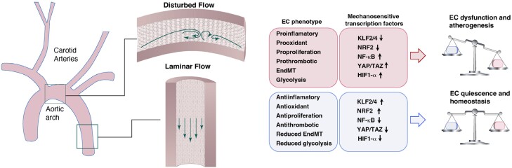
Cardiovascular disease (CVD) is a serious health challenge, causing more deaths worldwide than cancer. The vascular endothelium, which forms the inner lining of blood vessels, plays a central role in maintaining vascular integrity and homeostasis and is in direct contact with the blood flow. Research over the past century has shown that mechanical perturbations of the vascular wall contribute to the formation and progression of atherosclerosis. While the straight part of the artery is exposed to sustained laminar flow and physiological high shear stress, flow near branch points or in curved vessels can exhibit 'disturbed' flow. Clinical studies as well as carefully controlled in vitro analyses have confirmed that these regions of disturbed flow, which can include low shear stress, recirculation, oscillation, or lateral flow, are preferential sites of atherosclerotic lesion formation. Because of their critical role in blood flow homeostasis, vascular endothelial cells (ECs) have mechanosensory mechanisms that allow them to react rapidly to changes in mechanical forces, and to execute context-specific adaptive responses to modulate EC functions. This review summarizes the current understanding of endothelial mechanobiology, which can guide the identification of new therapeutic targets to slow or reverse the progression of atherosclerosis.

Keywords: Antiatherogenic drugs; Cardiovascular disease; Endothelial cells; Mechanotransduction; Shear stress.


Conflict of interest statement

Conflict of interest: Dr. Munn has received equity from Bayer AG.

Figures

Figure 1
Figure 1
Hemodynamic SS and its role in vessel pathophysiology. The straight part of the artery is exposed to sustained laminar flow and physiological high SS, while flow near branch points or in curved vessels can exhibit disturbed flow. ECs subjected to laminar flow show anti-inflammatory, antioxidant, and antiproliferative phenotypes accompanied by reduced EndMT and glycolysis, thereby maintaining EC quiescence and vascular homeostasis. Corresponding flow-responsive mechanisms include KLF2, KLF4, NRF2, and other protective pathways. By contrast, ECs in regions of disturbed flow show a proinflammatory, prooxidant, proproliferative response and an enhanced endothelial to EndMT phenotype. As a consequence, disturbed flow leads to EC dysfunction and atherogenesis. The mechanisms responsible for disturbed flow-mediated endothelial dysfunction involves activation of NF-κB, YAP/TAZ, and HIF-1 among other pathways. ↑: upregulation; ↓: downregulation.
Figure 2
Figure 2
Methods for investigating endothelial SS in vitro. (A) Modified cone and plate viscometer system for applying spatially uniform SS. (B) Shear ring device constructed from petri dishes, with flow driven by placement on a shaker plate. (C) Parallel plate flow chamber; flow is controlled by a syringe pump (not shown). (D) Step flow chamber. In this modification of the parallel plate flow chamber, a step expansion is introduced to produce localized flow separation/disturbance at the EC surface. (E) Bifurcating channel in a microfluidic device. (F) Cylindrical channel cast in a hydrogel (e.g. collagen I) and then coated with ECs.
Figure 3
Figure 3
In vivo methods for modulating endothelial SS. (A) In the SS–modifying cuff model, a restrictive cuff is placed around the right carotid artery (RCA) to modify blood flow. The resulting changes in SS can be estimated using CFD (at right). There is decreased laminar SS upstream and oscillatory SS downstream. (B) The partial ligation model. Three branches of the left carotid artery (LCA) are ligated, thus reducing SS in the LCA. (C) AVF model. A connection is made between the common carotid artery (CCA) and the external jugular vein (EJV) causing disturbed flow in multiple regions near the anastomosis. Right panel in part A is adapted from Mohri; right panel in part (B) is adapted from Mitra; right panel in part (C) is adapted from Bai..
Figure 4
Figure 4
Endothelial response to SS. ECs exposed to laminar flow are spindle shaped, aligned, and elongated parallel to the flow direction, with characteristics of lower turnover rate, increased anticoagulant activity, hypopermeability and less lipid accumulation, reduced leukocyte adhesion and migration, and inhibited EndMT, hence maintaining endothelial barrier function, while disturbed flow leads to a more polygonal morphology of ECs with random orientation and endothelial barrier disruption, which is characterized by accelerated turnover, decreased anticoagulant activity, hyperpermeability and lipid accumulation, enhanced leukocyte adhesion to endothelium, and the occurrence of EndMT.
Figure 5
Figure 5
Mechanosensors in the endothelium that sense and convert biomechanical cues into biological signals. The glycocalyx, primary cilia, ion channels, cell–cell and cell–substrate adhesion complexes, G-protein–coupled sensors, caveolae, and NE proteins (e.g. LINC complexes) have been implicated in the transduction of fluid forces.
Figure 6
Figure 6
EC mechanotransduction pathways involved in FSS–mediated endothelial function.

References

    1. Xu S, Ilyas I, Little PJ, Li H, Kamato D, Zheng X, Luo S, Li Z, Liu P, Han J, Harding IC, Ebong EE, Cameron SJ, Stewart AG, Weng J. Endothelial dysfunction in atherosclerotic cardiovascular diseases and beyond: from mechanism to pharmacotherapies. Pharmacol Rev 2021;73:924–967. - PubMed
    1. Souilhol C, Serbanovic-Canic J, Fragiadaki M, Chico TJ, Ridger V, Roddie H, Evans PC. Endothelial responses to shear stress in atherosclerosis: a novel role for developmental genes. Nat Rev Cardiol 2020;17:52–63. - PubMed
    1. He L, Zhang C-L, Chen Q, Wang L, Huang Y. Endothelial shear stress signal transduction and atherogenesis: from mechanisms to therapeutics. Pharmacol Therapeut 2022;235:108152. - PubMed
    1. Fung YC. Biomechanics: circulation. Shock 1998;9:155.
    1. Fung YC. Stress, strain, growth, and remodeling of living organisms. Z Angew Math Phys 1995;46:S469–S482.

Publication types