Severe Complications after Corneal Collagen Cross-Linking (CXL)
- PMID: 37164391
- PMCID: PMC10129411
- DOI: 10.1055/a-2040-4290
Severe Complications after Corneal Collagen Cross-Linking (CXL)
Abstract
Purpose: To present a case series of rare and severe complications after corneal collagen cross-linking (CXL) of keratoconus patients.
Methods: Single-center descriptive case series covering the period of 2012 to 2022 at the Department of Ophthalmology at the University Hospital, Zurich, Switzerland.
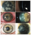
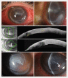
Results: We present four eyes of four patients that showed severe unusual complications within the first month after CXL. Three patients had been treated with the classical epithelium-off "Dresden" protocol. One patient had been treated with the accelerated epithelium-off protocol. One patient presented with extensive corneal edema due to rubbing the eye after treatment. Two patients showed a bacterial infectious keratitis: one due to Streptococcus pneumoniae and the other due to Staphylococcus hominis, Micrococcus luteus, and Streptococcus epidermidis. The latter of the two patients exhibited extensive infectious crystalline keratopathy. The fourth patient showed a severe ulcerative lesion where no infectious cause could be found. Therefore, an autoimmune keratolytic process had to be suspected. Apart from the corneal edema, which resolved ad integrum, the other complications resulted in permanent corneal scarring and thinning. One patient needed an emergency amniotic transplant.
Conclusion: Severe complications after CXL remain rare. Most common causes are complications that are not directly associated with the treatment as such. Those indirect complications occur after the treatment during the healing course of the epithelium. Associations with bandage contact lenses, topical steroids, atopic disease, and inappropriate patient behavior are often suspected. Correctly performed corneal scrapings with repeated microbiological analysis and a detailed patient history are essential for establishing the correct diagnosis, especially in complicated cases that do not respond to a standard therapeutic regimen. This case series supports the efforts that are currently taken to improve the CXL technique in a way that postoperative complications are further reduced. A more efficient epithelium-on technique might be a step in that direction.
ZIEL: Darstellung einer Fallserie von seltenen, schwerwiegenden Komplikationen nach kornealem Kollagen-Crosslinking (CXL) von Patienten mit Keratokonus.
Methoden: Monozentrische, beschreibende Fallserie über einen Zeitraum von 2012 bis 2022 an der Augenklinik des Universitätsspitals Zürich, Schweiz.
Ergebnisse: Wir zeigen 4 Augen von 4 Patienten, die mit ungewöhnlichen, schwerwiegenden Komplikationen innerhalb des 1. Monats nach CXL an unserer Klinik vorstellten. Drei der Patienten waren mit dem klassischen Epithelium-off-„Dresden“-Protokoll behandelt worden, ein Patient mit einem beschleunigten Epithelium-off-Protokoll. Ein Patient stellte sich mit einem ausgeprägten Hornhautstromaödem aufgrund von Reiben des Auges vor. Zwei Patienten zeigten eine infektiöse, bakterielle Keratitis mit Streptococcus pneumoniae bzw. Staphylococcus hominis, Micrococcus luteus, Streptococcus epidermidis. Der 4. Patient zeigte eine schwere ulzerative Läsion ohne nachweisbare infektiöse Genese, weshalb ein autoimmuner, keratolytischer Prozess vermutet wurde. Außer dem Hornhautödem, das sich vollständig zurückbildete, resultierten die anderen Komplikationen in einer bleibenden Vernarbung und Ausdünnung der Hornhaut. Ein Patient benötigte eine notfallmäßige Amnionmembrantransplantation.
Schlussfolgerung: Schwerwiegende Komplikationen nach CXL bleiben selten. Die häufigsten Ursachen hierfür sind Komplikationen, die nicht direkt mit der eigentlichen Prozedur zusammenhängen, sondern erst im postoperativen Heilungsverlauf des Hornhautepithels auftreten. Ein Zusammenhang mit therapeutischen Kontaktlinsen und unangemessenem Patientenverhalten wird in vielen Fällen angenommen. Korrekt durchgeführte Hornhaut-Scrapings mit wiederholter mikrobiologischer Aufarbeitung und eine detaillierte Patientenanamnese sind unerlässlich für eine korrekte Diagnosestellung, insbesondere in komplizierten Fällen, die nicht auf die üblichen Therapieregime ansprechen. Diese Fallserie unterstützt die aktuellen Bemühungen, die CXL-Technik dahingehend zu verbessern, sodass postoperative Komplikationen weiter reduziert werden. Eine effizientere Epithelium-on-Technik wäre ein Schritt in diese Richtung.
The Author(s). This is an open access article published by Thieme under the terms of the Creative Commons Attribution-NonDerivative-NonCommercial License, permitting copying and reproduction so long as the original work is given appropriate credit. Contents may not be used for commecial purposes, or adapted, remixed, transformed or built upon. (https://creativecommons.org/licenses/by-nc-nd/4.0/).
Conflict of interest statement
The authors declare that they have no conflict of interest.
Figures


References
-
- Feldmann B H, Bernfeld E, Reddy V.Corneal Collagen Cross-Linking. 2021. In: Shafer B, ed. https://eyewiki.aao.org/Corneal_Collagen_Cross-LinkingAccessed March 15, 2023 at:https://eyewiki.aao.org/Techniques_for_Corneal_Collagen_Crosslinking:_Ep...
MeSH terms
LinkOut - more resources
Full Text Sources

