

Case Reports
doi: 10.1055/a-2045-7314.
Epub 2023 Apr 25.
Posterior Lamellar Corneal Graft (DSAEK) in an Aphakic and Congenital Aniridic Single Eye: A Case Report Presenting a New Surgical Procedure
Affiliations
- PMID: 37164417
- PMCID: PMC10129408
- DOI: 10.1055/a-2045-7314
Item in Clipboard
Case Reports
Posterior Lamellar Corneal Graft (DSAEK) in an Aphakic and Congenital Aniridic Single Eye: A Case Report Presenting a New Surgical Procedure
Klin Monbl Augenheilkd.
2023 Apr.
No abstract available
Conflict of interest statement
The authors declare that they have no conflict of interest.
Figures

a
Right eye in 2007, 14 years before posterior lamellar graft was performed. Aniridia and aphakia were not associated with AAK (aniridia-associated keratopathy). The
pachymetry was 479 microns. Light scattering with the slit lamp showing corneal transparency with no vessels in the corneal stroma.
b
Right eye in 2021, just before the surgery.
Pachymetry has increased up to 920 microns at the thinnest point measured in the central 8 mm of the cornea.
c
Light scattering with the slit lamp just before the surgery showing
that AKK is still absent.

a
Shape of the DSAEK with the two “ears” prepared to stitch the graft in the corneal stroma and stabilize it during postoperative 24 hours tamponade.
b
In
green, the DSAEK and the two “ears” placed in the corneal stroma. In yellow, sagittal cut of a cornea.
c
Cutting the donor on a Moria anterior artificial anterior chamber with a
Moria Microkeratome.
d
Putting the donor endothelial cells side up on a 7.75 Moria trephine.
e
Breaking the trephine blade in two spots to create the “ears”.
f
Cutting
the donor with the trephine.
g
Creating the two “ears” on the donor with an 11 blade.
h
Creating the two “ears” on the donor with an 11 blade.
i
Creating the two
“ears” on the donor with an 11 blade.
j
Removing the graft from the corneal ring.
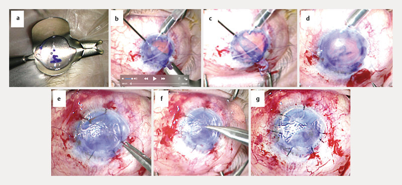
a
Placing the graft on the Busin glider.
b
Virgin silk stitches (7.0) were placed to hold the two “ears” through two side ports placed on a 9-mm diameter
circle face to face.
c
The graft is slid into the eye and held with intraocular forceps in one hand and with the glider and the stitch in the other hand.
d
The graft is held
through the two side ports with the stitches. The number “2” that we can read on the graft through the corneal stroma of the recipient confirms the good position of the DSAEK (stromal
side, up).
e
Side port is closed with a 10.0 nylon stitch.
f
Second side port is closed with a 10.0 nylon stitch.
g
Tamponade is performed with a mix of air and
SF
6
(50/50%) for 24 hours in the supine position.
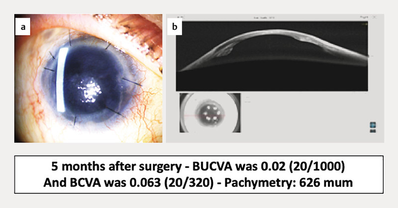
a
Five months after the surgery, the graft is attached to the posterior stroma of the recipient. Corrected vision increased up to 0.063 (20/320) and the thinnest
central corneal thickness was 626 microns.
b
Anterior segment OCT (OPTOVUE SOLIX, Visionix SA, Paris, France) exhibits the posterior lamellar graft attached to the posterior corneal
stroma of the recipient with one “ear” slipped into the corneal stroma through a side port.
Similar articles
-
[Effect of donor graft characteristics on clinical outcomes in Descemet stripping automated endothelial keratoplasty (DSAEK)].J Fr Ophtalmol. 2017 Jan;40(1):36-43. doi: 10.1016/j.jfo.2016.09.018. Epub 2017 Jan 6. J Fr Ophtalmol. 2017. PMID: 28069281 French.
-
Long-term follow-up of deep anterior lamellar keratoplasty after Descemet stripping automated endothelial keratoplasty.Graefes Arch Clin Exp Ophthalmol. 2018 Sep;256(9):1669-1677. doi: 10.1007/s00417-018-3997-6. Epub 2018 May 8. Graefes Arch Clin Exp Ophthalmol. 2018. PMID: 29737416
-
Comparison of posterior lamellar keratoplasty techniques to penetrating keratoplasty.Ophthalmology. 2008 Sep;115(9):1525-33. doi: 10.1016/j.ophtha.2008.02.010. Epub 2008 Apr 28. Ophthalmology. 2008. PMID: 18440638
-
New perspectives on lamellar keratoplasty.Adv Ther. 2014 May;31(5):494-511. doi: 10.1007/s12325-014-0121-0. Epub 2014 May 21. Adv Ther. 2014. PMID: 24846543 Review.
-
The progress and future of corneal endothelial transplantation.Jpn J Ophthalmol. 2024 Sep;68(5):429-442. doi: 10.1007/s10384-024-01083-1. Epub 2024 Jul 31. Jpn J Ophthalmol. 2024. PMID: 39083145 Free PMC article. Review.
References
Publication types
MeSH terms
LinkOut - more resources
Full Text Sources
Medical