Review
. 2023 May;9(2):e003135.
doi: 10.1136/rmdopen-2023-003135.

Bilateral swelling of the salivary glands and sicca symptoms: an unusual differential diagnosis-Kimura's disease, a rare allergic condition with a high IgE serum level-a case report and review of the literature

Affiliations
Review

Bilateral swelling of the salivary glands and sicca symptoms: an unusual differential diagnosis-Kimura's disease, a rare allergic condition with a high IgE serum level-a case report and review of the literature

Jüergen Braun et al. RMD Open. 2023 May.

Abstract

A 68-year-old woman presented with bilateral swelling of the salivary glands, sicca symptoms of eyes and mouth, itching, fatigue and weight gain of about 5 kg in the last 2-3 years. As part of a careful diagnostic work up including lab tests for antinuclear antibodies (ANA), antibodies to extractable nuclear antigens (ENA), anti-neutrophilic cytoplasmatic antiobodies (ANCA), immunoglobulin (Ig)G4, a whole body computed tomography (CT) and a parotid biopsy several rheumatic diseases such as Sjoegren's syndrome, IgG4-related disease and sarcoidosis were ruled out and, considering a very high titre of IgE, Kimura's disease was diagnosed. The case and a short review of the literature are presented.

Keywords: inflammation; sarcoidosis; sjogren's syndrome.


Conflict of interest statement

Competing interests: None declared.

Figures

Figure 1
Figure 1
The patient.
Figure 2
Figure 2
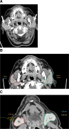
CT of the head showing bilaterally enlarged parotid glands (A) overview (arrows), (B) parotid glands, (C) submandibular glands. Red left, green right. The CT scan shows slightly enlarged parotid and submandibular glands (arrows) on both sides, with some differences in volume between sites.
Figure 3
Figure 3
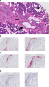
(A) Biopsy from the parotid gland: HE staining showing eosinophilic and lymphocytic infiltrates and fibrotic changes. The biopsy of the parotid gland of the patient described shows acini composed of serous cells with darkly staining granules (thin white arrows) and some admixed fat cells (black arrowheads), intercalated ducts lined by cuboidal epithelium (white arrowheads). Cellular infiltrates contain lymphocytes (thick white arrows) and eosinophilic granulocytes (black arrows). Bands of fibrous tissue (thick black arrow). Surrounds gland and separates lobules. (B) Biopsy from the parotid gland: staining for CD 3+T cells. The CD 3 staining demonstrates T cell infiltrates close to the ducts. (C) Biopsy from the parotid gland: staining for CD 20+B cells. The CD 20 staining demonstrates B cell infiltrates close to the ducts. (D) Staining for Mum 1 (plasma cells). The Mum1 staining shows that there are plasma cells in the tissue of the parotid gland at different sites.

References

    1. Rudwaleit M, Andermann B, Alten R, et al. . Atopic disorders in ankylosing spondylitis and rheumatoid arthritis. Ann Rheum Dis 2002;61:968–74. 10.1136/ard.61.11.968 - DOI - PMC - PubMed
    1. Vitte J, Vibhushan S, Bratti M, et al. . Allergy, anaphylaxis, and nonallergic hypersensitivity: IgE, mast cells, and beyond. Med Princ Pract 2022;31:501–15. 10.1159/000527481 - DOI - PMC - PubMed
    1. Ishizaka K, Ishizaka T. Identification of γE-antibodies as a carrier of reaginic activity. J Immunol 1967;99:1187–98. 10.4049/jimmunol.99.6.1187 - DOI - PubMed
    1. Smith SA, Chruszcz M, Chapman MD, et al. . Human monoclonal IgE antibodies-a major milestone in allergy. Curr Allergy Asthma Rep 2023;23:53–65. 10.1007/s11882-022-01055-w - DOI - PMC - PubMed
    1. Sjögren H. On knowledge of keratoconjunctivitis sicca: keratitis filiformis due to lacrimal gland hypofunction. Acta Ophthalmol 1933;2:1–151.

Substances