

Comparison of diabetic retinopathy severity grading on ETDRS 7-field versus ultrawide-field assessment
- PMID: 37165011
- PMCID: PMC10517125
- DOI: 10.1038/s41433-023-02445-8
Comparison of diabetic retinopathy severity grading on ETDRS 7-field versus ultrawide-field assessment
Abstract
Objectives: To compare the diabetic retinopathy (DR) severity level determined when considering only the ETDRS 7-field region versus the entire ultrawidefield (UWF) image.
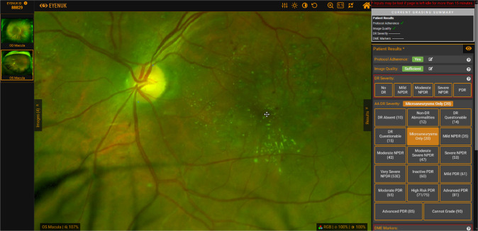
Methods: In this retrospective, cross-sectional study, UWF pseudocolor images were graded on the Eyenuk image viewing, grading, and annotation platform for the severity of DR considering only the regions within the ETDRS 7-fields as well as the entire UWF image using two different protocols: 1) the simple International Classification of Diabetic Retinopathy (ICDR) scale and 2) the more complex DRCR.net Protocol AA grading scale.
Results: A total of 250 eyes from 157 patients were included in this analysis. Six eyes (2.4%) demonstrated a discrepancy in severity level between the ETDRS 7-field region and the entire UWF image when using the ICDR classification system. The discrepancies were due to the presence of lesions [intraretinal haemorrhage (n = 2), neovascular disease (n = 4)] in the peripheral fields which were not identified in the ETDRS 7-fields. Fourteen eyes (5.6%) had a discrepancy in severity level between the ETDRS 7-field region and the entire UWF image when using the ETDRS DRSS Protocol AA grading scale. The discrepancies were due to the presence of a higher level of disease [intraretinal haemorrhage (n = 4), neovascularization (n = 4), preretinal haemorrhage (n = 2), scatter laser scars (n = 4)] in the peripheral fields.
Conclusion: Although considering regions outside of the ETDRS 7-fields altered the DR severity level assessment in <5% of cases in this cohort, significant and potentially vision-threatening lesions including neovascularization and preretinal haemorrhage were identified in these peripheral regions. This highlights the importance of evaluating the entire UWF region when assessing patients with diabetic retinopathy.
© 2023. The Author(s).
Conflict of interest statement
YA, MGN, SBV (None), CR: Eyenuk, Inc. (E), SB: Eyenuk, Inc. (E), KS: Eyenuk, Inc. (E), CJ (None), NC: Abbvie (C,S) Apellis ©, Bayer (C,S,R), Novartis (C), Bausch & Lomb (C), Alcon (C), Optos PLC (S,C,R), Topcon (S,C,R), Hoffman La Roche (C), SriniVas R Sadda: Amgen (C), Allergan (C), Genentech/Roche (C), Iveric (C), Oxurion (C), Novartis (C), Regeneron (C), Bayer (C), 4DMT (C), Centervue (C,R), Heidelberg (C,R), Optos (C,R), Merck (C), Apellis (C), Astellas (C), Carl Zeiss Meditec (R), Nidek (R), Topcon (R). The authors declare no competing interests.
Figures

References
-
- Wilkinson CP, Ferris FL, 3rd, Klein RE, Lee PP, Agardh CD, Davis M, Global Diabetic Retinopathy Project Group et al. Proposed international clinical diabetic retinopathy and diabetic macular edema disease severity scales. Ophthalmology. 2003;110:1677–82. doi: 10.1016/S0161-6420(03)00475-5. - DOI - PubMed
-
- Aiello LP, Odia I, Glassman AR, Melia M, Jampol LM, Bressler NM, et al. Diabetic Retinopathy Clinical Research Network. Comparison of early treatment diabetic retinopathy study standard 7-field imaging with ultrawide-field imaging for determining severity of diabetic retinopathy. JAMA Ophthalmol. 2019;137:65–73. doi: 10.1001/jamaophthalmol.2018.4982. - DOI - PMC - PubMed
MeSH terms
Grants and funding
LinkOut - more resources
Full Text Sources
Medical
