Review
. 2023 Jul;18(7):2283-2312.
doi: 10.1038/s41596-023-00827-6. Epub 2023 May 10.

A distal lung organoid model to study interstitial lung disease, viral infection and human lung development

Affiliations
Review

A distal lung organoid model to study interstitial lung disease, viral infection and human lung development

Ivana Matkovic Leko et al. Nat Protoc. 2023 Jul.

Abstract

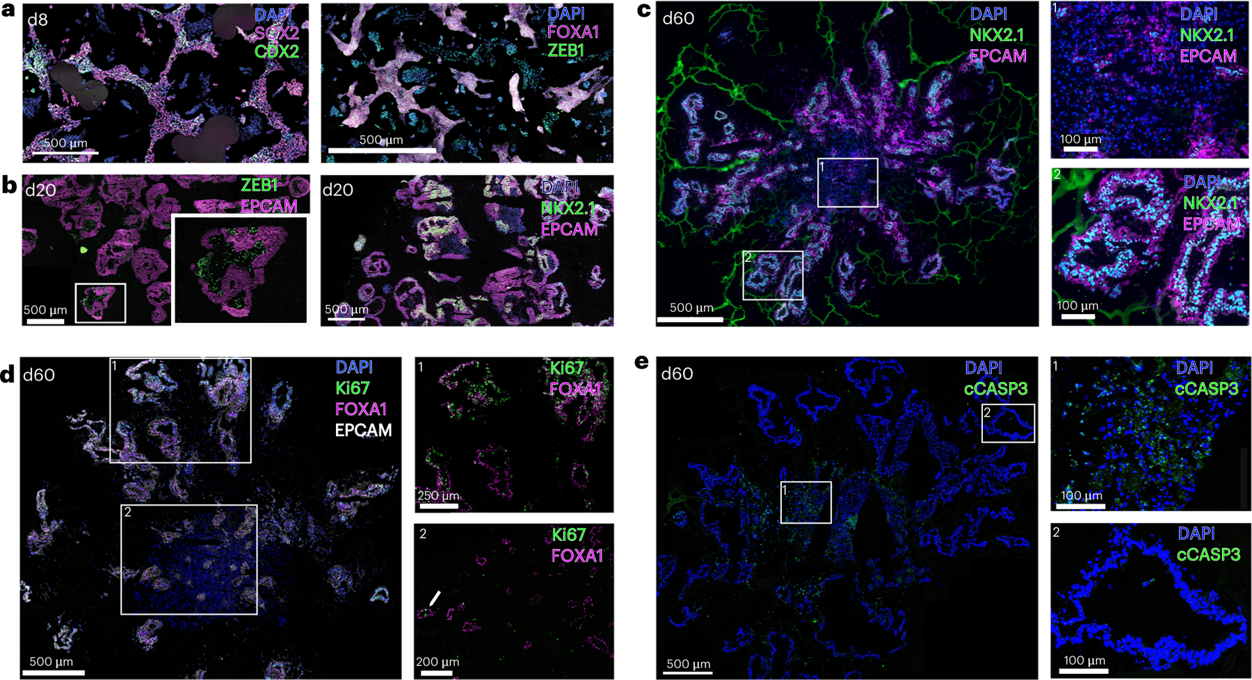
Organoids have been an exciting advancement in stem cell research. Here we describe a strategy for directed differentiation of human pluripotent stem cells into distal lung organoids. This protocol recapitulates lung development by sequentially specifying human pluripotent stem cells to definitive endoderm, anterior foregut endoderm, ventral anterior foregut endoderm, lung bud organoids and finally lung organoids. The organoids take ~40 d to generate and can be maintained more than 180 d, while progressively maturing up to a stage consistent with the second trimester of human gestation. They are unique because of their branching morphology, the near absence of non-lung endodermal lineages, presence of mesenchyme and capacity to recapitulate interstitial lung diseases. This protocol can be performed by anyone familiar with cell culture techniques, is conducted in serum-free conditions and does not require lineage-specific reporters or enrichment steps. We also provide a protocol for the generation of single-cell suspensions for single-cell RNA sequencing.


Conflict of interest statement

Competing interests

H.-W.S. and Y.-W.C. hold patents pertaining to the lung organoid technologies described. The other authors declare no competing interests.

Figures

Fig. 1 |
Fig. 1 |. Overview of the protocol.
a, Schematic representation of the distinct stages of the protocol. b, Representative brightfield images at indicated days of the protocol. Scale bars, 200 μm.
Fig. 2 |
Fig. 2 |. FBS-containing media does not maintain organoid cultures.
Tile scan of brightfield images showing the morphological changes of a d47 organoid switched to FBS-containing media. Scale bars, 1 mm.
Fig. 3 |
Fig. 3 |. Maturation of organoids and selection of suspension organoids for embedding.
a, Induction of select AT2 markers in RNAseq studies (data from ref. , mean ± s.e.m.). RPKM, reads per kilobase of transcript per million mapped reads. b, Representative images of d20 LBOs. The upper row shows organoids that we prefer not to pick due to the dense center and hollowing structures. The lower row shows ‘good’ organoids with folding epithelial structures and clear and transparent center.
Fig. 4 |
Fig. 4 |. Morphology of lung organoids embedded in Col I gel.
Tile scan of a brightfield image showing the morphology of d50 organoids grown in the Col I gel. Scale bar, 1 mm.
Fig. 5 |
Fig. 5 |. IF analysis of organoids at different stages.
a, Tile scan image at d8 (ventralization) stained for indicated markers. b, Tile scan images and higher magnification fields (insets) of culture LBOs at d20 of the culture protocol. c–e, Confocal tile scans of ~d60 organoids stained for the indicated markers, and higher magnification images (right) corresponding to numbered boxes. All scale bars in a–e, 500 μm, except for the insets of d (250 μm upper, 200 μm lower) and the insets of c and e (100 μm).
Fig. 6 |
Fig. 6 |. RT–qPCR analysis of the core and tip areas of a lung organoid.
a, Upper image shows the morphology of organoids before isolation of core (dashed area) and tips. Bottom image shows the punched out core (left) and tip (right) of an organoid. b, RT–qPCR for select lung and non-lung endoderm markers in the centers relative to the periphery of the organoids (n = 3 independent experiments at ~d60, two-way ANOVA).
Fig. 7 |
Fig. 7 |. Integrated scRNAseq analysis.
a, UMAP clusters after integrated analysis of organoids collected at d25, d40, d60 and d80. b, Dot plots of dynamic expression of endodermal (FOXA1), lung (NKX2.1, CPM), distal lung (SFTPB, NAPSA, SLC34A2, LPCAT), distal airway (SCGB3A2), mid and hindgut (GATA4, CDX2), mesenchymal (THY1, COL1A2, ZEB1, TBX4, WNT2B), proliferating cell (Ki67) and housekeeping genes (ACTA2) genes over time. c, UMAP feature plots of genes shown in b.
Fig. 8 |
Fig. 8 |. Merged scRNAseq analysis.
a, UMAP clusters after merged (as opposed to integrated) analysis of organoids collected at d25, d40, d60 and d80, showing expression of each marker on top of each graph. Clusters corresponding to each timepoint are shown in the upper left plot. b, scVelo trajectory analysis. Clusters are identified by colors. Pseudotime analysis was performed using scVelo on the merged datasets,.
Fig. 9 |
Fig. 9 |. Troubleshooting of LBO formation.
a, Effect of different plating densities on the morphology of the ventralized AFE stage (d8), before lifting the cells to generate LBOs. b, Proliferation as measured by staining for Ki67 and 7-AAD at different stages of the protocol indicated on top of the panels. Single-cell suspension at d35 generated as described in Box 2. c, Abortive organoids generated from LBOs cultured in suspension for more than 25 d. Scale bars in c, 250 μm.

References

    1. Clevers H Modeling development and disease with organoids. Cell 165, 1586–1597 (2016). - PubMed
    1. McCauley HA & Wells JM Pluripotent stem cell-derived organoids: using principles of developmental biology to grow human tissues in a dish. Development 144, 958–962 (2017). - PMC - PubMed
    1. Miller AJ & Spence JR In vitro models to study human lung development, disease and homeostasis. Physiology 32, 246–260 (2017). - PMC - PubMed
    1. Basil MC et al. The cellular and physiological basis for lung repair and regeneration: past, present, and future. Cell Stem Cell 26, 482–502 (2020). - PMC - PubMed
    1. Herriges M & Morrisey EE Lung development: orchestrating the generation and regeneration of a complex organ. Development 141, 502–513 (2014). - PMC - PubMed

Publication types