. 2023 May 10;13(1):7618.
doi: 10.1038/s41598-023-34820-6.

Extracellular vesicles of trypomastigotes of Trypanosoma cruzi induce changes in ubiquitin-related processes, cell-signaling pathways and apoptosis

Affiliations

Extracellular vesicles of trypomastigotes of Trypanosoma cruzi induce changes in ubiquitin-related processes, cell-signaling pathways and apoptosis

Alberto Cornet-Gomez et al. Sci Rep. .

Erratum in

Abstract

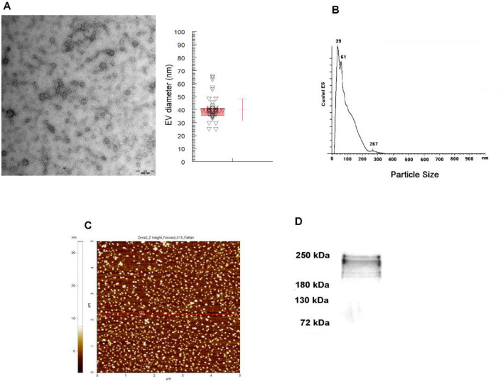
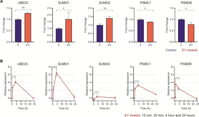
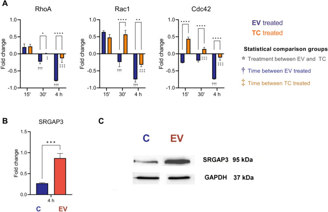
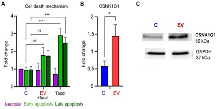
Chagas disease is caused by the protozoan parasite Trypanosoma cruzi. The disease has an acute and a chronic phase in which approximately 30% of the chronic patients suffer from heart disease and/or gastrointestinal symptoms. The pathogenesis of the disease is multifactorial and involves the virulence of the strains, immunological factors and extracellular vesicles (EV) shed by the parasite which participate in cell-cell communication and evasion of the immune response. In this work, we present a transcriptomic analysis of cells stimulated with EV of the trypomastigote stage of T. cruzi. Results after EV-cell incubation revealed 322 differentially expressed genes (168 were upregulated and 154 were downregulated). In this regard, the overexpression of genes related to ubiquitin-related processes (Ube2C, SUMO1 and SUMO2) is highlighted. Moreover, the expression of Rho-GTPases (RhoA, Rac1 and Cdc42) after the interaction was analyzed, revealing a downregulation of the analyzed genes after 4 h of interaction. Finally, a protective role of EV over apoptosis is suggested, as relative values of cells in early and late apoptosis were significantly lower in EV-treated cells, which also showed increased CSNK1G1 expression. These results contribute to a better understanding of the EV-cell interaction and support the role of EV as virulence factors.


Conflict of interest statement

The authors declare no competing interests.

Figures

Figure 1
Figure 1
Isolation of extracellular vesicles of trypomastigotes of T. cruzi Pan4 strain: (A) transmission electron microscopy image of EV (scale bar: 200 nm, mean size: 39.9 nm, mode: 38.8 nm); (B) nanoparticle tracking analysis size distribution of EV (mean size: 94 nm ± 57 nm, the largest peak of number of particles corresponding to a size of 39 nm); (C) atomic force microscopy image of EV and, (D) Western blot analysis for the confirmation of trans-sialidase in EV of trypomastigotes.
Figure 2
Figure 2
Transcriptomics analysis of cells incubated with extracellular vesicles of trypomastigotes of T. cruzi. Enrichment analyses suggest that EV of trypomastigotes downregulate genes that belong to processes related to vesicle formation, deubiquitylation/SUMOylation, as well as cell cycle control. (A) Top-ranked GO processes detected by enrichment analyses on downregulated DEGs, followed by a topological representation of the common genes in the top four ranked processes (B), as determined by ClusterProfiler. (C) Selected GO processes detected by enrichment analyses using the highly modulated DEGs, as determined against the DAVID database and (D) respective relevant genes validated by RT-qPCR. Gene expression was calculated as fold induction caused by the respective treatment/control as compared to the expression of GAPDH. Data are presented as shown above, with mean ± S.D. (n = 3). Differences to this value were analyzed by one sample Tukey multiple comparison test signed rank tests (asterisks), where ** p < 0.01; *** p < 0.001.
Figure 3
Figure 3
Expression analysis of genes involved in SUMOylation in Vero cells incubated with extracellular vesicles for 4 h (A) and cell cultures of Vero cells incubated with cell derived trypomastigotes of T. cruzi at different times during the process of infection (B). Gene expression was calculated as fold induction caused by the respective treatment/control as compared to the expression of GAPDH. Data are presented as shown above, with mean ± S.D. (n = 3). Differences to this value were analyzed by one sample Tukey multiple comparison test signed rank tests (asterisks), where * p < 0.05; ** p < 0.01; *** p < 0.001.
Figure 4
Figure 4
Changes in the expression of Rho-GTPase genes (RhoA, Rac1 and Cdc42) induced by extracellular vesicles secreted by trypomastigotes of T. cruzi. (A) An overexpression of RhoA and Rac1 was observed 15 min post-incubation of Vero cells with EV, while the expression of Cdc42 was downregulated. After this time- point, a downregulation in the expression of the 3 analyzed genes appeared at 30 min, being more evident 4 h post-interaction. The incubation of cells with trypomastigotes revealed an overexpression of the 3 genes both 15 and 30 min post-interaction, while the same behavior of EV in downregulating the expression of the 3 genes was observed 24 h post-interaction. Gene expression was calculated as fold induction caused by the respective treatment/control as compared with the expression of GAPDH. Data are presented as shown above, with mean ± S.D. (n = 3). Differences to this value were analyzed by one-way ANOVA using the Tukey’s post correction as rank tests (asterisks), where * p < 0.05; ** p < 0.01; **** p < 0.0001, comparing within the same time stamp the trypomastigotes (TC, orange) and the EV controls (blue). Comparison of each time point against the 15 min time are represented as †, for the TC groups, and ‡ for the EV groups. Expression levels of SRGAP3, an activating protein of Rho-GTPases, in cells incubated with EV of trypomastigotes of T. cruzi for 4 h, derived from the mRNA levels (B) and on protein levels (C). Expression was calculated as fold induction caused by the respective treatment/control as compared with the expression of GAPDH. Data are presented as shown above, with mean ± S.D. (n = 3). Differences to this value were analyzed by one sample Tukey multiple comparison test signed rank tests (asterisks), where *** p < 0.001.
Figure 5
Figure 5
“Protective” effect of extracellular vesicles of trypomastigotes of T. cruzi over apoptosis of Vero cells. (A) Significant lower relative values of cells in early and late apoptosis were observed in cells incubated with EV and then treated with taxol for 72 h. The values are represented as the mean percentages ± SEM. Tukey–Kramer test, where *** p < 0.001; **** p < 0.0001 and ns: non-significant differences. mRNA expression levels (B) and Western blot analysis (C) of casein kinase protein CSNK1G1 that shows an increase in protein synthesis after the interaction of cells with EV of the parasite. Expression was calculated as fold induction caused by the respective treatment/control as compared with the expression of GAPDH. Data are presented as shown above, with mean ± S.D. (n = 3). Differences to this value were analyzed by one sample Wilcoxon signed rank tests (asterisks), where * p < 0.05.

References

    1. WHO. Chagas disease (American trypanosomiasis) (2021).
    1. WHO Chagas disease in Latin America: An epidemiological update based on 2010 estimates. Wkly. Epidemiol. Rec. 2015;90:33–43. - PubMed
    1. Pecoul B, et al. The BENEFIT trial: Where do we go from here? PLoS Negl. Trop. Dis. 2016;10:e0004343. doi: 10.1371/journal.pntd.0004343. - DOI - PMC - PubMed
    1. Tyler KM, Engman DM. The life cycle of Trypanosoma cruzi revisited. Int. J. Parasitol. 2001;31:472–481. doi: 10.1016/S0020-7519(01)00153-9. - DOI - PubMed
    1. Fernandes MC, Andrews NW. Host cell invasion by Trypanosoma cruzi: A unique strategy that promotes persistence. FEMS Microbiol. Rev. 2012;36:734–747. doi: 10.1111/j.1574-6976.2012.00333.x. - DOI - PMC - PubMed

Publication types