. 2023 Sep;39(9):1363-1374.
doi: 10.1007/s12264-023-01062-5. Epub 2023 May 10.

The Antinociceptive Effect of Sympathetic Block is Mediated by Transforming Growth Factor β in a Mouse Model of Radiculopathy

Affiliations

The Antinociceptive Effect of Sympathetic Block is Mediated by Transforming Growth Factor β in a Mouse Model of Radiculopathy

Debora Denardin Lückemeyer et al. Neurosci Bull. 2023 Sep.

Abstract

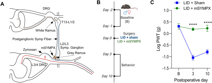
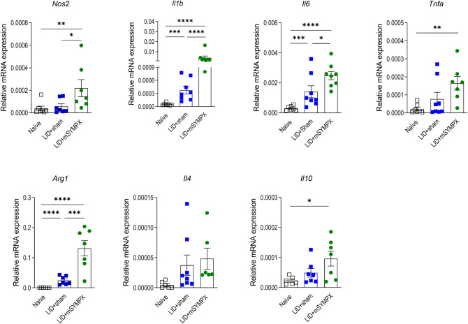
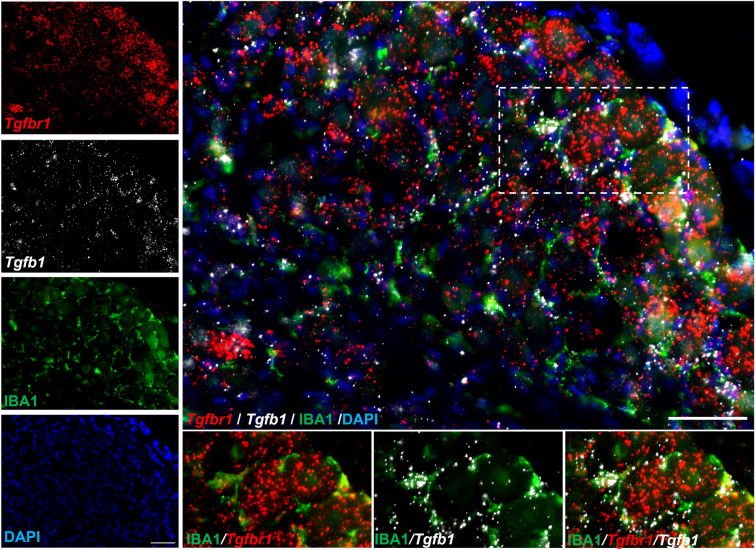
Although sympathetic blockade is clinically used to treat pain, the underlying mechanisms remain unclear. We developed a localized microsympathectomy (mSYMPX), by cutting the grey rami entering the spinal nerves near the rodent lumbar dorsal root ganglia (DRG). In a chemotherapy-induced peripheral neuropathy model, mSYMPX attenuated pain behaviors via DRG macrophages and the anti-inflammatory actions of transforming growth factor-β (TGF-β) and its receptor TGF-βR1. Here, we examined the role of TGF-β in sympathetic-mediated radiculopathy produced by local inflammation of the DRG (LID). Mice showed mechanical hypersensitivity and transcriptional and protein upregulation of TGF-β1 and TGF-βR1 three days after LID. Microsympathectomy prevented mechanical hypersensitivity and further upregulated Tgfb1 and Tgfbr1. Intrathecal delivery of TGF-β1 rapidly relieved the LID-induced mechanical hypersensitivity, and TGF-βR1 antagonists rapidly unmasked the mechanical hypersensitivity after LID+mSYMPX. In situ hybridization showed that Tgfb1 was largely expressed in DRG macrophages, and Tgfbr1 in neurons. We suggest that TGF-β signaling is a general underlying mechanism of local sympathetic blockade.

Keywords: Cytokine; Inflammation; Radiculopathy; Sympathetic; TGF-β.


Conflict of interest statement

All authors claim that there are no conflicts of interest.

Figures

Fig. 1
Fig. 1
Effect of microsympathectomy (mSYMPX) on LID-induced mechanical allodynia. A Schematic of the mSYMPX surgery which consisted of cutting the gray rami from the L2 and L3 postganglionic sympathetic ganglia. Blue indicates a postganglionic sympathetic cell in the paravertebral ganglia, which is innervated by preganglionic neurons (brown). Red indicates a sensory neuron. B Schematic of the experiment showing the timeline of local inflammation of the DRG (LID), mSYMPX, and behavior. C Time course of von Frey (static allodynia test) in LID and LID+mSYMPX mice [n = 8 males and females per group, the behavior was measured at baseline (“B”; an average of 2 days)], on postoperative day 3 and postoperative day 10. LID and mSYMPX were performed on day 0, after the baseline days. ****P <0.0001, a significant difference between the two groups at the indicated time points (two-way repeated-measures ANOVA, Bonferroni post hoc test, overall group effect and group × time interaction, both P <0.0001). PWT, paw withdrawal threshold. Data separated by sex are in Fig. S1.
Fig. 2
Fig. 2
Effect of mSYMPX on cytokine profiles after local inflammation of the DRG (LID). n = 6 male mice per group. Protein samples were obtained from naïve mice 3 days after LID or LID+mSYMPX, and the indicated cytokines were measured with the Luminex method. *P <0.05, **P <0.01, ***P <0.001, and ****P <0.0001, a significant difference between the indicated two groups (ANOVA with Tukey’s post-test). The post-test significance values are only indicated for assays in which the overall summary P value was also significant. IL-4, the overall P value was 0.11. See Table S2 for the full names of the cytokines.
Fig. 3
Fig. 3
mRNA expression of cytokines and macrophage markers. DRG mRNA samples were obtained from naïve mice 3 days after LID or LID+mSYMPX. Expression is normalized to GAPDH. *P <0.05, **P <0.01, ***P <0.001, and ****P <0.0001, a significant difference between the indicated two groups at the indicated points (ANOVA with Tukey’s post-test on log-transformed values). The post-test significance values are only indicated for assays in which the overall summary P value was also significant. IL-4 overall P value was 0.13. See Table S2 for the full names of the cytokines and macrophage markers. n = 7 in the naïve group, 4 males/3 females; n = 8, 4 males/4 females per group in the LID and LID+mSYMPX groups. Data separated by sex are in Fig. S2.
Fig. 4
Fig. 4
Effect of LID and mSYMPX on TGF-β isoform expression. Protein samples were obtained from naïve mice or 3 days after LID or LID+mSYMPX, and measured with the Luminex method. n = 6 male mice per group in all groups, the same animals as Fig. 2, but separate Luminex assays. *P <0.05, **P <0.01, ***P <0.001, a significant difference between the indicated two groups (ANOVA with Tukey’s post-test). The overall P value for the ANOVA was also significant for each isoform (β1 and β2, P <0.001; β3, P = 0.0155). Note the difference in scales for TGF-β2 and TGF-β3 that were expressed at much lower levels than TGF-β1.
Fig. 5
Fig. 5
Interaction of local inflammation of the DRG (LID) and microsympathectomy (mSYMPX) with anti-inflammatory transforming growth factor-β (TGF-β) signaling. A Transcriptional increase of Tgfb1 and Tgfbr1 in DRGs (L3 and L4) on POD 3 after LID and LID+mSYMPX (n = 7 in naïve group, 4 males/3 females; n = 8, 4 males/4 females per group in LID and LID+mSYMPX). *P <0.05, **P <0.01, ****P <0.0001 between the indicated 2 groups, using one-way ANOVA with Tukey’s post-test on log-transformed values; overall P value <0.0001 for both transcripts. B Antinociceptive effect of intrathecal injection of TGF-β1 on behavior (von Frey threshold) induced by LID. C The mSYMPX effect on the LID von Frey threshold was abolished by intrathecal injection of the TGF-βR1 inhibitor SB431542. n = 8/group, 4 males/4 females. *P <0.05, **P <0.01, *** P <0.001, a significant difference between the two groups at the indicated time points (two-way repeated-measures ANOVA, Bonferroni post hoc test). Overall p values for treatment factor and time × treatment, respectively, were 0.01 and P <0.001 (B) and 0.001 and <0.0001 (C). “B” is the average of two baselines before surgery; PWT, paw withdrawal threshold. Data separated by sex are in Fig. S3.
Fig. 6
Fig. 6
mSYMPX increases Tgfb1 and Tgfbr1 in mouse DRGs 3 days after local inflammation of the DRG (LID). A Representative images of mouse DRGs labeled with RNAScope in situ hybridization for Tgfbr1 (red) and Tgfb1 (white) mRNA and co-stained with DAPI (blue). Upper: LID only. Lower: LID+mSYMPX. Scale bar, 50 µm. B Intensity quantification normalized by area using images of mouse DRGs labeled with RNAScope in situ hybridization for Tgfbr1 and Tgfb1 mRNA. Representative of results from 3 male mice for LID and 4 male mice for LID+mSYMPX. Data were analyzed using an unpaired t-test, comparing LID and LID+mSYMPX, *P <0.05. C Tgfb1 is expressed primarily by non-neuronal cells and Tgfbr1 by sensory neurons in mouse DRGs 3 days after LID and LID+mSYMPX. The percentage of intensity normalized by area was measured using images of mouse DRGs labeled with RNAScope in situ hybridization for Tgfbr1 and Tgfb1 mRNA. Representative results from 3 male mice for LID and 4 male mice for LID+mSYMPX. Data were analyzed using one-way ANOVA with Tukey’s post-test comparing neurons and non-neuronal cells, ***P <0.001 and ****P <0.0001. D Tgfbr1 distribution by neuron means diameter size. Intensity normalized by diameter was calculated using images of mouse DRGs labeled with RNAScope in situ hybridization for Tgfbr1 mRNA. Representative of results in 4 male mice with LID+mSYMPX. Data were analyzed using one-way ANOVA and the Bonferroni post hoc test comparing the different neuron diameters, ****P <0.0001.
Fig. 7
Fig. 7
Distribution of Tgfb1 and Tgfbr1 in mouse DRGs 3 days after local inflammation of the DRG (LID) and microsympathectomy (mSYMPX). Representative images of mouse DRGs labeled with RNAScope in situ hybridization for Tgfbr1 (red) and Tgfb1 (white) mRNA. DRGs were co-stained for the general macrophage marker ionized Ca2+-binding adaptor molecule 1 (IBA1) protein (green) and nuclear staining is shown in blue (DAPI). Scale bar, 50 µm. Representative of results in 4 male mice.
Fig. 8
Fig. 8
Low expression levels of Tgfb1 and Tgfbr1 mRNA in satellite glial cells (SGCs) in mouse DRGs 3 days after local inflammation of the DRG (LID) and microsympathectomy (mSYMPX). A Representative image of a mouse DRG labeled with RNAScope in situ hybridization for Tgfbr1 (red) and Tgfb1 (white) mRNA. The DRG was co-stained for the satellite glial cell marker fatty acid binding protein 7 (FABP7) (green). Nuclear staining is shown in blue (DAPI). Scale bar, 50 µm. Representative of results from 4 male mice. B Tgfbr1 and Tgfb1 were primarily expressed by non-SGCs in mouse DRGs 3 days after LID and LID+mSYMPX. The percentage intensity normalized by area was calculated using images of mouse DRGs labeled with RNAScope in situ hybridization for Tgfbr1 and Tgfb1 mRNA and co-staining with FABP7 (green). Representative of results from 3 male mice for LID and 3 male mice for LID+mSYMPX. Data were analyzed using one-way ANOVA with Tukey’s post-test comparing SGCs and non-SGCs, **P <0.01, ***P <0.001, and ****P <0.0001.

References

    1. Borchers AT, Gershwin ME. Complex regional pain syndrome: A comprehensive and critical review. Autoimmun Rev. 2014;13:242–265. doi: 10.1016/j.autrev.2013.10.006. - DOI - PubMed
    1. Schlereth T, Drummond PD, Birklein F. Inflammation in CRPS: Role of the sympathetic supply. Auton Neurosci. 2014;182:102–107. doi: 10.1016/j.autneu.2013.12.011. - DOI - PubMed
    1. Scadding JW. Complex regional pain syndrome. In: Textbook of Pain. 4th ed. Churchill Livingstone, 1999: 835–849.
    1. Mantyh PW. The neurobiology of skeletal pain. Eur J Neurosci. 2014;39:508–519. doi: 10.1111/ejn.12462. - DOI - PMC - PubMed
    1. Drummond PD. Sensory-autonomic interactions in health and disease. Handb Clin Neurol. 2013;117:309–319. doi: 10.1016/B978-0-444-53491-0.00024-9. - DOI - PubMed

MeSH terms

Substances

LinkOut - more resources