Clinical Trial
. 2023 Jun;618(7963):144-150.
doi: 10.1038/s41586-023-06063-y. Epub 2023 May 10.

Personalized RNA neoantigen vaccines stimulate T cells in pancreatic cancer

Affiliations
Clinical Trial

Personalized RNA neoantigen vaccines stimulate T cells in pancreatic cancer

Luis A Rojas et al. Nature. 2023 Jun.

Abstract

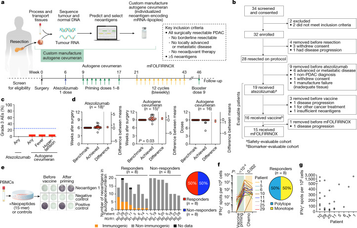
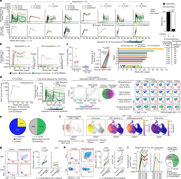
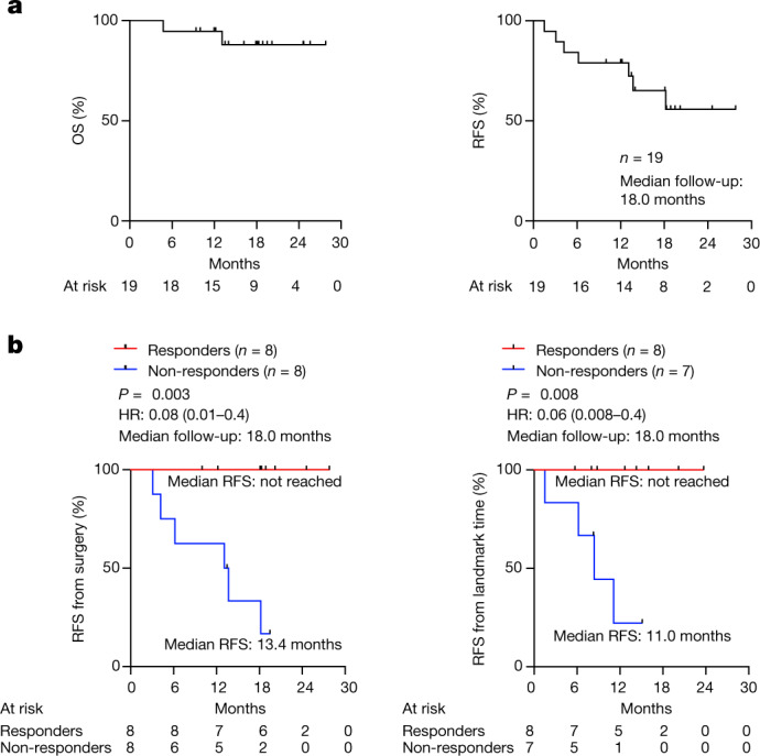
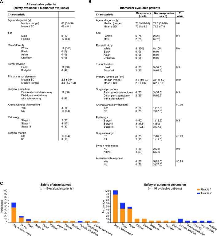
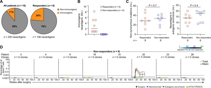
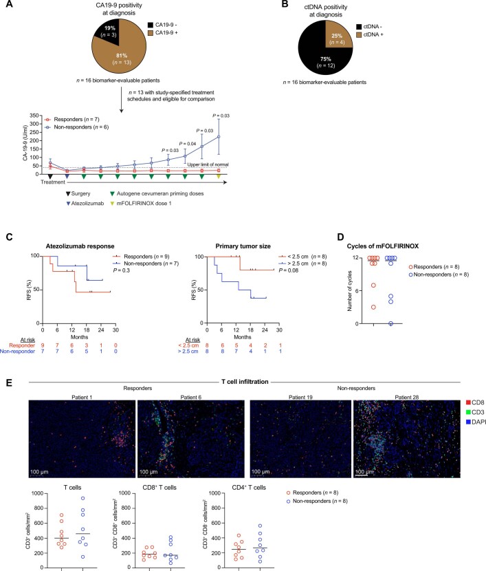
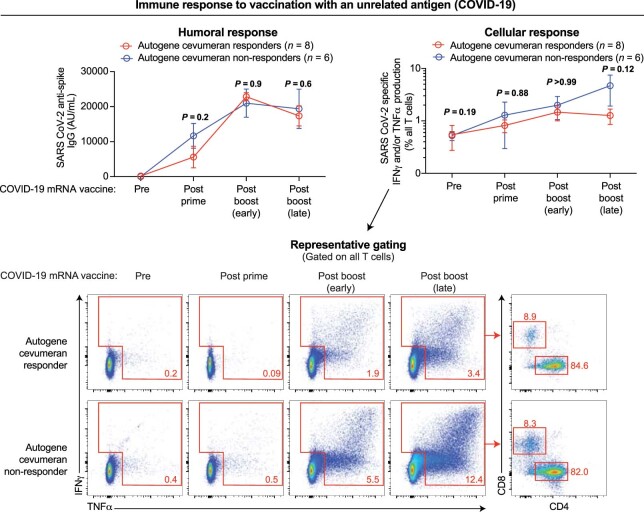
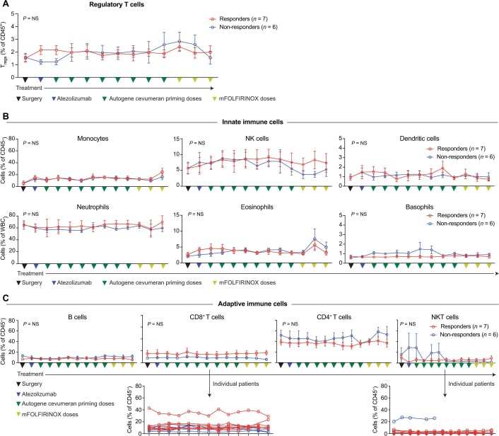
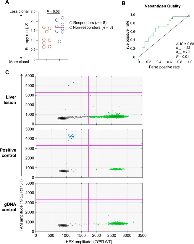
Pancreatic ductal adenocarcinoma (PDAC) is lethal in 88% of patients1, yet harbours mutation-derived T cell neoantigens that are suitable for vaccines 2,3. Here in a phase I trial of adjuvant autogene cevumeran, an individualized neoantigen vaccine based on uridine mRNA-lipoplex nanoparticles, we synthesized mRNA neoantigen vaccines in real time from surgically resected PDAC tumours. After surgery, we sequentially administered atezolizumab (an anti-PD-L1 immunotherapy), autogene cevumeran (a maximum of 20 neoantigens per patient) and a modified version of a four-drug chemotherapy regimen (mFOLFIRINOX, comprising folinic acid, fluorouracil, irinotecan and oxaliplatin). The end points included vaccine-induced neoantigen-specific T cells by high-threshold assays, 18-month recurrence-free survival and oncologic feasibility. We treated 16 patients with atezolizumab and autogene cevumeran, then 15 patients with mFOLFIRINOX. Autogene cevumeran was administered within 3 days of benchmarked times, was tolerable and induced de novo high-magnitude neoantigen-specific T cells in 8 out of 16 patients, with half targeting more than one vaccine neoantigen. Using a new mathematical strategy to track T cell clones (CloneTrack) and functional assays, we found that vaccine-expanded T cells comprised up to 10% of all blood T cells, re-expanded with a vaccine booster and included long-lived polyfunctional neoantigen-specific effector CD8+ T cells. At 18-month median follow-up, patients with vaccine-expanded T cells (responders) had a longer median recurrence-free survival (not reached) compared with patients without vaccine-expanded T cells (non-responders; 13.4 months, P = 0.003). Differences in the immune fitness of the patients did not confound this correlation, as responders and non-responders mounted equivalent immunity to a concurrent unrelated mRNA vaccine against SARS-CoV-2. Thus, adjuvant atezolizumab, autogene cevumeran and mFOLFIRINOX induces substantial T cell activity that may correlate with delayed PDAC recurrence.


Conflict of interest statement

L.A.R. is an inventor of a patent related to oncolytic viral therapy (US20170051022A1). L.A.R., Z.S., B.D.G. and V.P.B. are inventors on patent applications related to work on antigen cross-reactivity (PCT/US2023/011643) and tracking vaccine-expanded T cell clones. M.Ł., B.D.G. and V.P.B. are inventors on a patent application on neoantigen quality modelling (63/303,500). B.D.G. has received honoraria for speaking engagements from Merck, Bristol Meyers Squibb and Chugai Pharmaceuticals; has received research funding from Bristol Meyers Squibb, Merck, and ROME Therapeutics; and has been a compensated consultant for Darwin Health, Merck, PMV Pharma, Shennon Biotechnologies and Rome Therapeutics of which he is a co-founder. R.K.D. received one-time consulting fees from GE Healthcare and Bayer Healthcare. A.S.E. received royalties from Up-To-Date. A.M.V. discloses the following relationships (provision of services, uncompensated): Bristol-Myers Squibb, GlaxoSmithKline, Lilly Oncology, OBI Pharma, and Silenseed. M.S. has collaborative research agreements with Atara Biotherapeutics, Fate Therapeutics, Mnemo Therapeutics, and Takeda Pharmaceuticals. A.C.W. is a consultant for Histosonics and Biosapien; received honorarium from Medtronic and AstraZeneca; and received travel support from Intuitive Surgical. T.P.K. received a one-time consulting fee from Olympus Surgical. J.D. owns stock in Alnylam Pharmaceuticals, Arrowroot Acquisition, and Ionis Pharmaceuticals. W.P. receives grant and research support from Astellas, Merck, NIH/NCI, Parker Institute for Cancer Immunotherapy, and Break Through Cancer. E.M.O. receives grant and research support from Genentech/Roche, Celgene/Bristol-Myers Squibb, BioNTech, AstraZeneca, Arcus, Elicio, Parker Institute, and the NIH/NCI. T.M. is a co-founder and holds equity in IMVAQ Therapeutics; he is a consultant for Immunos Therapeutics, ImmunoGenesis and Pfizer; he has research support from Bristol-Myers Squibb, Surface Oncology, Kyn Therapeutics, Infinity Pharmaceuticals, Peregrine Pharmaceuticals, Adaptive Biotechnologies, Leap Therapeutics and Aprea; he has patents on applications related to work on oncolytic viral therapy, alphavirus-based vaccine, neoantigen modelling, CD40, GITR, OX40, PD-1 and CTLA-4. J.D.W. is a consultant for Apricity, CellCarta, Ascentage Pharma; AstraZeneca, Bicara Therapeutics, Boehringer Ingelheim, Bristol-Myers Squibb, Daiichi Sankyo, Dragonfly, Georgiamune, Imvaq, Larkspur, Psioxus, Recepta, Tizona, and Sellas. J.D.W. receives grant and research support from Bristol-Myers Squibb and Sephora. J.D.W. has equity in Apricity, Arsenal IO, Ascentage, Imvaq, Linneaus, Georgiamune, Maverick, and Tizona Therapeutics. V.P.B. has received honoraria for speaking engagements from Genentech, and research support from Bristol-Myers Squibb and Genentech. Ö.T. and U.S. are co-founders, management board members and employees at BioNTech. E.D. and F.M. are employees at BioNTech. I.R., M.Y. and I.M. are employees at Genentech. The other authors (K.C.S., C.O., N.P., E.P., J.L., N. Ceglia, P.G., A.C., R.Y., A.K.C., T.W., J.R., M.A., A.Z., Z.O., G.P., A. Dobrin, N. Cohen, L.T., O.B., M.G., S.K., P.M., E.W., R.S., A. Desai, M.I.D. and W.R.J.) declare no competing interests.

Figures

Fig. 1
Fig. 1. Individualized mRNA neoantigen vaccines are safe, feasible and immunogenic in patients with PDAC.
a,b, Trial design (a) and consolidated standards of reporting trials diagram (b). c, Percentage of grade 3 AEs attributable to atezolizumab and autogene cevumeran (vaccine) in atezolizumab (n = 19) and vaccine (n = 16) safety-evaluable patients. Blue line indicates the study-defined safety threshold (25%). d, Achieved and benchmarked times to atezolizumab (left) and first vaccine dose (middle), and number of vaccine doses (right). Red line indicates the median, error bars are 95% confidence intervals and dotted lines the zone of clinical indifference. Asterisks indicate patients on study-specified treatment sequence. eg, PBMCs collected after atezolizumab and before vaccine administration, 1–3 weeks after vaccine priming, and 5–6 weeks after mFOLFIRINOX were analysed for IFNγ+ T cells specific to all individual vaccine neoantigens by ex vivo IFNγ ELISpot in n = 16 patients in the biomarker-evaluable cohort. Patients were classified as responders if ELISpot detected IFNγ+ T cell induction against at least one vaccine neoantigen. e, Left, schematic and representative image of ex vivo IFNγ ELISpot. Middle, Number of vaccine neoantigens per patient that induced IFNγ+ T cells in PBMCs collected after vaccine priming. R0/R1 indicates the surgical margin status. For patient 25, 2 out of 5 ELISpot responses were detected against 2 neoantigen pools (pool 1 with 2 neoantigens, pool 2 with 5 neoantigens). Right, Proportion of vaccine responders and non-responders. f,g, Normalized ex vivo IFNγ ELISpot counts for vaccine neoantigens that induced a de novo response (n = 25 neoantigens in 8 patients): longitudinal (f, left); after priming (g). Spot counts of the non-stimulated controls were subtracted. Proportion of patients with monotope compared with polytope responses to all vaccine neoantigens (f, right). n indicates individual patients. Chemo, chemotherapy (mFOLFIRINOX). P values calculated using two-tailed unpaired t-test (d) or Wilcoxon matched-pairs signed-rank test (f). Source Data
Fig. 2
Fig. 2. mRNA vaccines expand polyclonal, polyfunctional effector CD8+ T cells.
a, Vaccine-expanded T cell clones assessed using CloneTrack (top), vaccine-induced IFNγ by ELISpot (bottom) and their correlation (right). b,c, Vaccine-expanded clones identified by CloneTrack: longitudinal aggregate percentage (b), number of unique clones (c, left), before vaccine and peak expansion aggregate percentage (c, middle), and final per patient assessment times (bar graph) with aggregate percentage and clonal fraction at final assessment (c, right). For b, inverted triangles indicate collection times for single-cell sequencing in f and circles indicate vaccine booster. d, Immunodominant vaccine neoantigen-specific clonal overlap with vaccine-expanded clones and specificity to immunodominant vaccine-neoantigens by TCR cloning in patient 1. e, Left, Percentage of patients with immunodominant vaccine neoantigen-specific clones in vaccine-expanded clones. Right, Percentage of vaccine-expanded clones specific to immunodominant vaccine neoantigens. f, Single-cell phenotypes of vaccine-expanded CD8+ T cells. Dots indicate blood CD8+ T cells. Coloured dots (far left) indicate vaccine-expanded clones in a. g,h, Percentage of IFNγ+, TNF+ and CD107a+CD8+ (g,h) and CD4+ T cells (g) in PBMCs after vaccine priming with ex vivo immunodominant long (g) or minimal (h) neopeptide rechallenge. Representative flow plots from patient 1 (g, h). Pregated on CD3+CD56CD8+ (g,h) or CD4+ (g) cells. i, Left, Aggregate percentage of vaccine-expanded clones with priming, chemotherapy and booster in peripheral blood. Right, Percentage of primed clones that re-expand with booster. n indicates individual clones or patients. In a and d, the green lines indicate individual clone trajectories; the black line, the geometric mean clonal trajectory (error bars are the geometric s.d.); the red line, the cumulative percentage of all expanded clones. In b,c, asterisks indicate altered treatment schedules. In b–d, rectangles indicate the treatment sequence. P values calculated using modified two-tailed Fisher’s exact test (a,d, left), two-sided Chi square test (a, right), two-tailed paired t-test (c), one-tailed binomial test with Bonferroni correction (d, middle) or two-tailed Wilcoxon matched-pairs signed-rank test (gi). Source Data
Fig. 3
Fig. 3. mRNA vaccine response correlates with delayed PDAC recurrence.
a, OS and RFS in n = 19 patients in the safety-evaluable cohort. b, RFS from surgery and from landmark time (date of the last vaccine priming dose) stratified by vaccine response in patients in the biomarker-evaluable cohort. n indicates individual patients. HR indicates hazard ratio with 95% CI. P values calculated using two-tailed log-rank test.
Fig. 4
Fig. 4. Vaccine-expanded T cells can infiltrate a micrometastasis.
Clinical and immunological snapshot of a disappearing intrahepatic lymphoid aggregate after vaccination in a patient who responded to the vaccine. a, Serial percentage of vaccine-expanded T cells in blood analysed using CloneTrack and serum CA19-9 (left), and abdominal MRI (right) before and after vaccination. b, Haematoxylin and eosin staining (left), multiplexed immunofluorescence (middle) and percentage of vaccine-expanded T cells measured using CloneTrack (right, grey bar) in a new liver lesion that developed after vaccination detected by MRI as in a. All 15 vaccine-expanded T cell clones (a, red line) were present in liver lesion (right, grey bar). c, Percentage of mutant TP53R175H reads by digital droplet PCR in the liver lesion. The bar indicates the median, the error bars are the s.e.m. d, Uniform manifold approximation and projection (UMAP) plots of single-cell phenotypes of all blood T cells (left) and vaccine-expanded clones (middle), with effector markers (right). n indicates the number of T cells detected in liver lesion (b) or technical replicates (c). Data represent analyses of a single patient. Source Data
Extended Data Fig. 1
Extended Data Fig. 1. Patient demographics, clinical characteristics, and toxicity.
(A, B) Demographics and clinical characteristics of all evaluable patients (n = 19) (A) and biomarker-evaluable patients (n = 16) stratified by autogene cevumeran responders and non-responders (B). (C) Frequency of grade 1 and 2 adverse events attributable to atezolizumab (left) and vaccine (right) in evaluable patients who received each drug. n = individual patients. Data are n (%) unless noted. P values by two-tailed Mann-Whitney test for numerical variables and Fisher’s exact test for categorical variables. Source Data
Extended Data Fig. 2
Extended Data Fig. 2. Immunogenic autogene cevumeran neoantigens.
(A) Frequency of immunogenic autogene cevumeran (vaccine) neoantigens in all patients (left) and in vaccine responders (right). (B) Number of immunogenic vaccine neoantigens per patient. (C) Number of non-synonymous mutations (left) and vaccine neoantigens (right) in vaccine responders and non-responders. (D) Vaccine-expanded T cell clones by CloneTrack in non-responders. n = neoantigens or patients as noted. P values by two-tailed Mann-Whitney test (C) and modified two-tailed Fisher’s exact test (D). Source Data
Extended Data Fig. 3
Extended Data Fig. 3. Autogene cevumeran–expanded T cell clones do not overlap with atezolizumab-expanded T cell clones.
(A) Cumulative percentage of atezolizumab-expanded T cells by CloneTrack in autogene cevumeran (vaccine) responders and non-responders. * = altered treatment schedules for patients 3, 4, 14 and 18. (B) Atezolizumab-expanded T cell clones by CloneTrack in vaccine responders and non-responders. Red box = atezolizumab responder. Blue line = trajectory of an individual atezolizumab-expanded clone. Black line = geometric mean trajectory of all atezolizumab-expanded clones; error bars = geometric standard deviation. Red line = cumulative percentage of all atezolizumab-expanded clones. Black rectangle/triangle = time of surgery; blue rectangle/triangle = time of atezolizumab; green rectangle/triangle = times of autogene cevumeran (vaccine); yellow bars = mFOLFIRINOX cycles. (C) Venn diagrams show overlap of vaccine-expanded clones (as identified in Fig. 2a) with atezolizumab-expanded clones in vaccine responders and non-responders. n = number of clones or individual patients as noted. P values by modified two-tailed Fisher’s exact test (B). Source Data
Extended Data Fig. 4
Extended Data Fig. 4. Autogene cevumeran–expanded T cell clones contain immunodominant neoantigen-specific T cells.
(A, B) Autogene cevumeran (vaccine) induced IFNγ production by ex vivo IFNγ ELISpot (assay schematic, Fig. 1e) and clonal expansion by CloneTrack (assay schematic, B, top). Briefly, for ELISpot, we analyzed each patient’s PBMCs for specific T cells against all individual vaccine neoantigens post-atezolizumab/pre-vaccine, 1-3 weeks post-vaccine priming doses, and 5-6 weeks post-mFOLFIRINOX. Each patient’s PBMCs were stimulated overnight with separate pools of overlapping peptides (15 amino acids long), each pool representing one of up to 20 neoantigens in vaccines, or with anti-CD3 antibody as a positive control, followed by measurement of IFNγ production by ELISpot. PBMCs incubated with media alone were used as a negative control. To track T cell clones, we identified vaccine-expanded T cell clones with CloneTrack. To identify if expanded clones contained immunodominant neoantigen-specific clones, in 4 of 8 responders, we stimulated vaccine-expanded PBMCs collected 1-3 weeks post-vaccine priming doses in vitro with computationally predicted minimal neopeptide pools (8-14 amino acids long) from 6 neoantigens that generated the highest per-patient magnitude response by ex vivo IFNγ ELISpot. We then purified CD8+ T cells that either expressed or did not express the degranulation marker CD107a, identified clones with greater proportion of CD107a+ versus CD107a- cells as in vitro neoepitope-activated clones, and examined overlap of in vitro neoepitope-activated to in vivo vaccine-expanded clones (Venn diagrams). For select patients, we further validated neoepitope-specificity by TCR cloning. (A) Flow cytometry gating strategies. (B) Ex vivo IFNγ ELISpot and T cell clonal expansion by CloneTrack in n = 3 of 4 patients tested (fourth patient in Fig. 2d). (B, left) Normalized ex vivo IFNγ ELISpot spot count per 1 x 10⁶ PBMCs for each immunogenic neoantigen in patients 10, 11 and 5. In patient 5, one neoantigen induced a high magnitude T cell response, while patients 10 and 11 had polytopic high magnitude responses against 8 and 3 vaccine neoantigens respectively. Black lines = individual neoantigens; coloured lines = neoantigen pools. (B, middle): T cell clonal expansion in patients 10, 11 and 5 by CloneTrack. Green line = trajectory of an individual expanded clone. Black line = geometric mean trajectory of all expanded clones; error bars = geometric standard deviation. Red line = cumulative percentage of all expanded clones. Black triangle = time of surgery; blue triangle = time of atezolizumab; green triangles = times of autogene cevumeran (vaccine); yellow bars = mFOLFIRINOX cycles. Dotted black line = detection threshold. (B, right): In vitro neoantigen-specific activation. Flow cytometry = CD107a expression on CD8+ T cells stimulated with neopeptides or control (DMSO). Dot plots = number of CD107a+ versus CD107a- cells per T cell clone. Each circle = individual T cell clone. Green/blue squares = clones also detected by CloneTrack. Diagonal: CD107a+ clone frequency = CD107a- clone frequency. Venn diagram = vaccine-expanded and in vitro neoantigen-specific clonal overlap. For patient 11, immunodominant neoantigen-specific clones resided in a lower magnitude vaccine-expanded clonal pool. (B, right, bottom) TCR cloning, patient 10: 4-1BB expression on putative neoantigen-specific TCR-transduced CD8+ T cells cultured with HLA-matched, neopeptide-pulsed antigen presenting cells (HLA-transduced K562 cells). n = number of clones. P values by modified two-tailed Fisher’s exact test (B, middle), and by one-tailed binomial test with Bonferroni correction (B, right). Source Data
Extended Data Fig. 5
Extended Data Fig. 5. Autogene cevumeran activates neoantigen-specific polyfunctional effector CD8+ T cells.
(A) Uniform manifold approximation and projection (UMAP) plots of single peripheral blood T cells by single-cell RNA/TCR sequencing in n = 4 patients (patients 1, 10, 11 and 29) stratified by lineage (CD8 vs. CD4, left), patient (middle), and vaccine-expanded clones (right; expanded clones identified in Fig. 2a). T cells purified post-vaccine priming doses at time points indicated in Fig. 2b (inverted triangles). (B, C) UMAP plots of single peripheral blood CD8+ T cells in patients 1, 10, 11 and 29 stratified by CD8+ T cell naïve (SELL, CCR7, IL7R, BCL2, PECAM1, TCF7, BACH2, LEF1), dysfunctional (TIGIT, TOX, LAG3, ENTPD1, CXCL13, HAVCR2, GZMB), memory (EOMES, GZMK, CXCR3, TCF1, ID3, STAT4, CCR7, SELL) and effector (PRF1, GZMB, GNLY, IFNγ, EOMES, ZEB2, E2F7, TBX21, PDCD1, CXCR3, FAS) transcriptional phenotypes (B) and select individual phenotype defining markers (C). (D) Cytokine (IFNγ, TNFα) production in CD8+ and CD4+ T cells after post-vaccine bulk PBMC ex vivo rechallenge with pools of overlapping long neopeptides. (E) Cytokine (IFNγ, TNFα) production and degranulation (CD107a) by CD8+ T cells after bulk PBMC ex vivo rechallenge with predicted minimal neopeptides. Flow cytometry gating strategies as in Extended Data Fig. 4a. (F) Peripheral blood CD8+ T cell proliferation (Ki67) and activation (PD-1, LAG-3, TIM-3, HLA-DR) pre- and serially post-atezolizumab, vaccine and mFOLFIRINOX by flow cytometry in n = 7 of 8 responders and n = 7 of 8 non-responders with available data. n = number of patients. Source Data
Extended Data Fig. 6
Extended Data Fig. 6. Post-mFOLFIRINOX autogene cevumeran booster re-expands primed T cell clones.
(A) CloneTrack-identified autogene cevumeran (vaccine) expanded T cell clones with vaccine priming, mFOLFIRINOX chemotherapy (chemo) and vaccine booster. Data shown for patients that had detectable vaccine-expanded clones and received the vaccine booster. (B) Percentage of vaccine-expanded T cell clones that are detectable and re-expand with vaccine booster in responders. n = number of patients or clones. Source Data
Extended Data Fig. 7
Extended Data Fig. 7. Autogene cevumeran responders evidence lower post-vaccination serum CA19-9, equivalent chemotherapy doses and comparable intratumoural T cells.
(A, B) Fraction of biomarker-evaluable patients with detectable CA19-9 (A) or circulating tumour DNA (ctDNA) (B) in the peripheral blood at diagnosis. Longitudinal serum CA19-9 levels in autogene cevumeran (vaccine) responders and non-responders (A, bottom). Circle = mean, error bars = standard error of the mean (C) Recurrence-free survival (RFS) stratified by atezolizumab response (left) and median primary tumour size (right). (D) Number of cycles of mFOLFIRINOX (chemotherapy) in vaccine responders and non-responders. (E) Intratumoural T cell infiltration in autogene cevumeran responders and non-responders. n = individual patients. P values by two-tailed Mann Whitney test (A), and log-rank test (C). Source Data
Extended Data Fig. 8
Extended Data Fig. 8. Autogene cevumeran responders and non-responders have equivalent humoral and cellular responses to an unrelated mRNA vaccine.
(Top left) Anti-SARS-CoV-2 spike IgG in sera of vaccine responders and non-responders before (pre), after priming (post-prime) and after booster (post-boost early and late) doses of COVID-19 mRNA vaccine (Pfizer-BioNTech BNT162b2 or Moderna Spikevax). (Top right) Serial PBMCs pre- and post-COVID-19 mRNA vaccination were stimulated with SARS CoV-2 peptide pools. SARS CoV-2-specific IFNγ and/or TNFα production by all T cells was measured by flow cytometry. Composite data (top right) with representative gating (bottom) are shown. Flow cytometry pre-gated on CD3+ CD56 cells. Circle = mean, error bars = standard error of the mean; n = individual patients; data shown for all patients with available samples. P values by two-tailed Wilcoxon matched-pairs signed rank test. Source Data
Extended Data Fig. 9
Extended Data Fig. 9. Autogene cevumeran responders and non-responders have equivalent frequencies of circulating immune cells.
(A-C) Longitudinal frequencies of regulatory T cells (A), innate (B) and adaptive (C) immune cells in the peripheral blood of autogene cevumeran responders and non-responders during vaccination. Analyses in n = 13 patients with identical study-specified treatment schedules and thus eligible for direct comparison. Circle = mean, error bars = standard error of the mean; n = individual patients. P values by two-tailed Mann Whitney test. Source Data
Extended Data Fig. 10
Extended Data Fig. 10. Tumour clonality and neoantigen quality in biomarker-evaluable patients, and mutated TP53 in a disappearing liver lesion.
(A) Shannon entropy (S) of tumour clones in autogene cevumeran (vaccine) responders and non-responders. (B) Receiver operating curve indicating the ability of neoantigen quality as a continuous variable to classify vaccine neoantigens as an inducer or non-inducer of an IFNγ ELISpot response. Dotted line = all neoantigens included in individualized mRNA vaccines. (C) Digital droplet PCR showing number of wild-type (WT) TP53, or R175H mutated TP53 droplets in liver lesion from patient 29 (Fig. 4), with positive and negative (gDNA) controls. n = individual patients (A) or neoantigens (B). P values by two-tailed Mann-Whitney test. Source Data

Comment in

Similar articles

  • RNA neoantigen vaccines prime long-lived CD8+ T cells in pancreatic cancer.
    Sethna Z, Guasp P, Reiche C, Milighetti M, Ceglia N, Patterson E, Lihm J, Payne G, Lyudovyk O, Rojas LA, Pang N, Ohmoto A, Amisaki M, Zebboudj A, Odgerel Z, Bruno EM, Zhang SL, Cheng C, Elhanati Y, Derhovanessian E, Manning L, Müller F, Rhee I, Yadav M, Merghoub T, Wolchok JD, Basturk O, Gönen M, Epstein AS, Momtaz P, Park W, Sugarman R, Varghese AM, Won E, Desai A, Wei AC, D'Angelica MI, Kingham TP, Soares KC, Jarnagin WR, Drebin J, O'Reilly EM, Mellman I, Sahin U, Türeci Ö, Greenbaum BD, Balachandran VP. Sethna Z, et al. Nature. 2025 Mar;639(8056):1042-1051. doi: 10.1038/s41586-024-08508-4. Epub 2025 Feb 19. Nature. 2025. PMID: 39972124 Free PMC article. Clinical Trial.
  • Autogene cevumeran with or without atezolizumab in advanced solid tumors: a phase 1 trial.
    Lopez J, Powles T, Braiteh F, Siu LL, LoRusso P, Friedman CF, Balmanoukian AS, Gordon M, Yachnin J, Rottey S, Karydis I, Fisher GA, Schmidt M, Schuler M, Sullivan RJ, Burris HA, Galvao V, Henick BS, Dirix L, Jaeger D, Ott PA, Wong KM, Jerusalem G, Schiza A, Fong L, Steeghs N, Leidner RS, Rittmeyer A, Laurie SA, Gort E, Aljumaily R, Melero I, Sabado RL, Rhee I, Mancuso MR, Muller L, Fine GD, Yadav M, Kim L, Leveque VJP, Robert A, Darwish M, Qi T, Zhu J, Zhang J, Twomey P, Rao GK, Low DW, Petry C, Lo AA, Schartner JM, Delamarre L, Mellman I, Löwer M, Müller F, Derhovanessian E, Cortini A, Manning L, Maurus D, Brachtendorf S, Lörks V, Omokoko T, Godehardt E, Becker D, Hawner C, Wallrapp C, Albrecht C, Kröner C, Tadmor AD, Diekmann J, Vormehr M, Jork A, Paruzynski A, Lang M, Blake J, Hennig O, Kuhn AN, Sahin U, Türeci Ö, Camidge DR. Lopez J, et al. Nat Med. 2025 Jan;31(1):152-164. doi: 10.1038/s41591-024-03334-7. Epub 2025 Jan 6. Nat Med. 2025. PMID: 39762422 Free PMC article. Clinical Trial.
  • Personalized mRNA Vaccine Immunogenic against PDAC.
    [No authors listed] [No authors listed] Cancer Discov. 2023 Jul 7;13(7):1504. doi: 10.1158/2159-8290.CD-NB2023-0038. Cancer Discov. 2023. PMID: 37163684
  • Neoantigen-based immunotherapy in pancreatic ductal adenocarcinoma (PDAC).
    Chen H, Yang G, Xiao J, Zheng L, You L, Zhang T. Chen H, et al. Cancer Lett. 2020 Oct 10;490:12-19. doi: 10.1016/j.canlet.2020.06.011. Epub 2020 Jun 23. Cancer Lett. 2020. PMID: 32590021 Review.
  • Advances in Vaccine-Based Therapies for Pancreatic Cancer.
    McMillan MT, Soares KC. McMillan MT, et al. J Gastrointest Cancer. 2025 Feb 12;56(1):62. doi: 10.1007/s12029-025-01165-4. J Gastrointest Cancer. 2025. PMID: 39939414 Free PMC article. Review.

Cited by

References

    1. Siegel RL, Miller KD, Wagle NS, Jemal A. Cancer statistics, 2023. CA Cancer J. Clin. 2023;73:17–48. doi: 10.3322/caac.21763. - DOI - PubMed
    1. Balachandran VP, et al. Identification of unique neoantigen qualities in long-term survivors of pancreatic cancer. Nature. 2017;551:512–516. doi: 10.1038/nature24462. - DOI - PMC - PubMed
    1. Łuksza M, et al. Neoantigen quality predicts immunoediting in survivors of pancreatic cancer. Nature. 2022;606:389–395. doi: 10.1038/s41586-022-04735-9. - DOI - PMC - PubMed
    1. Park W, Chawla A, O’Reilly EM. Pancreatic cancer. JAMA. 2021;326:851–862. doi: 10.1001/jama.2021.13027. - DOI - PMC - PubMed
    1. Sung H, et al. Global cancer statistics 2020: GLOBOCAN estimates of incidence and mortality worldwide for 36 cancers in 185 countries. CA Cancer J. Clin. 2020;71:209–249. doi: 10.3322/caac.21660. - DOI - PubMed

Publication types

MeSH terms