

Published Erratum
doi: 10.1186/s40659-023-00433-6.
Correction: Disseminated intravascular coagulation phenotype is regulated by the TRPM7 channel during sepsis
Affiliations
- PMID: 37165417
- PMCID: PMC10173557
- DOI: 10.1186/s40659-023-00433-6
Item in Clipboard
Published Erratum
Correction: Disseminated intravascular coagulation phenotype is regulated by the TRPM7 channel during sepsis
Biol Res.
.
No abstract available
Figures
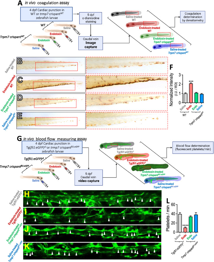
Administration of endotoxin induces coagulation in zebrafish vasculature mediated by TRPM7. (A) WT and trpm7 crispantWT zebrafish larvae were subjected to o-dianisidine staining to evaluate in vivo coagulation. Larvae were injected with 20 nL sterile saline solution (NaCl 0,09%), or endotoxin (LPS (O55:B5 Sigma, USA) 100 ng). Thrombus formation was analyzed 24 h post injection in the caudal vein by o-dianisidine staining. (B–E) Representative images of saline-injected WT (B) endotoxin-injected WT (C), endotoxin-injected trpm7 crispantWT (D) and saline-treated trpm7 crispantWT conditions (E). Doted red box depicts o-dianisidine staining. F Quantification of o-dianisidine staining in caudal vein of Zebrafish larvae in saline-injected WT (grey bars), endotoxin-injected WT (red bars), endotoxin-injected trpm7 crispantWT (green bars) and saline-treated trpm7 crispantWT (blue bars) conditions. Results of the total pixel intensity (I.U.) in a defined region of interest (ROI), were normalized with the median value of saline condition. Tg(fli1:eGFP)y1 and trpm7 crispantfli1:eGFP zebrafish larvae, having the vasculature and thrombocytes fluorescently green labeled, were subjected to time lapse analysis to evaluate blood flow in vivo coagulation. Blood flow time lapse analysis was determined as the number of platelets observed in 60 s in a section of the caudal vein (doted red box) were performed by time lapse analysis, in saline- and endotoxin-injected conditions (G). H–K Representative images of Tg(fli1:eGFP)y1 larvae saline-injected Tg(fli1:eGFP)y1 (H), endotoxin-injected Tg(fli1:eGFP)y1 (I), endotoxin-injected trpm7 crispantfli1:eGFP (J), and saline-treated trpm7 crispantfli1:eGFP conditions (K). L Quantification of blood flow time lapse analysis in a section of the caudal vein of Tg(fli1:eGFP)y1
larvae in saline-injected Tg(fli1:eGFP)y1 (grey bars), endotoxin-injected Tg(fli1:eGFP)y1 (red bars), endotoxin-injected trpm7 crispantfli1:eGFP (green bars) and saline-treated trpm7 crispantfli1:eGFP (blue bars) conditions. Statistical differences were assessed by a one-way analysis of variance (ANOVA) (Kruskal–Wallis) followed by Dunn's post hoc test. ***p < 0.001, ****p < 0.0001, compared with the saline-treated WT or Tg(fli1:eGFP)y1 conditions. Results showed as mean ± SEM
Erratum for
-
Disseminated intravascular coagulation phenotype is regulated by the TRPM7 channel during sepsis.Biol Res. 2023 Mar 3;56(1):8. doi: 10.1186/s40659-023-00419-4. Biol Res. 2023. PMID: 36869357 Free PMC article.
References
-
- Jiménez-Dinamarca I, Prado Y, Tapia P, Gatica S, Alt C, Lin CP, Reyes-Martínez C, Feijóo CG, Aravena C, González-Canacer A, Correa S, Varela D, Cabello-Verrugio C, Simon F. Disseminated intravascular coagulation phenotype is regulated by the TRPM7 channel during sepsis. Biol Res. 2023;56:8. doi: 10.1186/s40659-023-00419-4. - DOI - PMC - PubMed
Publication types
LinkOut - more resources
Full Text Sources
Miscellaneous
