Review
. 2023 Sep;101(9):1409-1432.
doi: 10.1002/jnr.25201. Epub 2023 May 11.

Functional neuroanatomy of monoaminergic systems in the basolateral nuclear complex of the amygdala: Neuronal targets, receptors, and circuits

Affiliations
Review

Functional neuroanatomy of monoaminergic systems in the basolateral nuclear complex of the amygdala: Neuronal targets, receptors, and circuits

Alexander Joseph McDonald. J Neurosci Res. 2023 Sep.

Abstract

This review discusses neuroanatomical aspects of the three main monoaminergic systems innervating the basolateral nuclear complex (BNC) of the amygdala (serotonergic, noradrenergic, and dopaminergic systems). It mainly focuses on immunohistochemical (IHC) and in situ hybridization (ISH) studies that have analyzed the relationship of specific monoaminergic inputs and their receptors to specific neuronal subtypes in the BNC in order to better understand the anatomical substrates of the monoaminergic modulation of BNC circuitry. First, light and electron microscopic IHC investigations identifying the main BNC neuronal subpopulations and characterizing their local circuitry, including connections with discrete PN compartments and other INs, are reviewed. Then, the relationships of each of the three monoaminergic systems to distinct PN and IN cell types, are examined in detail. For each system, the neuronal targets and their receptor expression are discussed. In addition, pertinent electrophysiological investigations are discussed. The last section of the review compares and contrasts various aspects of each of the three monoaminergic systems. It is concluded that the large number of different receptors, each with a distinct mode of action, expressed by distinct cell types with different connections and functions, should offer innumerable ways to subtlety regulate the activity of the BNC by therapeutic drugs in psychiatric diseases in which there are alterations of BNC monoaminergic modulatory systems, such as in anxiety disorders, depression, and drug addiction. It is suggested that an important area for future studies is to investigate how the three systems interact in concert at the neuronal and neuronal network levels.

Keywords: dopamine; immunohistochemistry; interneurons; norepinephrine; pyramidal neurons; serotonin.


Conflict of interest statement

Conflict of Interest Statement

The author has no conflict of interest to declare.

Figures

Fig. 1.
Fig. 1.
Coronal Nissl-stained sections through the amygdala of the rat (A) and monkey (B). Medial is to the right. (A) Nuclei of the rat amygdala (nomenclature of Paxinos and Watson, 1997): BLa, anterior basolateral nucleus; BLp, posterior basolateral nucleus; BLv, ventral basolateral nucleus; BMp, posterior basomedial nucleus; CL, lateral central nucleus; CM, medial central nucleus; Copl, posterolateral cortical nucleus; IN, intercalated nucleus; Ldl, dorsolateral lateral nucleus; Lvl, ventrolateral lateral nucleus; Lvm, ventromedial lateral nucleus; M, medial nucleus. (B) Nuclei of the monkey amygdala (nomenclature ofPrice et al., 1987): ABmg, magnocellular accessory basal nucleus; ABpc, parvicellular accessory basal nucleus; ABvm, ventromedial accessory basal nucleus; Bmg, magnocellular basal nucleus; Bpc, parvicellular basal nucleus; CL, lateral central nucleus; CM, medial central nucleus; Ldm, dorsomedial lateral nucleus; Lvl, ventrolateral lateral nucleus; LV, inferior horn of the lateral ventricle; M, medial nucleus; PAC, periamygdaloid cortex. The nuclear configuration of the human amygdala is similar to that of the monkey, but the BNC nuclei are rotated slightly in the counter-clockwise direction. Scale bar (see A) = 250 μm for A and 750 μm for B. Reproduced from McDonald, 2020, with permission.
Fig. 2.
Fig. 2.
Drawings of a Golgi-stained pyramidal neuron (left) and nonpyramidal neuron (right) in the rat BNC. Note the dense local axonal arborization (thin processes) of the nonpyramidal neuron. Scale bar = 50 μm. Modified from McDonald, 1982, with permission.
Fig. 3.
Fig. 3.
Left: Venn diagrams showing the overlap and relative proportions of IN subpopulations containing calcium-binding proteins and neuropeptides in the rat BLa and LA. The relative sizes of the rectangles representing these subpopulations are depicted in relation to the total population of GABA+ neurons in each nucleus. Note that there are two separate subpopulations of CCK+ INs in both nuclei. The subpopulation exhibiting partial overlap with CB are large CCK+ neurons (CCKL), whereas the subpopulation exhibiting partial overlap with CR and VIP are small CCK+ neurons (CCKS). Right: Photomicrographs of representative IN subpopulations in the rat. (A) Colocalization of PV (green) and GABA (magenta) in BL INs (white indicates colocalization). (B) Colocalization of SOM (green) and GABA (magenta) in LA INs (white indicates colocalization). (C) Lack of colocalization of CR (green) with CB (magenta) in BL INs. (D) Colocalization of VIP (magenta) with CCK (green) in small CCK+ INs (CCKS), but not in large CCK+ INs (CCKL), in the BL. Asterisks in A and B indicate several of the GABA-negative PNs in these fields. Scale bars = 25μm. Modified from McDonald, 2020, with permission.
Fig. 4.
Fig. 4.
The main cell types of the BNC and their interconnections. The dendritic tree of PNs (black) is represented by a single apical dendrite (the dense array of spines on PN dendrites have not been drawn). Different types of GABAergic INs are shown in different colors; they inhibit distinct PN compartments and other specific INs. There are two main types of PV INs: basket cells that innervate the cell bodies and proximal dendrites of PNs, and axo-axonic “chandelier” cells that innervate axon initial segments of PNs. PV neurons also inhibit SOM INs. SOM INs (many of which also express NPY) mainly innervate the distal dendritic compartment of PNs, including dendritic shafts and spines). Small VIP INs (many of which also express CR and/or CCK) also innervate the distal dendritic compartment of PNs, as well as other INs. Large CCK INs (CCKL) are mainly basket cells. PNs innervate different INs (not shown in this diagram), in addition to providing outputs to different brain areas or other amygdalar nuclei.
Fig. 5.
Fig. 5.
Photomicrographs of 5-HT+ axons in the basolateral amygdala. A) Low-power photomicrograph of the rostral portion of the BNC. Note high density of axons as well as diffuse reaction product surrounding the axons. At these rostral levels there appears to be an equal density of axons in the LA and BLa, but at more caudal levels the density of axons in the LA is much less. BLa, anterior subdivision of the basolateral nucleus; Ce, central nucleus; Cx, deep layers of the piriform cortex; La, lateral nucleus. B) Higher power photomicrograph of 5-HT+ axons in the BLa. Scale bars = 200 μm in A; 20 μm in B. Reproduced from Muller et al., 2007, with permission.
Fig. 6.
Fig. 6.
5-HT+ axons innervate INs in the BLa. A and B) Photomicrographs of 5-HT+ axon terminals (black) forming apparent contacts with (brown) parvalbumin (PV)+ (A) and calretinin (CR)+ (B) IN subpopulations in the BLa (two-color immunoperoxidase technique). Arrows indicate axon terminals forming contacts. C) Electron micrograph of a PV+ perikaryon (Ppk) receiving synaptic input from a 5-HT+ terminal (St, arrow) followed in serial thin sections. The particulate PV immunoreactivity was visualized using a Vector VIP peroxidase substrate kit. Insert: Higher power view of the synapse taken from a neighboring thin section. Scale bars = 10 μm in A (also applies to B) and 1 μm in C. Modified from Muller et al., 2007, with permission.
Fig. 7.
Fig. 7.
5-HT+ axons innervate PN dendritic structures in the BLa. A) A 5-HT+ terminal (St), in a preparation also labeled for PV, in appositional contact with a spine neck (sp) emerging from an unlabeled (PV-negative) dendrite (Ud). The spine head receives an asymmetrical (excitatory) synapse from an unlabeled terminal (Ut). Only PNs are characterized by such long dendritic spines receiving excitatory inputs. B. A small-caliber CaMK+ dendrite (Cd) receives an asymmetrical synaptic contact (arrow) from a 5-HT+ terminal (St), where three dense-core vesicles are visible in this section. The particulate CaMK immunoreactivity was visualized using a Vector VIP peroxidase substrate kit. Scale bars = 0.5 μm in A and B. Modified from Muller et al., 2007, with permission.
Fig. 8.
Fig. 8.
Dual localization of 5-HT2AR immunoreactivity and IN marker immunoreactivity in the rat BLa using immunofluorescence confocal microscopy. (A1–A3) Dual localization of 5-HT2AR and PV in the BLa. (A1) 5-HT2AR+ neurons (green). (A2) PV+ neurons (magenta) in the same field. (A3) Merging of the magenta and green channels indicates that there is extensive colocalization of 5-HT2AR and PV immunoreactivity (white), but there are also neurons that are single-labeled for 5-HT2AR (green) or PV (magenta). (B) Higher power merged image of 5-HT2AR (green) and PV (magenta) in the BLa. In addition to several PV+ neurons with variable amounts of 5-HT2AR immunoreactivity (white), there is also a single-labeled PV+ neuron (magenta arrow) and a single-labeled 5-HT2AR+ neuron (green arrow). (C, D) Dual localization of 5-HT2AR (green) and SOM (magenta) in the BLa. Double-labeled 5-HT2AR/SOM+ neurons (white) and single-labeled 5-HT2AR neurons (green) are observed in these two fields. All scale bars =25 μm. Reproduced from McDonald and Mascagni, 2007, with permission.
Fig. 9.
Fig. 9.
Dual-localization of 5-HT3AR immunoreactivity (green) and IN marker immunoreactivity (magenta) in the BNC using immunofluorescence confocal microscopy. Dual-labeled neurons are white. All images are merged images. (A–C) Dual localization of 5-HT3AR (green) and GABA (magenta) immunoreactivity in the Lvm (A, C) and BLa (B). Note that all 5-HT3AR+ neurons in these fields are double-labeled (white). (D–F) Dual localization of 5-HT3AR (green) and CCK (magenta) immunoreactivity in the Lvm (D, E) and BLa (F). Note that most CCK+ neurons are either large (type L neurons) or small (type S neurons). Most single-labeled 5-HT3AR+ neurons (green) are medium-sized or small, whereas double-labeled 5-HT3AR+/CCK+ neurons (white) are larger. All scale bars = 25 μm. Reproduced from Mascagni and McDonald, 2007, with permission.
Fig. 10.
Fig. 10.
NET immunoreactivity in the rat BNC. (A) Low power immunofluorescence micrograph illustrating NET+ axons (green) in the BLa, lateral nucleus (La), and lateral subdivision of the central nucleus (CL) at rostral levels of the BNC. (B) High power micrograph illustrating the morphology of NET+ axons in the BLa using nickel-enhanced DAB as a chromogen in an immunoperoxidase preparation. Arrows point to NET+ varicosities. Scale bars = 100 μm in A, 10 μm in B. Reproduced from Zhang et al., 2013, with permission.
Fig. 11.
Fig. 11.
An NET+ terminal (Nt) forms a symmetrical synapse (arrow) with a large-caliber CaMK+ PN dendrite (Cd). Arrowheads indicate particulate label for CaMK. The particulate label for CaMK is easily distinguished from the diffuse DAB label for NET. Scale bars=500 nm. Reproduced from Zhang et al., 2013, with permission.
Fig. 12.
Fig. 12.
(A and B) Two neighboring thin sections in a series through three NET+ terminals (Nt1–3) forming synapses (arrows) with spines (Sp). Arrowheads show particulate label for CAMK in a myelinated CaMK+ axon (Cax), and a CaMK+ dendrite (Cd)., Although almost all spines arise from PNs, in contrast to PN dendritic shafts only about half exhibit CAMK-ir (McDonald et al., 2002). Scale bars =500 nm. Reproduced from Zhang et al., 2013, with permission.
Fig. 13.
Fig. 13.
Photomicrographs of TH-ir in the rat BNC. (A) Low-power micrograph illustrating TH-ir in the BLa and lateral nuclei (La) at rostral levels of the BNC (medial is to the right). Ce: central amygdalar nucleus; asterisks indicate intercalated nuclei. (B) Higher power micrograph illustrating the morphology of TH+ axons in the BLa. Scale bars = 200 μm in A; 20 μm in B. Reproduced from Pinard et al., 2008, with permission.
Fig. 14.
Fig. 14.
TH+ axon terminals contact dendritic structures of PNs in the BLa. A and B A) Two views, three serial sections apart, of a broad spine head (sp) with particulate CaMK-ir (A) and the thin dendrite that gives rise to it (Cd, double arrows in B indicate beginning of spine neck). The spine receives a contact, possibly synaptic, from a small TH+ terminal (THt, arrow in A). This spine also receives a perforated asymmetrical synapse from a CaMK+ terminal (Ct, arrowheads in a) and an oblique symmetrical synapse from an adjacent unlabeled terminal (Ut, asterisk in B). Its dendrite of origin (Cd) also receives an asymmetrical synaptic contact from a CaMK+ terminal (Ct, arrowheads in B). C) A small-caliber CaMK+ dendrite (Cd) receives synaptic input from a TH+ terminal (THt, arrow). D) A small-caliber CaMK+ dendrite seen in cross-section (Cd, top) receives synaptic input from a TH+ terminal (THt, arrow). The TH+ terminal was also found to form a synapse with the CaMK+ small-caliber dendrite seen in longitudinal section (Cd, left) several sections further, and both this dendrite and the TH+ terminal were apposed to a CaMK+ terminal (Ct). Scale bars = 0.5 μm. D is at the same magnification as C. Modified from Muller et al., 2009 (A and B) and Pinard et al., 2008 (C and D), with permission.
Fig. 15.
Fig. 15.
TH+ terminals contact CR+ and PV+ dendrites in the BLa. (A) TH+ terminal (THt) forms a synaptic contact (arrow with large arrowhead) with a small unlabeled dendrite (Ud) and an apposition (arrow with small arrowhead) with a large dendrite with particulate CR-ir (CRd). (B) A thin CR+ dendrite (CRd) receives synaptic input (arrow) from a TH+ terminal (THt). (C, D) PV+ dendrites with particulate label (PVd) receive synaptic input (arrows) from TH+ terminals (THt). Scale bars= 500 nm. Reproduced from Pinard et al., 2008, with permission.
Fig. 16.
Fig. 16.
Drawing of a PV+ IN in the posterior subdivision of the BL (BLp) that is innervated by varicose TH+ axons (black). Note that axons form baskets around the soma and also run along the dendrites making multiple contacts (arrows). Scale bar = 10 μm. Insert: Photomicrograph of the soma of this neuron. Modified from Brinley-Reed et al., 1999, with permission.
Fig. 17.
Fig. 17.
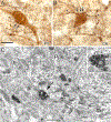
A PV+ perikaryon is enveloped by TH+ axons in a basket-like configuration. (A) Light micrograph of a PV+ perikaryon (PVpk) receiving contacts from TH+ terminals (boutons b1–b5). (B) Electron micrograph of the PV+ perikaryon shown in A. (C) Higher power view of the contact (large arrow) formed by TH+ terminal b1. This contact was considered to be a synapse with a narrow synaptic cleft. Small arrows indicate examples of the particulate PV-ir. (D) Higher power view of the contact (large arrow) formed by TH+ terminal b2. This contact was considered to be a small synapse. A cluster of synaptic vesicles adjacent to the presynaptic membrane was more obvious in an adjacent thin section. Small arrows indicate examples of the particulate PV-ir. Scale bars+ 5 μm (A, B); 500 nm (C, D). Reproduced from Pinard et al., 2008, with permission.
Fig. 18.
Fig. 18.
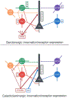
Main connections of monoaminergic inputs to BNC neuronal populations and the receptors expressed by these neurons (as revealed in IHC and ISH studies). The strength of the inputs to different cell types is indicated by the thickness of the lines; the main targets of all three monoaminergic systems are PN dendritic shafts and spines. A) Main connections of 5-HT inputs to BNC neuronal populations and the receptors expressed by these neurons, PNs and all types of INs are innervated by serotonergic axons that arise from the dorsal raphe nucleus (DRN; “starburst” lines emanating from the DRN indicate that all neuronal populations receive inputs, but the input to the dendritic domain of PNs is the most dense). The purple IN expresses 5-HT3Rs but does not contain an of the peptides/proteins investigated. B) Main connections of catecholamine inputs to BNC neuronal populations and the receptors expressed by these neurons. PNs and several IN subtypes, especially PV INs, have been shown to be innervated by dopaminergic axons that arise from the ventral tegmental area and substantia nigra (VTA/SN). Noradrenergic axons that arise from the locus coeruleus (LC) mainly target the dendritic domain of PNs (inputs to separate subpopulations of INs have not been investigated). For both A and B the main catecholamine receptors expressed by each neuronal population are indicated, although in some cases separate subpopulations of each neuron type may express some, but not all, of the receptors indicated. Since the expression of some of the receptors have only been documented using ISH, it remains to be determined to what extent these mRNAs are translated into proteins.

References

    1. Abi-Dargham A, Laruelle M, Wong DT, Robertson DW, Weinberger DR, Kleinman JE (1993) Pharmacological and regional characterization of [3H]LY278584 binding sites in human brain. J Neurochem 60:730–737. - PubMed
    1. Abraham PA, Xing G, Zhang L, Yu EZ, Post R, Gamble EH, Li H (2008) Beta1- and beta2-adrenoceptor induced synaptic facilitation in rat basolateral amygdala. Brain Res 1209:65–73. - PubMed
    1. Agnati LF, Fuxe K, Zoli M, Ozini I, Toffano G, Ferraguti F (1986) A correlation analysis of the regional distribution of central enkephalin and beta-endorphin immunoreactive terminals and of opiate receptors in adult and old male rats. Evidence for the existence of two main types of communication in the central nervous system: the volume transmission and the wiring transmission. Acta Physiol Scand 128:201–207. - PubMed
    1. Agnati LF, Guidolin D, Guescini M, Genedani S, Fuxe K (2010) Understanding wiring and volume transmission. Brain Res Rev 64:137–159. - PubMed
    1. Amaral DG, Price JL, Pitkanen A, and Carmichael ST (1992) Anatomical organization of the primate amygdala. In: The Amygdala. Neurobiological aspects of emotion, memory, and mental dysfunction. (Aggleton JP, ed), pp. 1–66. New York: Wiley-Liss

Publication types

LinkOut - more resources