Review
. 2023 Dec 1;74(22):6933-6949.
doi: 10.1093/jxb/erad174.

Auxin as an architect of the pectin matrix

Affiliations
Review

Auxin as an architect of the pectin matrix

François Jobert et al. J Exp Bot. .

Abstract

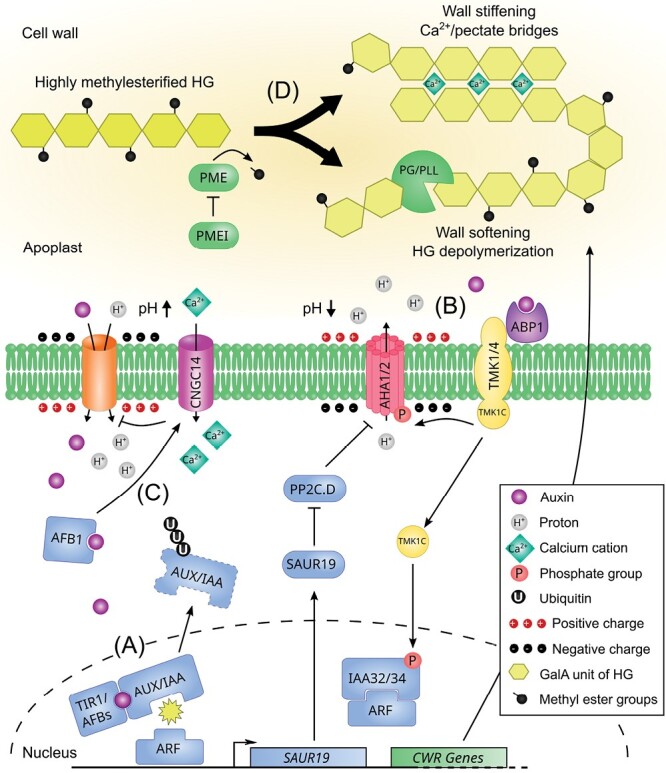
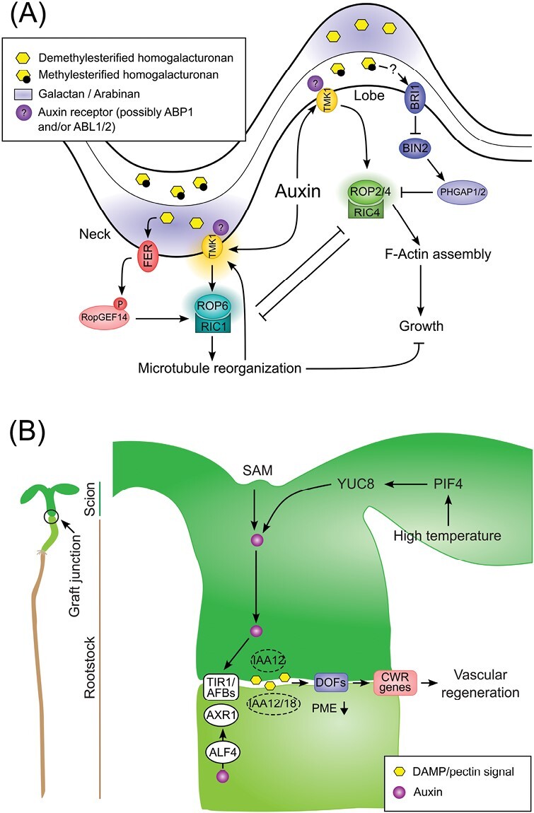
Auxin is a versatile plant growth regulator that triggers multiple signalling pathways at different spatial and temporal resolutions. A plant cell is surrounded by the cell wall, a complex and dynamic network of polysaccharides. The cell wall needs to be rigid to provide mechanical support and protection and highly flexible to allow cell growth and shape acquisition. The modification of the pectin components, among other processes, is a mechanism by which auxin activity alters the mechanical properties of the cell wall. Auxin signalling precisely controls the transcriptional output of several genes encoding pectin remodelling enzymes, their local activity, pectin deposition, and modulation in different developmental contexts. This review examines the mechanism of auxin activity in regulating pectin chemistry at organ, cellular, and subcellular levels across diverse plant species. Moreover, we ask questions that remain to be addressed to fully understand the interplay between auxin and pectin in plant growth and development.

Keywords: Auxin; calcium (Ca2+); cell wall; microdomains; pH; pectin; pectin methylesterase.


Conflict of interest statement

The authors have no conflicts of interest to declare.

Figures

Fig. 1.
Fig. 1.
An updated view of the auxin-triggered signalling cascades. (A) Nuclear auxin signalling pathway. Auxin is perceived intracellularly by the TIR1/AFB–AUX/IAA co-receptor complex, resulting in the polyubiquitinylation and degradation of AUX/IAAs, which releases the inhibition of the ARF transcription factors in the nucleus. This triggers the expression of auxin responsive genes, among which SAUR19 whose protein inhibits the phosphatase PP2C.D, preventing the deactivation of the plasma membrane H+-ATPase AHA1/2 through dephosphorylation of the penultimate residue. This cascade results in gradual apoplast acidification and hyperpolarization of the plasma membrane. In the meantime, auxin regulates the expression of cell wall remodelling enzymes modulating pectin properties (see (D)). (B) TMK-based auxin extracellular signalling pathway. Auxin is perceived in the apoplast by the secreted receptor ABP1 and its plasma membrane partners from the TMK family. This activates the TMKs, which in turn phosphorylate AHA1/2, allowing a net efflux of H+ and the rapid hyperpolarization of the plasma membrane. In the context of apical hook growth cessation, the TMK1 C-terminal domain is processed by an unknown molecular actor and translocates to the nucleus where it phosphorylates and stabilizes the non-canonical IAA32/34 repressors. (C) TIR1/AFB auxin extranuclear signalling pathway. Auxin enters the cell via AUX1-based IAA–H+ symport, triggering rapid plasma membrane alkalinization and depolarization. IAA perception by the cytoplasmic AFB1 complex elicits Ca2+ influx dependent on CNGC14. The high [Ca2+]cyt inhibits AUX1-mediated IAA influx in a negative feedback loop through an unknown mechanism. (D) The main pectin component, homogalacturonan, is secreted into the cell wall in a highly methylesterified form. Demethylesterification is catalysed by PME enzymes, which are actively regulated by PME proteinaceous inhibitors. Depending on the PME processivity, blockwise demethylesterified HG galacturonic acid chains can form Ca2+/pectate bridges and organize into an ‘egg-box’-like structure thought to stiffen the cell wall. In contrast, non-blockwise or random demethylesterification results in HG depolymerization through pectinolytic enzyme activity (PG/PLL) and softening of the cell wall.
Fig. 2.
Fig. 2.
Overview of the auxin and pectin interplay in Arabidopsis epidermal pavement cell shape acquisition and hypocotyl graft interface. (A) Epidermal pavement cell shape working model. Cell surface auxin sensitive TMK1-based module, possibly involving the ABP1 extracellular auxin receptor or its close paralogue ABL, activates the ROP2/4-RIC4 module to trigger F-Actin filament assembly and lobe expansion. At the neck location, auxin promotes TMK1 nanoclusters activating the ROP6–RIC1 module to induce cortical microtubule reorganization and growth restriction. The convex side of the cell wall is enriched in galactan and arabinan pectin components and in demethylesterified homogalacturonans. Demethylesterified pectin are sensed by the extracellular domain of FER triggering phosphorylation of RopGEF14, in turn activating ROP6 signalling. At the neck location, high BRI1-mediated brassinosteroid signalling supresses BIN2-triggered phosphostabilization of PHGAP resulting in the activation of ROP2 signalling. On the concave side of the cell wall, the highly methylesterifified pectin signal is possibly transduced to BRI1 by an unknown mechanism. (B) Hypocotyl graft interface working model. Auxin is synthesized in the shoot apical meristem (SAM) or in the leaves under high temperature through a PIF4/YUC8 module, moves across the grafting interface, and elicits the TIR1/AFBs cascade degrading IAA12/18 and activating unknown ARFs to promote vascular regeneration. AXR1 and ALF4 are required in the root stock for vascular reconnection but not in the scion. Wounding at the graft interface produces a damage associated molecular pattern (DAMP), possibly resulting from pectin degradation products, and activates DOF transcription factors that regulate the expression of cell wall remodeling (CWR) genes to allow vascular regeneration. Reduced PME activity below the graft junction is a prerequisite for plant healing.

Similar articles

Cited by

References

    1. Aalen RB, Wildhagen M, Stø IM, Butenko MA.. 2013. IDA: a peptide ligand regulating cell separation processes in Arabidopsis. Journal of Experimental Botany 64, 5253–5261. 10.1093/jxb/ert338. - DOI - PubMed
    1. Altartouri B, Bidhendi AJ, Tani T, Suzuki J, Conrad C, Chebli Y, Liu N, Karunakaran C, Scarcelli G, Geitmann A.. 2019. Pectin chemistry and cellulose crystallinity govern pavement cell morphogenesis in a multi-step mechanism. Plant Physiology 181, 127–141. 10.1104/pp.19.00303. - DOI - PMC - PubMed
    1. Amsbury S, Hunt L, Elhaddad N, Baillie A, Lundgren M, Verhertbruggen Y, Scheller HV, Knox JP, Fleming AJ, Gray JE.. 2016. Stomatal function requires pectin de-methyl-esterification of the guard cell wall. Current Biology 26, 2899–2906. 10.1016/j.cub.2016.08.021. - DOI - PMC - PubMed
    1. Andika IB, Sun L, Xiang R, Li J, Chen J.. 2013. Root-specific role for Nicotiana benthamiana RDR6 in the inhibition of Chinese wheat mosaic virus accumulation at higher temperatures. Molecular Plant-Microbe Interactions 26, 1165–1175. 10.1094/MPMI-05-13-0137-R. - DOI - PubMed
    1. Atmodjo MA, Hao Z, Mohnen D.. 2013. Evolving views of pectin biosynthesis. Annual Review of Plant Biology 64, 747–779. 10.1146/annurev-arplant-042811-105534. - DOI - PubMed

Publication types

MeSH terms