. 2023 Jul 3;133(13):e165988.
doi: 10.1172/JCI165988.

SELENOP modifies sporadic colorectal carcinogenesis and WNT signaling activity through LRP5/6 interactions

Affiliations

SELENOP modifies sporadic colorectal carcinogenesis and WNT signaling activity through LRP5/6 interactions

Jennifer M Pilat et al. J Clin Invest. .

Abstract

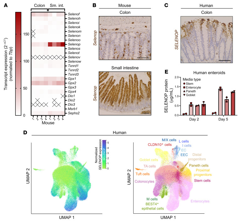
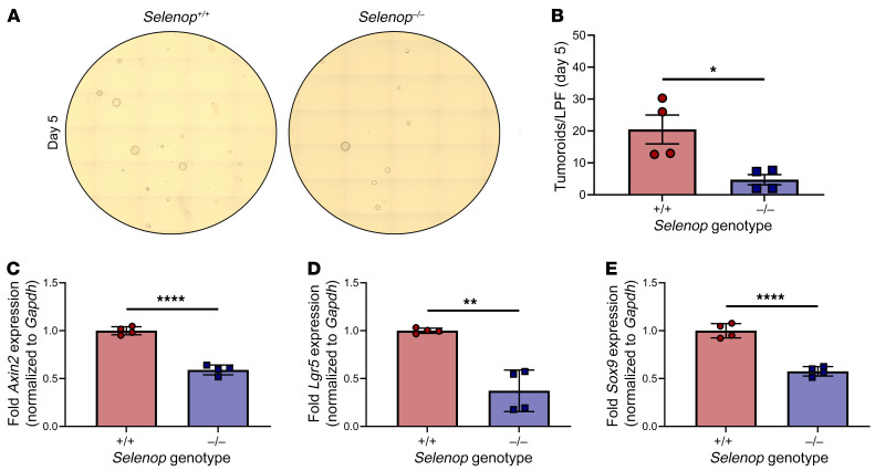
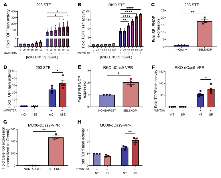
Although selenium deficiency correlates with colorectal cancer (CRC) risk, the roles of the selenium-rich antioxidant selenoprotein P (SELENOP) in CRC remain unclear. In this study, we defined SELENOP's contributions to sporadic CRC. In human single-cell cRNA-Seq (scRNA-Seq) data sets, we discovered that SELENOP expression rose as normal colon stem cells transformed into adenomas that progressed into carcinomas. We next examined the effects of Selenop KO in a mouse adenoma model that involved conditional, intestinal epithelium-specific deletion of the tumor suppressor adenomatous polyposis coli (Apc) and found that Selenop KO decreased colon tumor incidence and size. We mechanistically interrogated SELENOP-driven phenotypes in tumor organoids as well as in CRC and noncancer cell lines. Selenop-KO tumor organoids demonstrated defects in organoid formation and decreases in WNT target gene expression, which could be reversed by SELENOP restoration. Moreover, SELENOP increased canonical WNT signaling activity in noncancer and CRC cell lines. In defining the mechanism of action of SELENOP, we mapped protein-protein interactions between SELENOP and the WNT coreceptors low-density lipoprotein receptor-related proteins 5 and 6 (LRP5/6). Last, we confirmed that SELENOP-LRP5/6 interactions contributed to the effects of SELENOP on WNT activity. Overall, our results position SELENOP as a modulator of the WNT signaling pathway in sporadic CRC.

Keywords: Colorectal cancer; Gastroenterology.


Conflict of interest statement

Conflict of interest: EL is a co-founder of StemSynergy Therapeutics.

Figures

Figure 1
Figure 1. SELENOP is predominantly expressed by differentiated epithelial cells in the normal colon and small intestine epithelium.
(A) RT-qPCR of mouse colon and small intestine (sm. int.) epithelial isolates for selenoproteins. n = 4 mice. (B) RNAscope of mouse colon and small intestine for Selenop. Representative images of colon (original magnification, ×20) and/or small intestine (original magnification, ×10). Scale bars: 100 μm. (C) RNAscope of human colon for SELENOP. Representative images (original magnification, ×20). Scale bar: 100 μm. (D) Gut Cell Atlas scRNA-Seq data from human colon and small intestine epithelium queried for SELENOP. EC, enterochromaffin; EEC, enteroendocrine; TA, transit-amplifying. n = 6 donors. (E) ELISA of conditioned media from human enteroids treated with the indicated media for SELENOP. Data were pooled from 2 independent experiments. Data are displayed as the mean ± SEM. EC, enterochromaffin cell; EEC, enteroendocrine cell; M; microfold; M/X, MLN+GHRL+; TA, transit-amplifying.
Figure 2
Figure 2. SELENOP expression progressively increases throughout conventional colorectal carcinogenesis.
(A and B) scRNA-seq data from human colorectal polyps and cancers. (A) SELENOP expression in cell clusters. n = 62 polyps; n = 7 cancers; n = 149,116 cells. UMAP, uniform manifold approximation and projection. (B) SELENOP expression versus stemness inferred from CytoTRACE analysis. n = 29 polyps; n = 5 cancers. (C) scRNA-Seq data from human colorectal polyps or cancers and normal colon tissues (27). SELENOP expression by cell type. AD, adenoma. n = 34 normal samples; n = 29 polyps; n = 5 cancers. (D) scRNA-Seq data from human colorectal cancers (27, 30). SELENOP expression by tumor type. MMR-d, mismatch repair deficient; MMR-p, mismatch repair proficient. n = 2 MSI-H cancers; n = 5 MSS cancers (left); n = 32 MMR-d cancers; n = 28 MMR-p cancers (right). ****P < 0.0001, by Spearman’s rank correlation (B), Kruskal-Wallis test with a 2-sided Mann-Whitney U test (C), and 2-sided Mann-Whitney U test (D). Data are displayed as the mean ± SD.
Figure 3
Figure 3. Selenop KO decreases colon tumor incidence and size in Apc-dependent tumorigenesis.
(A) Schematic of murine tumorigenesis protocol. TAM, tamoxifen. (B) Colon tumor incidence, (C) colon tumor volume, (D) cumulative survival, (E) colon tumor numbers, (F) colon tumor dysplasia scores (HGD, high-grade dysplasia, LGD, low-grade dysplasia), and (G) histology of colon tumors from ApcΔIE/+ Selenop+/+ (n = 9), Selenop+/– (n = 10), and Selenop–/– (n = 8) mice. Original magnification, ×20. Scale bars: 100 μm. Data were pooled from 2 independent experiments. *P < 0.05, by Freeman-Halton test (B and F), Kruskal-Wallis test (C and E) with 2-sided Dunn’s multiple-comparison test (C), and log-rank test (D).
Figure 4
Figure 4. Selenop KO decreases tumoroid-forming capacity and WNT target gene expression.
(A and B) ApcΔIE/+ Selenop+/+ or Selenop–/– tumoroids 5 days after enzymatic dissociation. (A) Representative ×10 tile scans. (B) Visible tumoroids per low-powered field (LPF). (CE) RT-qPCR for (C) Axin2, (D) Lgr5, and (E) Sox9 of ApcΔIE/+ Selenop+/+ or Selenop–/– tumoroids. Data were pooled from 2 independent experiments with 2 mice per genotype. *P < 0.05, **P < 0.01, and ****P < 0.0001, by 2-sided, unpaired t test. Data are displayed as the mean ± SEM.
Figure 5
Figure 5. SELENOP restoration increases tumoroid-forming capacity and WNT target gene expression.
(A) RT-qPCR for Selenop of ApcΔIE/+ Selenop+/+-dCas9-VP64-NONTARGET or SELENOP tumoroids. (B and C) ApcΔIE/+ Selenop+/+-dCas9-VP64-NONTARGET or SELENOP tumoroids 5 days after enzymatic dissociation. (B) Representative ×10 tile scans. (C) Visible tumoroids per LPF. (DF) RT-qPCR for (D) Axin2, (E) Lgr5, and (F) Sox9 in ApcΔIE/+ Selenop+/+-dCas9-VP64-NONTARGET or SELENOP tumoroids. Data were pooled from 4 independent experiments. *P < 0.05, **P < 0.01, and ***P < 0.001, by 2-sided, paired t test. Data are displayed as the mean ± SEM.
Figure 6
Figure 6. SELENOP increases canonical WNT signaling activity in noncancer and colon cancer cell lines.
(A and B) TOPFlash activity of (A) 293 STF and (B) RKO STF cells treated or not with rhWNT3A and the indicated concentrations of hSELENOP. (C) ELISA for SELENOP of 293 STF-mCherry or hSELENOP conditioned media. (D) TOPFlash activity of 293 STF-mCherry or hSELENOP cells treated or not with rhWNT3A. hSE, hSELENOP; mCh, mCherry. (E) ELISA for SELENOP of RKO-dCas9-VPR-NONTARGET or SELENOP conditioned media. (F) TOPFlash activity of RKO-dCas9-VPR-NONTARGET (NT) or SELENOP (SP) cells treated or not with rhWNT3A. (G) RT-qPCR for Selenop of MC38-dCas9-VPR-NONTARGET or SELENOP cells. (H) TOPFlash activity of MC38-dCas9-VPR-NONTARGET or SELENOP cells treated or not with rmWNT3A. Data were pooled from 3–4 independent experiments. *P < 0.05, **P < 0.01, and ****P < 0.0001, by 2-way, repeated-measures ANOVA with 2-sided Dunnett’s multiple-comparison test (A and B), 2-sided, paired t test (C, E, and G), and 2-way, repeated-measures ANOVA with 2-sided Šidák’s multiple-comparison test (D, F, and H). Data are displayed as the mean ± SEM.
Figure 7
Figure 7. SELENOP interacts with LRP6.
(A) Western blot for FLAG and SELENOP of FLAG IPs from 293T or 293T-FLAG-LRP6 cells. (B) Western blot for FLAG and SELENOP of FLAG IPs from 293T or 293T-FLAG-LRP6 cells treated or not with sodium chlorate (NaClO3). (C) Western blot for FLAG and SELENOP of FLAG IPs from 293T or 293T-FLAG-LRP6 cells treated or not with heparin. (D) Western blot for LRP6, Na+/K+-ATPase (plasma membrane loading control), and β-tubulin (whole-cell loading control) of cell-surface biotinylation and isolation from 293T cells treated or not with SELENOP-conditioned media. Data are representative of 3 independent experiments.
Figure 8
Figure 8. Longer SELENOP isoforms interact with LRP6.
(A) Schematic of mouse SELENOP truncation (t) constructs. (B) Western blot for LRP6 and SELENOP of FLAG IPs from 293T cells cotransfected with FLAG-mLRP6 and full-length or truncated (at selenocysteine [U] number) mSELENOP. (C) Schematic of V5-tagged mouse SELENOP truncation constructs. (D) Western blot for LRP6 and V5 of FLAG IPs from 293T cells cotransfected with FLAG-mLRP6 and full-length or truncated (at U number) V5-mSELENOP. Data are representative of 3 independent experiments. F, full-length; HBS, heparin-binding site; His-rich, histidine-rich region; LRP8 BS, LRP8-binding site.
Figure 9
Figure 9. SELENOPU258–U299 mediates the SELENOP-LRP6 interaction and SELENOP-induced WNT signaling augmentation.
(A) Schematic of V5-tagged mouse SELENOP deletion constructs. ΔA, Δ258-267; ΔB, Δ268-277; ΔC, Δ278-287; ΔD, Δ288-299; ΔE, Δ258-299. (B) Western blot for LRP6 and V5 of FLAG IPs from 293T cells cotransfected with FLAG-mLRP6 and full-length or mutant (A–E) V5-mSELENOP. (C) Western blot for V5 and (D) TOPFlash activity of YAMC STF cells transduced with full-length or LRP5/6-uncoupling (E) V5-mSELENOP. Representative (B and C) or pooled (D) data from 3–4 independent experiments. *P < 0.05, **P < 0.01, and ***P < 0.001, by 2-way, repeated-measures ANOVA with 2-sided Tukey’s multiple-comparison test. Data are displayed as the mean ± SEM.

Comment in

Similar articles

Cited by

References

    1. Short SP, et al. Roles for selenium and selenoprotein P in the development, progression, and prevention of intestinal disease. Free Radic Biol Med. 2018;127:26–35. doi: 10.1016/j.freeradbiomed.2018.05.066. - DOI - PMC - PubMed
    1. Davis CD, et al. Selenium-enriched broccoli decreases intestinal tumorigenesis in multiple intestinal neoplasia mice. J Nutr. 2002;132(2):307–309. doi: 10.1093/jn/132.2.307. - DOI - PubMed
    1. Barrett CW, et al. Dietary selenium deficiency exacerbates DSS-induced epithelial injury and AOM/DSS-induced tumorigenesis. PLoS One. 2013;8(7):e67845. doi: 10.1371/journal.pone.0067845. - DOI - PMC - PubMed
    1. Duffield-Lillico AJ, et al. Baseline characteristics and the effect of selenium supplementation on cancer incidence in a randomized clinical trial: a summary report of the Nutritional Prevention of Cancer Trial. Cancer Epidemiol Biomarkers Prev. 2002;11(7):630–639. - PubMed
    1. Lippman SM, et al. Effect of selenium and vitamin E on risk of prostate cancer and other cancers: the Selenium and Vitamin E Cancer Prevention Trial (SELECT) JAMA. 2009;301(1):39–51. doi: 10.1001/jama.2008.864. - DOI - PMC - PubMed

Publication types

MeSH terms