. 2023 May 11;18(5):e0285257.
doi: 10.1371/journal.pone.0285257. eCollection 2023.

Pan-genome survey of the fish pathogen Yersinia ruckeri links accessory- and amplified genes to virulence

Affiliations

Pan-genome survey of the fish pathogen Yersinia ruckeri links accessory- and amplified genes to virulence

Andreas Riborg et al. PLoS One. .

Abstract

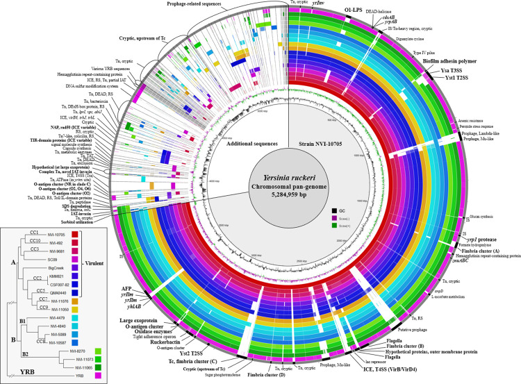
While both virulent and putatively avirulent Yersinia ruckeri strains exist in aquaculture environments, the relationship between the distribution of virulence-associated factors and de facto pathogenicity in fish remains poorly understood. Pan-genome analysis of 18 complete genomes, representing established virulent and putatively avirulent lineages of Y. ruckeri, revealed the presence of a number of accessory genetic determinants. Further investigation of 68 draft genome assemblies revealed that the distribution of certain putative virulence factors correlated well with virulence and host-specificity. The inverse-autotransporter invasin locus yrIlm was, however, the only gene present in all virulent strains, while absent in lineages regarded as avirulent. Strains known to be associated with significant mortalities in salmonid aquaculture display a combination of serotype O1-LPS and yrIlm, with the well-documented highly virulent lineages, represented by MLVA clonal complexes 1 and 2, displaying duplication of the yrIlm locus. Duplication of the yrIlm locus was further found to have evolved over time in clonal complex 1, where some modern, highly virulent isolates display up to three copies.


Conflict of interest statement

The authors have declared that no competing interests exist.

Figures

Fig 1
Fig 1. Maximum likelihood tree based on 2,387 genes, 49,389 core gene SNPs (16,762 SNPs were identified with the YRB lineage excluded; S2 Fig), with presence/absence data for specific systems or genes.
MLVA clonal complexes (CC) and lineages are indicated on branches. Bootstrap values as percentages of 200 replicates are indicated if < 75 percent. Branch lengths between the YRB lineage and remaining genomes are truncated (see S1 Fig). Presence of genetic determinants indicated by green shading for Ysa Type 3 secretion system, Yst1 Type 2 secretion system, virB/virD4 Type 4 secretion system (VirB), Toxin complex (Tc), the putative invasins yrIlm, yrInv and undescribed invasin WP_042527435, sorbitol utilization genes (gut/srl), the alkyl sulphatase associated with SDS degradation (yarS), and the serotype O1-LPS synthesis cluster. Partial deletions of Ysa, Yst1 and the O1-LPS cluster are shown as absent if genes regarded as critical for function are not present. Serotype NR indicates no reaction with available antisera (O1, O2, O5).
Fig 2
Fig 2
Maximum likelihood core-gene phylogenetic tree (bottom left) and circular representation of genomic comparisons generated with the BLAST Ring Image Generator (BRIG) from complete Y. ruckeri genome assemblies. Plasmid sequences were removed from the data prior to analysis. Lineages and MLVA clonal complexes (CC) are indicated on the phylogenetic tree tree branches, with strains regarded as virulent labelled as such. The branch length of YRB is truncated (see S1 Fig). Coloured rings represent genomic sequences used as BLAST queries, corresponding to the colour labelling on tree nodes. Ordering top-bottom on the tree and inner-outter on the circular representation is identical (virulent strains in innermost rings). Solid colour in each respective ring represents ≥95% nucleotide identity to the pan-genome reference. Cut-off identity for BLAST was 85%. The innermost ring (black line with nucleotide distances indicated) represents the pan-genome reference sequence, consisting of the chromosome of strain NVI-10705 for the first 3,818,566 bp, followed by additional Y. ruckeri chromosomal sequences with each segment separated by a 800 bp artificial gap with black fill, and ordered by commonality or general association. Prominent features within the NVI-10705 chromosome are labelled by black fill on the outermost ring (grey). Annular graphs towards the centre represent GC percent (black) and GC skew (purple and green). All chromosomal sequences from additional complete genomes belonging to CC1 (see Table 1) are represented, and vary from NVI-10705 only in prophage-related sequences (not shown) and plasmids (Table 1). Hemagglutinin repeat-containing protein is present in all sequences (n = 86) but divergent in YRB (82% nucleotide identity). Abbreviations used: Restriction system (RS), toxin-antitoxin (TAT), insertion sequence (IS), transposon (Tn), integrative and conjugative elements (ICE), Toxin Complex (Tc), inverse autotransporter (IAT), no reaction (NR), DEAD-box helicase (DEAD), Anti-feeding prophage (AFP).
Fig 3
Fig 3. Genetic organization of the Ysa-Yts1 locus, between mutS and fumA in the Y. ruckeri core genome.
Each gene is colored according to association with a system or other specific role as indicated by the panel on the left. Nomenclature for Ysa (ysa) and Yst1 (gsp) is according to Venecia and Young [64] and Iwobi et al. [63]. The putative O-antigen cluster consists of protein sequences (green) WP_045844464–67 and WP_042527525. Genotypes, labelled A to H, represent the following sequences: (A) NVI-10705 and typical configuration of serotype O1 sequences that possess intact Ysa and Yst1; (B) NVI-9681 and typical configuration of serotype O2 with a O-antigen cluster in green; (C) SC09; (D) NVI-492 with partial Ysa deletion characteristic of CC10; (E) NVI-11073 with Ysa deletion; (F) NVI-5635 with partial Ysa and Yst1 deletion and a putative ICE inserted between mutS and the partial Ysa; (G) NVI-4479 with complete deletion of Ysa and Yst1; (H) NVI-11065 with complete deletion of Ysa and Yst1 including DUF 1996. The dotted line indicates a contig split (incomplete genome sequence).
Fig 4
Fig 4. Genetic organization of the accessory VirB/VirD4 ICE and associated genes.
Genes are coloured according to function or association with each variant. Genes indicated by number represent virB2virB11. virB3 and virB4 are fused. Genotypes A to D represent the following sequences: (A) NVI-10705 and typical configuration in CC1 isolates with downstream genes exclusive to CC1; (B) SC09 with downstream genes exclusive to this strain; (C) CFS007-82 with typical configuration of CC2 isolates; (D) NVI-11076 (CC7) with some of the downstream genes present in CC2.
Fig 5
Fig 5. Genetic organization of the yrIlm locus, between yhlA and AFP18 in the Y. ruckeri core genome and maximum likelihood core gene phylogeny of complete CC1 genomes indicating year of isolation, yrIlm copy number and variant (bottom right).
The complete sequence of strain NVH-3758 (CP023184; not included in this tree), isolated in Norway in 1987, contains a single copy of yrIlm [21]. Locus variants, labelled A to K, represent the following complete genomes: (A) NVI-6614 and NVI-10571 (CC1); (B) most modern CC1; (C) CFS007-82 (CC2); (D) KMM821 (CC2); (E) NVI-701 (CC1) and NVI-9681 (CC3); (F) NVI-1176 and NVI-1292 (CC1), and QMA0440 (CC5); (G) BigCreek74; (H) NVI-492 (CC10); (I) SC09; (J) empty site with partial IS256 in lineage B2; (K) empty site with no IS256 in lineages B1 and YRB, and MLVA clonal complexes CC7 and CC9.

References

    1. Sparboe O, Koren C, Håstein T, Poppe TT, Stenwig H. The first isolation of Yersinia ruckeri from farmed Norwegian salmon. Bull Eur Assoc Fish Pathol. 1986;6, 41–42.
    1. Rintamäki P, Valtonen ET, Frerichs GN. Occurrence of Yersinia ruckeri infection in farmed whitefish, Coregonus peled Gmelin and Coregonus muksun Pallas, and Atlantic salmon, Salmo salar L., in northern Finland. J Fish Dis. 1986;9: 137–140. 10.1111/j.1365-2761.1986.tb00993.x - DOI
    1. Costa AA, Leef MJ, Bridle AR, Carson J, Nowak B F. Effect of vaccination against yersiniosis on the relative percent survival, bactericidal and lysozyme response of Atlantic salmon, Salmo salar. Aquaculture. 2011;315, 201–206. 10.1016/j.aquaculture.2011.02.031 - DOI
    1. Bastardo A, Bohle H, Ravelo C, Toranzo AE, Romalde JL. Serological and molecular heterogeneity among Yersinia ruckeri strains isolated from farmed Atlantic salmon Salmo salar in Chile. Dis Aquat Organ. 2011. Feb 22;93(3):207–14. doi: 10.3354/dao02296 - DOI - PubMed
    1. Wheeler RW, Davies RL, Dalsgaard I, Garcia J, Welch TJ, Wagley S et al.. Yersinia ruckeri biotype 2 isolates from mainland Europe and the UK likely represent different clonal groups. Dis Aquat Organ. 2009. Mar 9;84(1):25–33. doi: 10.3354/dao02039 - DOI - PubMed

Publication types