Review
. 2023 May 12;132(10):1387-1404.
doi: 10.1161/CIRCRESAHA.122.321882. Epub 2023 May 11.

Multimodality Cardiac Imaging in COVID

Affiliations
Review

Multimodality Cardiac Imaging in COVID

S Neil Holby et al. Circ Res. .

Abstract

Infection with SARS-CoV-2, the virus that causes COVID, is associated with numerous potential secondary complications. Global efforts have been dedicated to understanding the myriad potential cardiovascular sequelae which may occur during acute infection, convalescence, or recovery. Because patients often present with nonspecific symptoms and laboratory findings, cardiac imaging has emerged as an important tool for the discrimination of pulmonary and cardiovascular complications of this disease. The clinician investigating a potential COVID-related complication must account not only for the relative utility of various cardiac imaging modalities but also for the risk of infectious exposure to staff and other patients. Extraordinary clinical and scholarly efforts have brought the international medical community closer to a consensus on the appropriate indications for diagnostic cardiac imaging during this protracted pandemic. In this review, we summarize the existing literature and reference major societal guidelines to provide an overview of the indications and utility of echocardiography, nuclear imaging, cardiac computed tomography, and cardiac magnetic resonance imaging for the diagnosis of cardiovascular complications of COVID.

Keywords: COVID-19; echocardiography; magnetic resonance imaging; myocardial infarction; tomography.


Conflict of interest statement

Disclosures None.

Figures

Figure 1.
Figure 1.
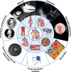
Cardiac complications of COVID. MIS-A/C indicates multisystem inflammatory syndrome in adults and children. Illustration credit: Sceyence Studios.
Figure 2.
Figure 2.
Relative utility of multimodality cardiac imaging in COVID. PET indicates positron emission tomography; and SPECT, single photon emission computed tomography. *Due to COVID infection or vaccination. **In the setting of acute infectious COVID.
Figure 3.
Figure 3.
Myocarditis in COVID. Twenty-four–year-old man experienced recurrent chest discomfort and palpitations after recovery from COVID. Top row: Initial cardiac magnetic resonance imaging (CMR) performed 2 months after diagnosis. A, Intramural/subepicardial late-gadolinium enhancement (LGE) of the inferolateral wall. B, Regional elevation of the native T1 (1125 ms, inferolateral wall). C, Regional elevation of the T2 (55 ms, inferolateral wall). Bottom row: Follow-up CMR performed 3 months later reveals a reduction in LGE volume and improvement in parametric map abnormalities.
Figure 4.
Figure 4.
Takotsubo cardiomyopathy in COVID. Seventy-one–year-old woman admitted with severe COVID reported shortness of breath and heart failure symptoms. Transthoracic echocardiography revealed basal hyperkinesis and apical ballooning. A, Parasternal long axis view at end-diastole. B, Apical 3-chamber view at end-systole. C, Coronary arteriography (LAO view) revealing no obstructive disease.
Figure 5.
Figure 5.
Computed tomography imaging of cardiovascular complications of COVID. A, Left atrial appendage thrombus with background pulmonary involvement of COVID. B, Left ventricular apical thrombus and right lower lobe pulmonary embolism (not pictured) and infarction. C. Acute pericarditis with pericardial effusion and diffuse pericardial thickening. D, Soft proximal left anterior descending coronary artery plaque with background COVID pneumonia.
Figure 6.
Figure 6.
Pericarditis in COVID. Seventy-four–year-old man with COVID complained of recurrent pleuritic chest pain. Cardiac magnetic resonance imaging phase-sensitive inversion-recovery images performed 10 minutes after gadolinium contrast administration revealed diffuse pericardial late-gadolinium enhancement. A, Four-chamber orientation. B, Two-chamber orientation.
Figure 7.
Figure 7.
Multisystem inflammatory syndrome in COVID complicated by left ventricular apical thrombus. Twenty-four–year-old man with COVID underwent cardiac magnetic resonance imaging to evaluate acute systolic heart failure and severe pleuritic chest pain. Inversion-recovery images with a prolonged inversion time (600 ms) highlight left ventricular apical thrombus (arrows) in 2-chamber (A) and short-axis (B) orientations. The native myocardial T1 (C) and T2 (D) times were elevated, consistent with a global inflammatory cardiomyopathy.

References

    1. Guo T, Fan Y, Chen M, Wu X, Zhang L, He T, Wang H, Wan J, Wang X, Lu Z. Cardiovascular implications of fatal outcomes of patients with coronavirus disease 2019 (COVID-19). JAMA Cardiol. 2020;5:811–818. doi: 10.1001/jamacardio.2020.1017 - PMC - PubMed
    1. Shi S, Qin M, Shen B, Cai Y, Liu T, Yang F, Gong W, Liu X, Liang J, Zhao Q, et al. . Association of cardiac injury with mortality in hospitalized patients with COVID-19 in Wuhan, China. JAMA Cardiol. 2020;5:802–810. doi: 10.1001/jamacardio.2020.0950 - PMC - PubMed
    1. Tian W, Jiang W, Yao J, Nicholson CJ, Li RH, Sigurslid HH, Wooster L, Rotter JI, Guo X, Malhotra R. Predictors of mortality in hospitalized COVID-19 patients: a systematic review and meta-analysis. J Med Virol. 2020;92:1875–1883. doi: 10.1002/jmv.26050 - PMC - PubMed
    1. Carfì A, Bernabei R, Landi F; Gemelli Against COVID-19 Post-Acute Care Study Group. Persistent symptoms in patients after acute COVID-19. JAMA. 2020;324:603–605. doi: 10.1001/jama.2020.12603 - PMC - PubMed
    1. Rudski L, Januzzi JL, Rigolin VH, Bohula EA, Blankstein R, Patel AR, Bucciarelli-Ducci C, Vorovich E, Mukherjee M, Rao SV, et al. ; Expert Panel From the ACC Cardiovascular Imaging Leadership Council. Multimodality imaging in evaluation of cardiovascular complications in patients with COVID-19: JACC scientific expert panel. J Am Coll Cardiol. 2020;76:1345–1357. doi: 10.1016/j.jacc.2020.06.080 - PMC - PubMed