. 2023 Jun 1;110(6):989-997.
doi: 10.1016/j.ajhg.2023.04.006. Epub 2023 May 10.

Bi-allelic variants in HMGCR cause an autosomal-recessive progressive limb-girdle muscular dystrophy

Affiliations

Bi-allelic variants in HMGCR cause an autosomal-recessive progressive limb-girdle muscular dystrophy

Joel A Morales-Rosado et al. Am J Hum Genet. .

Abstract

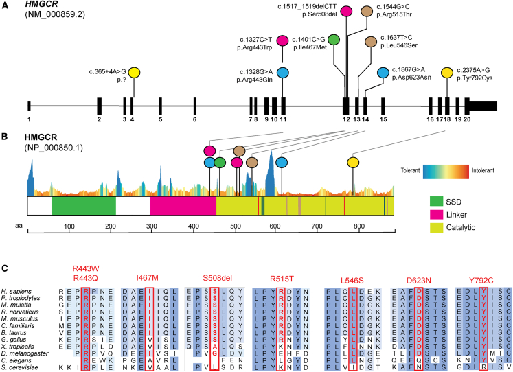
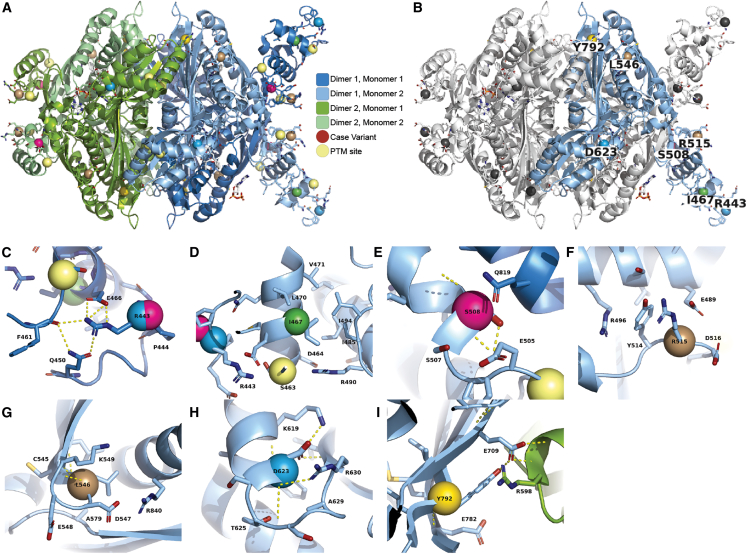
Statins are a mainstay intervention for cardiovascular disease prevention, yet their use can cause rare severe myopathy. HMG-CoA reductase, an essential enzyme in the mevalonate pathway, is the target of statins. We identified nine individuals from five unrelated families with unexplained limb-girdle like muscular dystrophy and bi-allelic variants in HMGCR via clinical and research exome sequencing. The clinical features resembled other genetic causes of muscular dystrophy with incidental high CPK levels (>1,000 U/L), proximal muscle weakness, variable age of onset, and progression leading to impaired ambulation. Muscle biopsies in most affected individuals showed non-specific dystrophic changes with non-diagnostic immunohistochemistry. Molecular modeling analyses revealed variants to be destabilizing and affecting protein oligomerization. Protein activity studies using three variants (p.Asp623Asn, p.Tyr792Cys, and p.Arg443Gln) identified in affected individuals confirmed decreased enzymatic activity and reduced protein stability. In summary, we showed that individuals with bi-allelic amorphic (i.e., null and/or hypomorphic) variants in HMGCR display phenotypes that resemble non-genetic causes of myopathy involving this reductase. This study expands our knowledge regarding the mechanisms leading to muscular dystrophy through dysregulation of the mevalonate pathway, autoimmune myopathy, and statin-induced myopathy.

Keywords: HMG-CoA reducatase; HMGCR; autoimmune myopathy; limb-girdle like muscular dystrophy; mevalonate pathway; rare genetic disease; statin-induced myopathy.


Conflict of interest statement

Declaration of interests The Department of Molecular and Human Genetics at Baylor College of Medicine receives revenue from clinical genetic testing from Baylor Genetics Laboratories. J.R.L. has stock ownership in 23andMe, is a paid consultant for Regeneron Pharmaceuticals, and is a co-inventor on multiple United States and European patents related to molecular diagnostics for inherited neuropathies, eye diseases, genomic disorders, and bacterial genomic fingerprinting.

Figures

None
Graphical abstract
Figure 1
Figure 1
Bi-allelic variants in HMGCR in five affected families results in an autosomal-recessive progressive limb-girdle muscular dystrophy (A) Five families with nine affected individuals and bi-allelic variant segregation. Filled symbols indicate affected individuals. Parents of consanguineous family are fourth cousins. (B) Spatial distribution of muscle weakness in six affected individuals (age during examination) utilizing MRC muscle strength scores generated by MuscleViz denoting: 0, paralysis; 1, trace or minute muscle contraction; 2, muscle movement is possible without gravity; 3, muscle movement is possible against gravity; 4, muscle strength is reduced, but movement against resistance is possible; and 5, preserved normal strength. (C) T1 axial MRI images (from family 5 II:I, at 31 years of age) show striking replacement of almost all muscles of the upper leg (top image) with fibroadipose tissue (signal artifact in right upper leg due to metallic implant). In the lower leg (bottom image), there is increased signaling in the soleus and gastrocnemius muscles with relative sparing of the anterior muscles (tibialis anterior, extensor hallucis longus, and extensor digitorum longus) and lateral muscles (peroneus longus and brevis). (D) Histopathologic findings from family 2 II:1 (top row) includes mild variation in myofiber size on H&E (left), increased subsarcolemmal oxidative enzyme activity on NADH stain (middle), and increased subsarcolemmal mitochondria on electron microscopy (right). Findings from family 2 II:2 (bottom row) includes dystrophic myopathic changes with marked variation in myofiber size with atrophic and hypertrophic myofibers, degenerating fibers with phagocytosis, endomysial fibrosis, and many rimmed vacuoles with basophilic granule deposition on H&E (left). Gomori trichrome (middle) highlights the endomysial fibrosis and stains the rimmed vacuoles stain red. Electron microscopy shows myofibrillar disruption and vacuoles containing lysosomal debris (right). (E) Ultrastructural findings of moderately increased subsarcolemmal mitochondria of the tibialis anterior muscle from family 5 II:I at 35 years of age.
Figure 2
Figure 2
Pathogenic variants in HMGCR cluster in linker and catalytic domains in highly conserved residues (A) Schematic representation of HMGCR transcript map and location of variants identified in cohort. Compound heterozygous variant combinations per family and affected probands are colored the same. (B) HMGCR domains are highlighted and reveal clustering of variants in the catalytic region and linker domain with a constraint plot by MetaDome. SSD, sterol-sensing domain. (C) Ortholog alignment of HMG-CoA reductase residues altered by missense or in-frame deletion variants are highlighted with a red rectangle.
Figure 3
Figure 3
3D molecular structure of HMGCR reveals likely molecular mechanisms (A) Each monomer of the tetramer is colored distinctly to illustrate the dimer-of-dimers architecture. Each dimer of the tetramer is a domain-swapped structure where the linker of one monomer is adjacent to the catalytic center of another (see Figure S1 for more details). (B) Three monomers have been colored white and one monomer remains colored as in (A). Sites of case variants are colored gray in three monomers and colored in one monomer with the WT amino acid labeled. (C and D) Arg443 is within the linker region, which is partially resolved in crystallographic experiments. Residues within the linker that are resolved participate in a hydrogen bond network with Arg443 (C) and moderately stabilizing effects on residue Ile467 (D). (E and F) Ser508 is a position directly at the monomer-monomer interface, and (F) Arg515 is at the monomer-monomer interface in the domain-swapped region (see Figure S1). Arg515 is flanked by negatively charged residues and may dynamically form inter- and intra-monomer interactions. (G) Leu546 is at the end of a small loop between the two beta strands of the domain-swapped sheet. It is surrounded by other hydrophobic residues and between the side chains of the adjacent alpha helix. (H) Asp623 is at the base of a loop that connects a beta sheet to an alpha helix, and its side chain forms multiple specific contacts with residues from both secondary structures. (I) Tyr792 is directly part of the cross-dimer interface of the tetramer. PTM, post translational modification.
Figure 4
Figure 4
Enzymatic activity, size, and stability of purified recombinant HMGCR (A and B) Enzymatic activity of the protein measured as a reduction in absorbance at 340 nm. The activity was quantified relative to WT HMGCR with all three variants showing significant reductions in activity compared to WT protein. Error bars are mean ± SD. (C) Fluorescence curves from thermal shift assays over increasing temperature from three missense variants constructs and WT. Thermal shift assay averaged across four replicates show the three missense variants cause significant reductions in stability compared to WT protein Tm estimates. (D) Estimation of the protein size using the S200 increase column. Two of the variant proteins, p.Asp623Asn and p.Tyr792Cys, eluted at the same volume as WT, while p.Arg443Gln eluted earlier. Acrylamide gels are depicted and stained with Coomassie blue showing only the p.Arg443Gln variant was different in size compared with WT with an approximately 20% increase in size. ∗∗p value 0.0041, ∗∗∗0.0002, and ∗∗∗∗<0.0001.

Similar articles

Cited by

  • Anti-Ku + myositis: an acquired inflammatory protein-aggregate myopathy.
    Holzer MT, Uruha A, Roos A, Hentschel A, Schänzer A, Weis J, Claeys KG, Schoser B, Montagnese F, Goebel HH, Huber M, Léonard-Louis S, Kötter I, Streichenberger N, Gallay L, Benveniste O, Schneider U, Preusse C, Krusche M, Stenzel W. Holzer MT, et al. Acta Neuropathol. 2024 Jul 16;148(1):6. doi: 10.1007/s00401-024-02765-3. Acta Neuropathol. 2024. PMID: 39012547 Free PMC article.
  • Expert panel curation of 31 genes in relation to limb girdle muscular dystrophy.
    Mohan S, McNulty S, Thaxton C, Elnagheeb M, Owens E, Flowers M, Nunnery T, Self A, Palus B, Gorokhova S, Kennedy A, Niu Z, Johari M, Maiga AB, Macalalad K, Clause AR, Beckmann JS, Bronicki L, Cooper ST, Ganesh VS, Kang PB, Kesari A, Lek M, Levy J, Rufibach L, Savarese M, Spencer MJ, Straub V, Tasca G, Weihl CC. Mohan S, et al. Ann Clin Transl Neurol. 2024 Sep;11(9):2268-2276. doi: 10.1002/acn3.52127. Epub 2024 Aug 30. Ann Clin Transl Neurol. 2024. PMID: 39215466 Free PMC article.
  • High-Density Lipoprotein-Associated Cholesterol Abnormalities in a Clinical Outcomes Study of Dysferlin-Deficient Limb-Girdle Muscular Dystrophy Type R2.
    White Z, Rufibach L, Dressman HG, Hilsden H, Cox D, Spuler S, Day JW, Jones KJ, Bharucha-Goebel DX, Salort-Campana E, Pestronk A, Walter MC, Paradas C, Stojkovic T, Mori-Yoshimura M, Bravver E, Diaz-Manera J, Pegoraro E, Mendell JR; Jain COS Consortium; Straub V, Bernatchez P. White Z, et al. J Cachexia Sarcopenia Muscle. 2025 Aug;16(4):e70042. doi: 10.1002/jcsm.70042. J Cachexia Sarcopenia Muscle. 2025. PMID: 40814795 Free PMC article.
  • Pathogenic autoantibody internalization in myositis.
    Pinal-Fernandez I, Muñoz-Braceras S, Casal-Dominguez M, Pak K, Torres-Ruiz J, Musai J, Dell'Orso S, Naz F, Islam S, Gutierrez-Cruz G, Cano MD, Matas-Garcia A, Padrosa J, Tobías-Baraja E, Garrabou G, Aldecoa I, Espinosa G, Simeon-Aznar CP, Guillen-Del-Castillo A, Gil-Vila A, Trallero-Araguas E, Christopher-Stine L, Lloyd TE, Liewluck T, Naddaf E, Stenzel W, Greenberg SA, Grau JM, Selva-O'Callaghan A, Milisenda JC, Mammen AL. Pinal-Fernandez I, et al. medRxiv [Preprint]. 2024 Jan 17:2024.01.15.24301339. doi: 10.1101/2024.01.15.24301339. medRxiv. 2024. Update in: Ann Rheum Dis. 2024 Oct 21;83(11):1549-1560. doi: 10.1136/ard-2024-225773. PMID: 38313303 Free PMC article. Updated. Preprint.
  • Drosophila melanogaster: A Model Organism in Muscular Dystrophy Studies.
    Zhao Y, Wang Y, Tulehalede A, Meng Z, Xu L, Bai H, Sha J, Xie W, Geng J. Zhao Y, et al. Int J Mol Sci. 2025 Feb 10;26(4):1459. doi: 10.3390/ijms26041459. Int J Mol Sci. 2025. PMID: 40003927 Free PMC article. Review.

References

    1. Mercuri E., Bönnemann C.G., Muntoni F. Muscular dystrophies. Lancet. 2019;394:2025–2038. doi: 10.1016/S0140-6736(19)32910-1. - DOI - PubMed
    1. Reddy H.M., Cho K.A., Lek M., Estrella E., Valkanas E., Jones M.D., Mitsuhashi S., Darras B.T., Amato A.A., Lidov H.G., et al. The sensitivity of exome sequencing in identifying pathogenic mutations for LGMD in the United States. J. Hum. Genet. 2017;62:243–252. doi: 10.1038/jhg.2016.116. - DOI - PMC - PubMed
    1. Töpf A., Johnson K., Bates A., Phillips L., Chao K.R., England E.M., Laricchia K.M., Mullen T., Valkanas E., Xu L., et al. Sequential targeted exome sequencing of 1001 patients affected by unexplained limb-girdle weakness. Genet. Med. 2020;22:1478–1488. doi: 10.1038/s41436-020-0840-3. - DOI - PMC - PubMed
    1. Karczewski K.J., Francioli L.C., Tiao G., Cummings B.B., Alföldi J., Wang Q., Collins R.L., Laricchia K.M., Ganna A., Birnbaum D.P., et al. The mutational constraint spectrum quantified from variation in 141,456 humans. Nature. 2020;581:434–443. doi: 10.1038/s41586-020-2308-7. - DOI - PMC - PubMed
    1. Istvan E.S., Palnitkar M., Buchanan S.K., Deisenhofer J. Crystal structure of the catalytic portion of human HMG-CoA reductase: insights into regulation of activity and catalysis. EMBO J. 2000;19:819–830. doi: 10.1093/emboj/19.5.819. - DOI - PMC - PubMed

Publication types

MeSH terms

Substances