. 2023 Jul 5;111(13):2012-2020.e4.
doi: 10.1016/j.neuron.2023.04.007. Epub 2023 May 10.

Abundant Aβ fibrils in ultracentrifugal supernatants of aqueous extracts from Alzheimer's disease brains

Affiliations

Abundant Aβ fibrils in ultracentrifugal supernatants of aqueous extracts from Alzheimer's disease brains

Andrew M Stern et al. Neuron. .

Abstract

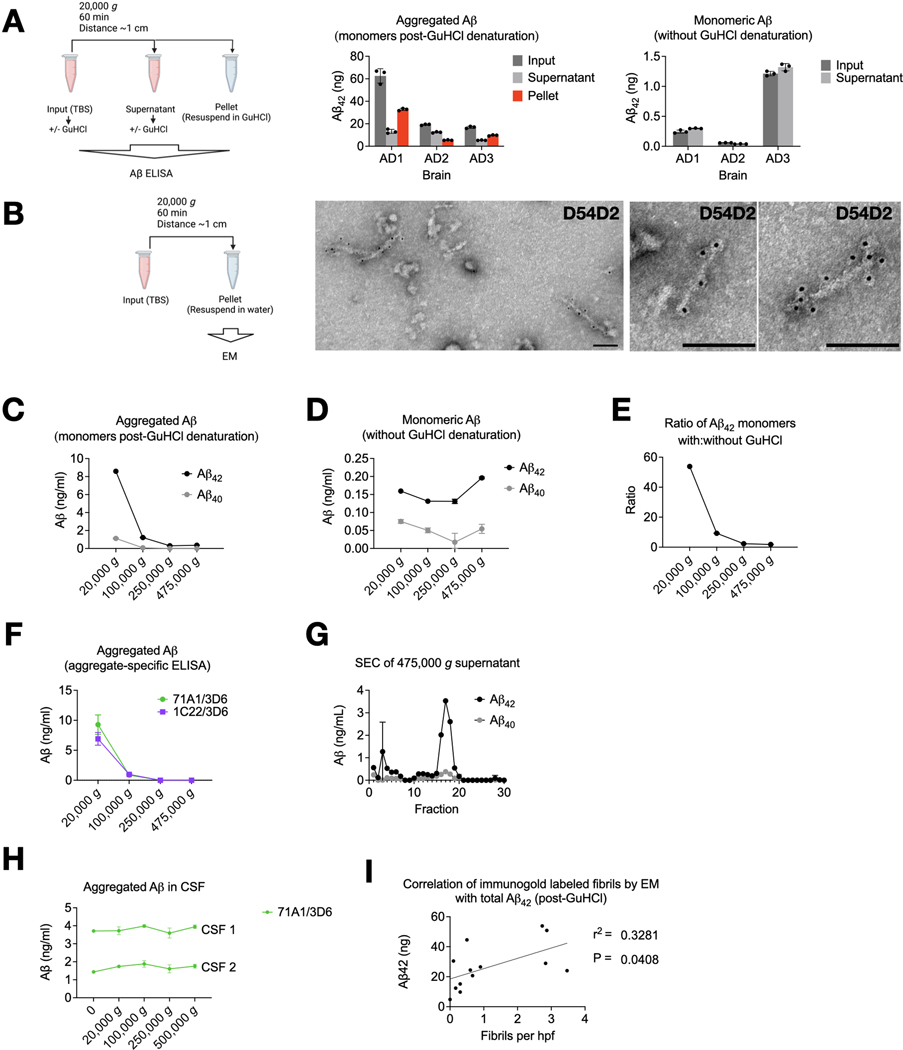
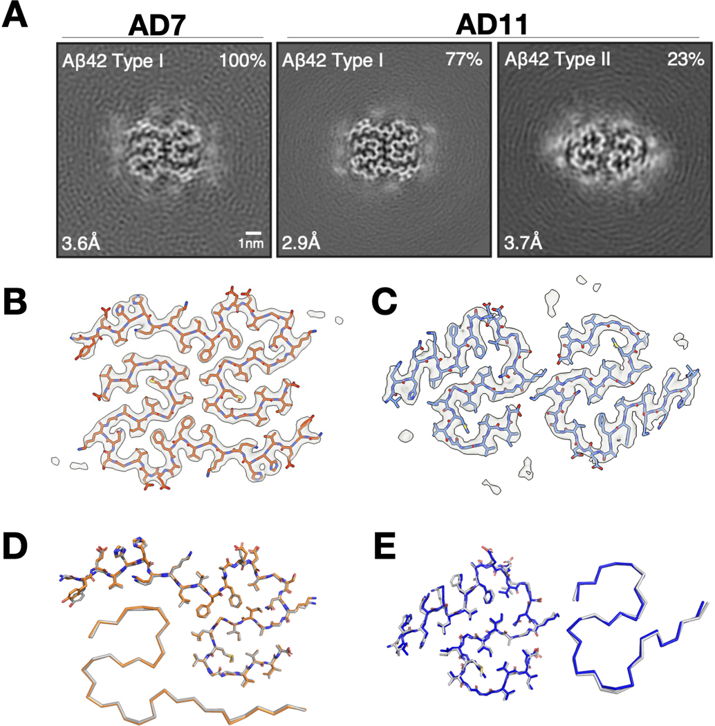
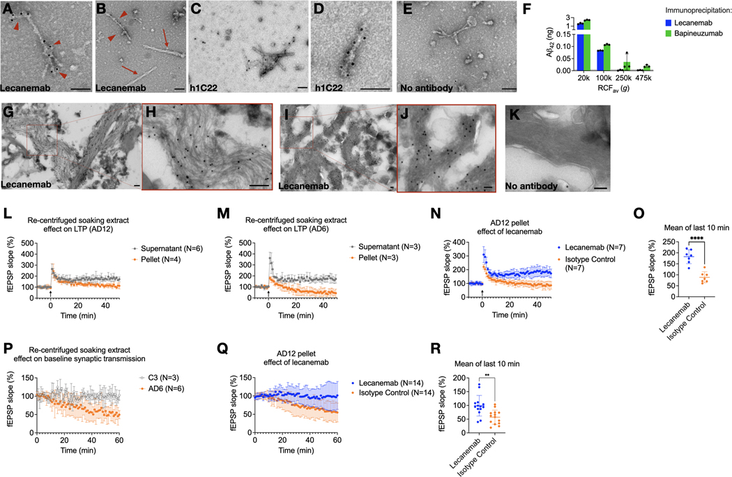
Soluble oligomers of amyloid β-protein (Aβ) have been defined as aggregates in supernatants following ultracentrifugation of aqueous extracts from Alzheimer's disease (AD) brains and are believed to be upstream initiators of synaptic dysfunction, but little is known about their structures. We now report the unexpected presence of Aβ fibrils in synaptotoxic high-speed supernatants from AD brains extracted by soaking in an aqueous buffer. The fibrils did not appear to form during preparation, and their counts by EM correlated with Aβ ELISA quantification. Cryo-EM structures of aqueous Aβ fibrils were identical to those from sarkosyl-insoluble homogenates. The fibrils in aqueous extracts were labeled by lecanemab, an Aβ aggregate-directed antibody reported to improve AD cognitive outcomes. Lecanemab provided protection against aqueous fibril synaptotoxicity. We conclude that fibrils are abundant in aqueous extracts from AD brains and have the same structures as those from plaques. These findings have implications for AD pathogenesis and drug design.

Keywords: Alzheimer’s disease; amyloid β; fibril; lecanemab; oligomer; protofibril; therapeutic antibody.


Conflict of interest statement

Declaration of interests D.J.S. is a founding director and consultant of Prothena Biosciences. L.L. is a consultant for Korro Bio.

Figures

Fig 1.
Fig 1.. Aqueous soaking extracts of AD brain contain insoluble Aβ fibrils.
(A) Aliquots of aqueous soaking extracts from ultracentrifugal supernatants of AD cortex (see Methods and Table 1) were thawed and spun at 20,000 g in a tabletop centrifuge, and Aβ was quantified by ELISA with (center) and without (right) 5 M GuHCl. Note different y-axis scales. GuHCl denatures Aβ aggregates and allows detection by a monomer-preferring ELISA. Aβ detectable without GuHCl denaturation is monomeric. ELISAs revealed that aggregated Aβ but not Aβ monomers could be re-pelleted. (B) Fibrils in the pellets of re-centrifuged aqueous soaking extracts of brain AD7 were labeled by anti-Aβ N-terminal antibody D54D2. Scale bar = 100 nm. (C-F) A TBS soaking extract of brain AD6 was prepared by mincing cortex, soaking in TBS for 30 min, and a 10 min spin at 2,000 g (Table 1, steps 1–4). This supernatant was divided and ultracentrifuged in a TLA100.3 rotor (pelleting distance ~2 cm) at indicated g forces, and Aβ quantified in the supernatant. Monomer-preferring ELISA performed with (C) or without (D) GuHCl denaturation showed that Aβ aggregates but not monomers were depleted as the g-force increased, as did the ratio of Aβ concentration with:without GuHCl (E). (F) Increasing g-force abolished signal from two aggregate-preferring Aβ ELISAs. (G) Superdex 200 Increase size exclusion chromatography of the 475,000 g supernatant revealed a low MW Aβ profile. (H) Unlike brain, CSF Aβ aggregates were not depleted as g-force increased. (I) The Aβ concentration in soaking extracts after GuHCl denaturation correlated with the number of fibrils in the pellet after centrifugation by manual blinded counting of immunogold EM. Line represents simple linear regression. Drawings made with Biorender.com.
Fig 2.
Fig 2.. Aβ fibrils from aqueous extracts of AD brains have the same cryo-EM structures as fibrils from sarkosyl-insoluble homogenates.
(A) XY-cross-sections of cryo-EM maps of Aβ fibrils from soaking extracts of cases AD7 and AD11. For each map, a sum of the reconstructed densities for several XY-slices, approximating one β-rung, is shown. Aβ fibril types I and II are indicated at top left; the percentages of Type I and Type II fibrils relative to the total of imaged fibrils are shown at top right. Resolutions at bottom left. Scale bar = 1 nm. (B, C) Cryo-EM density maps (grey) and atomic models for Aβ Type I (orange), Aβ Type II (blue). (D, E), Comparison of the cryo-EM structures of Aβ Type I (B) fibrils and Aβ Type II (C) fibrils extracted from aqueous soaking extracts vs. sarkosyl-insoluble homogenates (in grey for Type I and Type II fibrils) from brains of AD patients. Structures are shown as sticks (above) for one protofilament and as ribbons (below) for the other.
Fig 3.
Fig 3.. Lecanemab decorates Aβ fibrils from Alzheimer’s disease brains and protects from synaptotoxicity.
(A, B) Lecanemab labels pelleted Aβ fibrils from the re-centrifugation of AD7 TBS soaking extract with protein A gold (arrowheads). PHFs in the pellet did not react with lecanemab (arrows). (C, D) Immunogold labeling with Aβ aggregate-preferring h1C22 similarly decorates soaking extract fibrils. (E) Protein A gold alone does not label fibrils. (F) ELISA quantification of Aβ42 immunoprecipitated from AD6 aqueous extract followed by washing, elution and denaturation in 5 M GuHCl. Bapineuzumab, but not lecanemab, immunoprecipitated Aβ from the 250,000 g and 475,000 g (TLA100.3, pelleting distance ~2 cm) supernatants. (G-I) Immunogold of paraformaldehyde-fixed ultrathin cryosections from AD11 (G,H) and AD7 (I,J) brains reveals labeling of Aβ fibrils by lecanemab. (K) Protein A gold alone (brain AD7) shows minimal background labeling. Scale bar = 100 nm. (L, M) Soaking extracts were centrifuged for 2h, and the pellet resuspended in an equivalent volume of TBS. Supernatants or pellets from two AD soaking extracts were applied to mouse hippocampal slices at 5% v:v perfusate, and LTP was induced (arrow at time 0). (N) Lecanemab reversed the impairment of LTP due to the AD 12 soaking extract pellet. (O) The average of the last 10 minutes of recording in (N) showed a mean difference of 94% baseline fEPSP (95% CI, 61% - 127%, P<0.001). (P) The pelleted fraction of AD but not control brain soaking extracts impaired the hippocampal synaptic baseline without LTP induction. (Q) Lecanemab reversed impairment of baseline fEPSP slope by AD12 soaking extract pellet by a mean of 42% compared to isotype control (95% CI, 17 – 67, P = 0.002), quantified in (R). Error bars represent SD.

References

    1. Shankar GM, Li S, Mehta TH, Garcia-Munoz A, Shepardson NE, Smith I, Brett FM, Farrell MA, Rowan MJ, Lemere CA, et al. (2008). Amyloid-β protein dimers isolated directly from Alzheimer’s brains impair synaptic plasticity and memory. Nat Med 14, 837–842. 10.1038/nm1782. - DOI - PMC - PubMed
    1. Hughes C, Choi ML, Yi J-H, Kim S-C, Drews A, George-Hyslop PS, Bryant C, Gandhi S, Cho K, and Klenerman D. (2020). Beta amyloid aggregates induce sensitised TLR4 signalling causing long-term potentiation deficit and rat neuronal cell death. Commun Biol 3, 79. 10.1038/s42003-020-0792-9. - DOI - PMC - PubMed
    1. Li S, Hong S, Shepardson NE, Walsh DM, Shankar GM, and Selkoe D. (2009). Soluble oligomers of amyloid Beta protein facilitate hippocampal long-term depression by disrupting neuronal glutamate uptake. Neuron 62, 788–801. 10.1016/j.neuron.2009.05.012. - DOI - PMC - PubMed
    1. Jin M, Shepardson N, Yang T, Chen G, Walsh D, and Selkoe DJ (2011). Soluble amyloid beta-protein dimers isolated from Alzheimer cortex directly induce Tau hyperphosphorylation and neuritic degeneration. Proc Natl Acad Sci U S A 108, 5819–5824. 10.1073/pnas.1017033108. - DOI - PMC - PubMed
    1. Jin M, O’Nuallain B, Hong W, Boyd J, Lagomarsino VN, O’Malley TT, Liu W, Vanderburg CR, Frosch MP, Young-Pearse T, et al. (2018). An in vitro paradigm to assess potential anti-Aβ antibodies for Alzheimer’s disease. Nat Commun 9, 2676. 10.1038/s41467-018-05068-w. - DOI - PMC - PubMed

Publication types

Substances

LinkOut - more resources