Review
. 2023 Apr 24:14:1176748.
doi: 10.3389/fphys.2023.1176748. eCollection 2023.

Myography of isolated blood vessels: Considerations for experimental design and combination with supplementary techniques

Affiliations
Review

Myography of isolated blood vessels: Considerations for experimental design and combination with supplementary techniques

Rudolf Schubert et al. Front Physiol. .

Abstract

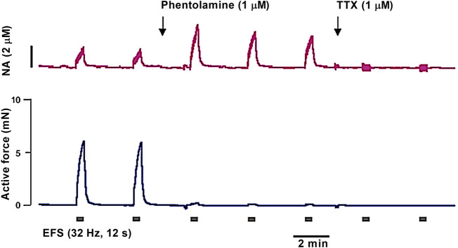
The study of the mechanisms of regulation of vascular tone is an urgent task of modern science, since diseases of the cardiovascular system remain the main cause of reduction in the quality of life and mortality of the population. Myography (isometric and isobaric) of isolated blood vessels is one of the most physiologically relevant approaches to study the function of cells in the vessel wall. On the one hand, cell-cell interactions as well as mechanical stretch of the vessel wall remain preserved in myography studies, in contrast to studies on isolated cells, e.g., cell culture. On the other hand, in vitro studies in isolated vessels allow control of numerous parameters that are difficult to control in vivo. The aim of this review was to 1) discuss the specifics of experimental design and interpretation of data obtained by myography and 2) highlight the importance of the combined use of myography with various complementary techniques necessary for a deep understanding of vascular physiology.

Keywords: artery; endothelium; innervation; intracellular calcium; membrane potential; myography; smooth muscle.


Conflict of interest statement

The authors declare that the research was conducted in the absence of any commercial or financial relationships that could be construed as a potential conflict of interest.

Figures

FIGURE 1
FIGURE 1
Control for similarity of initial contractility. As an example, the protocol of an experiment is shown in which 4 groups of blood vessels will be studied in myograph units 1–4. After the initial distension (i.e., normalization) and the starting procedure (i.e., viability testing) the effect of the contractile agonist methoxamine is tested by determining its concentration-response relationship (CRR) (protocol scheme shown in the left panel). As shown in the right panel, vessel tension in the 4 different experimental groups (CONTROL1—CONTROL4) were not different (n = 7; p = 0.94 based on the area under the concentration-response relationships). Reproduced and modified with permission from (Schmid et al., 2018).
FIGURE 2
FIGURE 2
Effect of NS19504 and IBTX on methoxamine-induced contractions of the rat A. saphena. (A) Normalized tension (normalized to the maximum response of the 1st CRR-see Figure 1) of A. Saphena at different methoxamine concentrations in the absence of BK channel active agents (Control), in the presence of IBTX (IBTX 10−7 M), in the presence of NS19504 (NS19504 6*10−6 M) and in the combined presence of NS19504 and IBTX (NS19504 and IBTX). (B) NS19504 anti-contractile effect in the absence (Control) and presence of IBTX (IBTX 10−7 M). N = 7; * - p < 0.05. Reproduced and modified with permission from (Ma et al., 2020).
FIGURE 3
FIGURE 3
Effect of GoSlo-SR 5-6 on spontaneous tone of isobaric vessel preparations of rat Gracilis muscle arteries at 80 mmHg. (A) Normalised vessel diameter (ratio of diameter/fully relaxed diameter at 80 mmHg) at different concentrations of GoSlo-SR 5-6 in the absence (control) and presence of 3*10–6 M XE991 (n = 10; p < 0.05); (B) vessel dilation at different concentrations of GoSlo-SR 5-6 in the absence (control) and presence of 3*10–6 M XE991 (n = 10; p < 0.05); (C) Normalised vessel diameter at different concentrations of GoSlo-SR 5-6 in the absence (control) and presence of 10–7 M IBTX (n = 7; p < 0.05); (D) vessel dilation in the absence (time control = application of GoSlo-SR 5-6 solvent DMSO) and presence of GoSlo-SR 5–6 (control) (n = 7; p < 0.05), vessel dilation at different concentrations of GoSlo-SR 5-6 in the absence (control) and presence of 10–7 M IBTX (n = 7; p = 0.94). Reproduced and modified with permission from (Zavaritskaya et al., 2020).
FIGURE 4
FIGURE 4
GoSlo-SR-5-6 causes concentration dependent relaxations of isometric preparations of rat Gracilis muscle arteries. (A) Example of the effect of GoSlo 5-6 on tension of an isometric vessel preparation at 10–6 M MX-induced tone. Application denotes the time point where GoSlo or vehicle was added. (B) Effect of GoSlo 5-6 on 10–6 M MX-induced contraction. Vessel tension in the absence (time control) and presence of GoSlo 5-6 at 3 × 10⁻⁶ M and at 10–5 M (repeated measures ANOVA: con vs. GoSlo 10–5 M - n = 11; p < 0.05; con vs. GoSlo 3 × 10⁻⁶ M—n = 8; p < 0.05; GoSlo 10–5 M vs. 3 × 10⁻⁶ M - p < 0.05). (C) Effect of GoSlo 5-6 on 10–5 M MX-induced contraction. Vessel tension in the absence (time control) and presence of GoSlo 5-6 at 10–5 M (repeated measures ANOVA: n = 8; p < 0.05). Reproduced and modified with permission from (Zavaritskaya et al., 2020).
FIGURE 5
FIGURE 5
Contribution of the BK channel to the anti-contractile effect of SNP depends on the conditions used for pre-contraction. (A) and (B) Vessel tension in the absence of IBTX and SNP (CONTROL), in the presence of IBTX alone (IBTX), in the presence of SNP alone (SNP), and in the combined presence of IBTX and SNP (IBTX + SNP) at 10–5 M SNP. (A) Selection of similar levels of submaximal pre-contraction in the absence and presence of IBTX when IBTX had almost no contractile effect (black curves, 10–6 M MX); relaxing effect of 10–5 M SNP (blue curves) (filled parts of the vertical bars) in the absence and presence of IBTX presented also in (C) together with the effect of other concentrations of SNP (* - two-way ANOVA control vs. IBTX: p < 0.05); (B) Selection of similar levels of submaximal pre-contraction in the absence and presence of IBTX when IBTX had a considerable contractile effect (black curves, 3*10–7 M and 10–6 M MX); relaxing effect of 10–5 M SNP (blue curves) (filled parts of the vertical bars) in the absence and presence of IBTX presented also in (D) together with the effect of other concentrations of SNP (* - two-way ANOVA control vs. IBTX: p < 0.05); Reproduced and modified with permission from (Schmid et al., 2018).
FIGURE 6
FIGURE 6
Schematic representation of the mechanism of BK channel contribution to the anti-contractile effect of SNP. (A) Conditions when NO produced only a small decrease in [Ca2+]i: NO induces a PKG-mediated activation of the BK channel that is stronger than the small [Ca2+]i-decrease-mediated deactivation of the BK channel. The overall effect of both is an activation of the BK channel contributing to the reduction of calcium influx; BK channels facilitate NO-induced vasodilation. (B) Conditions when NO produced a considerable decrease in [Ca2+]i: NO induces a PKG-mediated activation of the BK channel that is weaker than the large [Ca2+]i-decrease-mediated deactivation of the BK channel. The overall effect of both is a deactivation of the BK channel opposing the reduction of calcium influx; BK channels limit NO-induced vasodilation. Reproduced and modified with permission from (Schmid et al., 2018).
FIGURE 7
FIGURE 7
Custom-made wire myography analogue. This device is equipped with wider heads to increase arterial segment length up to 10 mm. It has a micrometer that allows to change the distance between the heads but has no force transducer.
FIGURE 8
FIGURE 8
Original trace obtained in an experiment with membrane potential registration in rat saphenous artery. Membrane potential recordings are accepted according to the following criteria: (i) a sharp drop of the potential on cell penetration; (ii) a stable level of the membrane potential recording for at least 30 s; (iii) a return to the zero-potential level after electrode removal; and (iv) a similar electrode resistance before and after the measurement (Nilsson et al., 1998; Rummery and Brock, 2011; Shvetsova et al., 2019).
FIGURE 9
FIGURE 9
Original traces of force and membrane potential simultaneously recorded in an experiment on rat tail artery. Incubation with the α1-adrenoceptor agonist methoxamine (0.4 µM) and the BKCa channel blocker Iberiotoxin (0.1 µM) caused the development of action potential-like spikes accompanied by phasic oscillations of tone.
FIGURE 10
FIGURE 10
Pitfalls to be considered in studies with synthetic calcium dyes. 1) dye precipitates stick to the adventitia adding a strong calcium-insensitive signal; 2) buffering of intracellular calcium by calcium dye affecting the calcium concentration as well as the kinetics of calcium concentration changes; 3) dye loss; 4) photobleaching; 5) compartmentalization of dyes; 6) screening or scatter of fluorescence by connective tissue; 7) phototoxicity and incomplete hydrolysis; 8) vasoactive substances alter fluorescence of the dye or contribute to the fluorescence signal; 9) inhibition of Na/K ATPase especially at high dye concentrations; 10) interaction with intracellular proteins..
FIGURE 11
FIGURE 11
Dependence of fluorescence intensity on calcium concentration. In the absence of calcium, the dye is free of calcium and minimum fluorescence is observed. With increasing calcium concentration the dye binds increasingly more calcium ions, but when all dye molecules are occupied by calcium ions a maximum fluorescence is achieved. There is a non-linear dependence of fluorescence intensity on calcium concentration. Consequently, similar changes in calcium concentration (ΔCa1 = ΔCa2), when evoked from different initial calcium concentrations, produce different changes in fluorescence intensity (ΔF1 > ΔF2).
FIGURE 12
FIGURE 12
Cumulative frequency–response relationships obtained for segments cut from the medial tarsal branch of the saphenous artery (small skin artery, normalized diameter 225 µm) and from the distal (intramuscular) section of the external sural artery (small skeletal muscle artery, normalized diameter 210 µm).
FIGURE 13
FIGURE 13
Representative recordings of the noradrenaline concentration (A) and active force (B) in response to EFS of sympathetic nerves by rectangular current pulses (85 mA, 0.1 ms) in an experiment on a segment of the rat mesenteric artery (normalized diameter 280 µm). Phentolamine greatly reduces contraction due to the blockade of postjunctional α1-adrenoceptors but increases the concentration of noradrenaline (NA) due to the blockade of prejunctional α2-adrenoceptors. In the presence of tetrodotoxin (TTX) contraction and oxidation current completely disappear.

References

    1. Aalkjær C., Nilsson H. (2005). Vasomotion: Cellular background for the oscillator and for the synchronization of smooth muscle cells. Br. J. Pharmacol. 144, 605–616. 10.1038/sj.bjp.0706084 - DOI - PMC - PubMed
    1. Aalkjær C., Nilsson H., De Mey J. G. R. (2021). Sympathetic and sensory-motor nerves in peripheral small arteries. Physiol. Rev. 101, 495–544. 10.1152/physrev.00007.2020 - DOI - PubMed
    1. Adrian R. H. (1956). The effect of internal and external potassium concentration on the membrane potential of frog muscle. J. Physiol. 133, 631–658. 10.1113/jphysiol.1956.sp005615 - DOI - PMC - PubMed
    1. Akopian A. N., Sivilotti L., Wood J. N. (1996). A tetrodotoxin-resistant voltage-gated sodium channel expressed by sensory neurons. Nature 379, 257–262. 10.1038/379257A0 - DOI - PubMed
    1. Anschütz S., Schubert R. (2005). Modulation of the myogenic response by neurogenic influences in rat small arteries. Br. J. Pharmacol. 146, 226–233. 10.1038/SJ.BJP.0706323 - DOI - PMC - PubMed

LinkOut - more resources