Review
. 2023 Feb 26;26(5):106276.
doi: 10.1016/j.isci.2023.106276. eCollection 2023 May 19.

An inventory of crosstalk between ubiquitination and other post-translational modifications in orchestrating cellular processes

Affiliations
Review

An inventory of crosstalk between ubiquitination and other post-translational modifications in orchestrating cellular processes

Haithem Barbour et al. iScience. .

Abstract

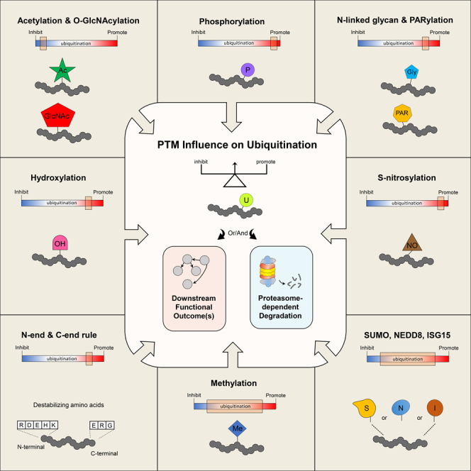
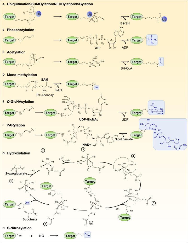
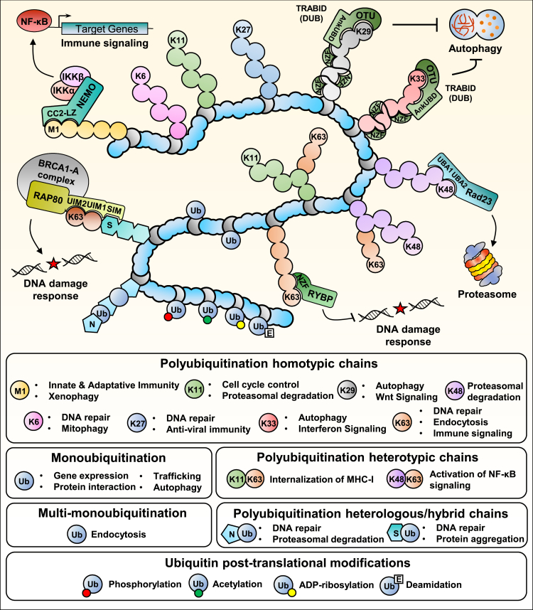
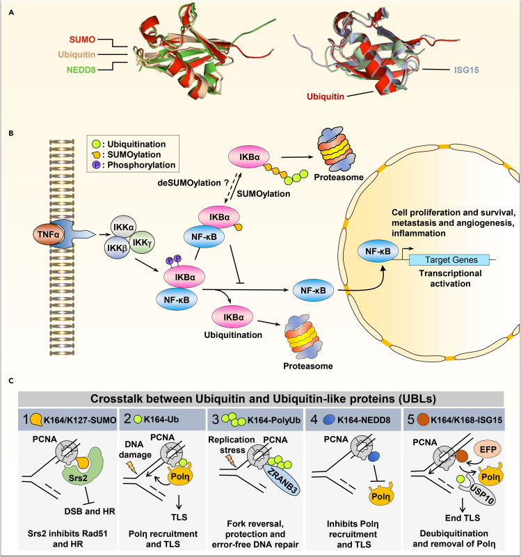
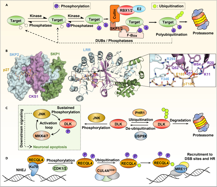
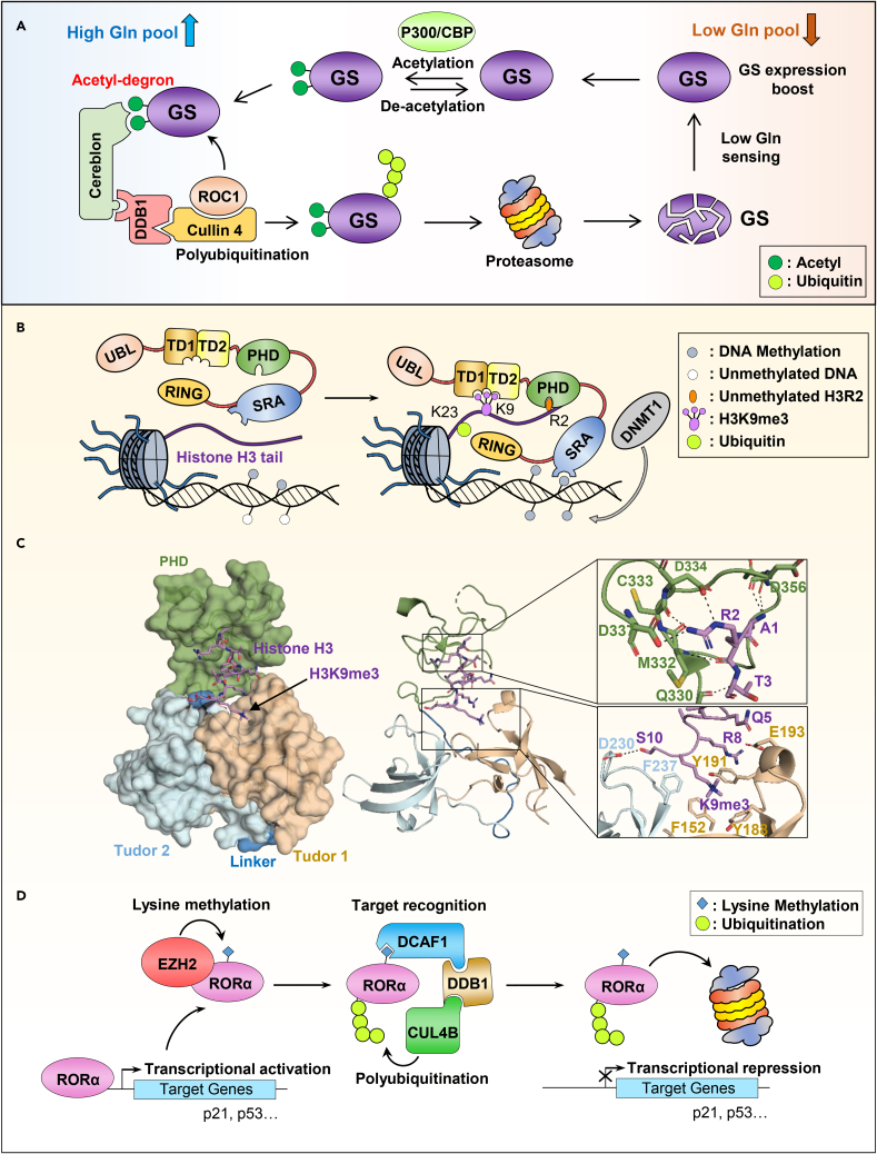
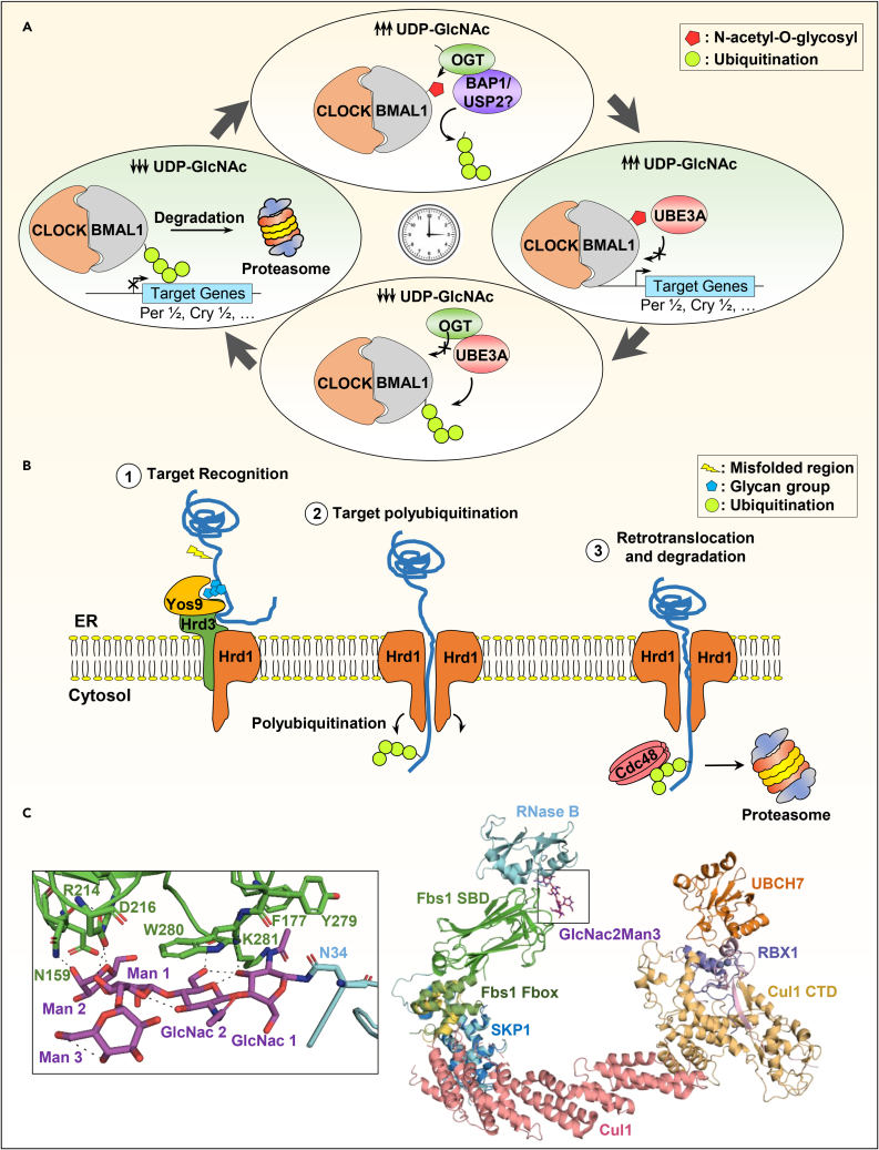
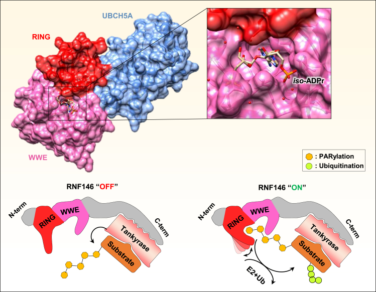
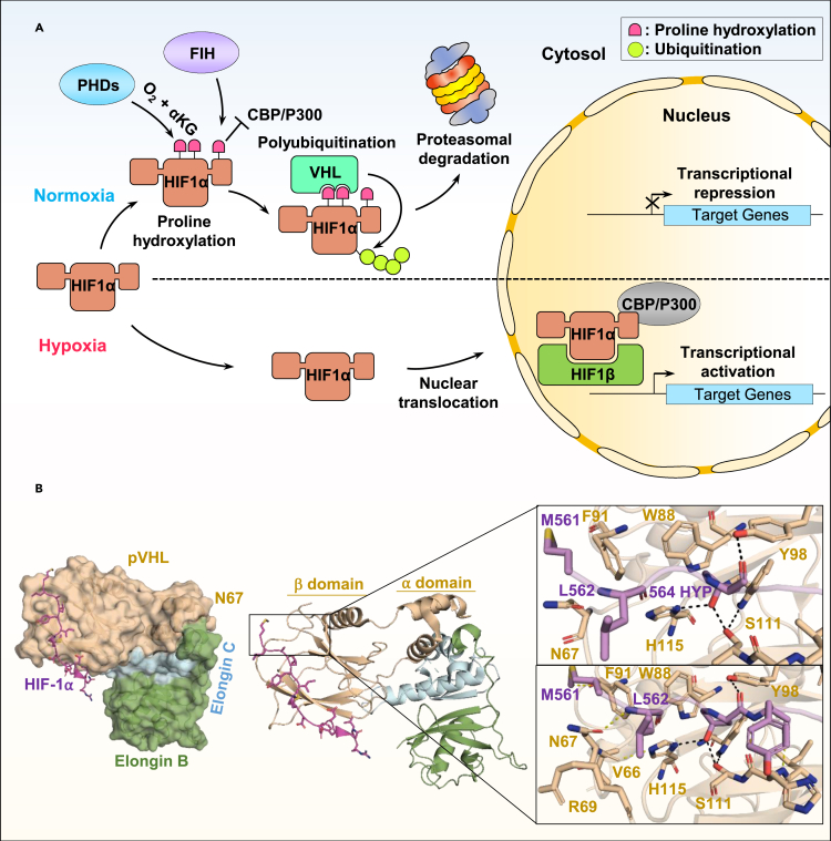
Ubiquitination is an important post-translational modification (PTM) that regulates a large spectrum of cellular processes in eukaryotes. Abnormalities in ubiquitin signaling underlie numerous human pathologies including cancer and neurodegeneration. Much progress has been made during the last three decades in understanding how ubiquitin ligases recognize their substrates and how ubiquitination is orchestrated. Several mechanisms of regulation have evolved to prevent promiscuity including the assembly of ubiquitin ligases in multi-protein complexes with dedicated subunits and specific post-translational modifications of these enzymes and their co-factors. Here, we outline another layer of complexity involving the coordinated access of E3 ligases to substrates. We provide an extensive inventory of ubiquitination crosstalk with multiple PTMs including SUMOylation, phosphorylation, methylation, acetylation, hydroxylation, prolyl isomerization, PARylation, and O-GlcNAcylation. We discuss molecular mechanisms by which PTMs orchestrate ubiquitination, thus increasing its specificity as well as its crosstalk with other signaling pathways to ensure cell homeostasis.

Keywords: Biochemistry; Biological sciences; Cell biology; Molecular biology.


Conflict of interest statement

The authors declare no competing interests with the content of this article.

Figures

None
Graphical abstract
Figure 1
Figure 1
Chemical mechanism or global reaction of various post-translational modifications (A) Ubiquitination mechanism on the lysine residue of a target protein. The deprotonated amino group of the lysine engages a nucleophilic attack on the carbonyl of the thioester group linking ubiquitin to the E2 ubiquitin-conjugating enzyme or E3 ubiquitin ligase. An isopeptide bond is therefore formed after rearrangement, attaching the ubiquitin to the target protein and freeing the E2. SUMOylation, NEDDylation, and ISGylation use the same mechanism. (B) Phosphorylation mechanism on the serine residue of a target protein. A phosphoserine is formed by nucleophilic substitution on the phosphorus of the gamma phosphate group of ATP (adenosine triphosphate) with the deprotonated alcohol as nucleophile which results in ADP release. (C) Acetylation mechanism on the lysine residue of a target protein. Acetyl-CoA is a common source of acetyl groups. The deprotonated amino group of the lysine makes a nucleophilic attack on the carbonyl group of the acetyl-CoA, fusing it to the target protein. (D) Mono-methylation mechanism on the lysine residue of a target protein. SAM (S-adenosyl methionine) is a common donor of methyl groups for the successive methylation of a target residue. By nucleophilic substitution, the methyl group of SAM is transferred to a target protein with a deprotonated amino group, resulting in SAH (S-adenosyl homocysteine) as a free product. The deprotonated amino group of the lysine residue acts as a nucleophile. (E) O-GlcNAcylation mechanism on the serine residue of a target protein. By nucleophilic substitution, the N-acetylglucosamine of the UDP-GlcNAc is attached to the target protein with the deprotonated alcohol as nucleophile, resulting in the formation of a β-glycosidic bond and the release of UDP. (F) PARylation mechanism on the serine residue of a target protein. By nucleophilic substitution, the ADP-ribose from the NAD+ (nicotinamide adenine dinucleotide) is attached to the target protein with the deprotonated alcohol as nucleophile. (G) Hydroxylation mechanism on the proline residue of a target protein. The shown dioxygenase has an iron cofactor and complex with two histidines and aspartic acid. In the presence of O2 (dioxygen) and 2-oxoglutarate, the proline residue is targeted for hydroxylation at the end of the catalytic cycle (step 7). (H) S-nitrosylation reaction on the cysteine residue of a target protein in the presence of NO. For each mechanism, only the reactive functional group of residues implicated is shown. The B group (for phosphorylation, PARylation, and O-GlcNAcylation) represents the enzyme base that transfer the proton to the substrate. The resulting modification is colored in blue. Chemical structures were drawn with ChemDraw.
Figure 2
Figure 2
Ubiquitin chains and their functions Ubiquitination of proteins is catalyzed by the action of E1 ubiquitin-activating, E2 ubiquitin-conjugating, and E3 ubiquitin-ligating enzymes. The concerted action of E2 and E3 enzymes dictates the nature of ubiquitin modification. Ubiquitination is reversed by deubiquitinases (DUBs). A protein target (in blue) can be monoubiquitinated, multi-monoubiquitinated, or polyubiquitinated. Monoubiquitination and multi-monoubiquitination are associated with diverse signaling pathways and processes including intracellular membrane trafficking, transcription regulation, DNA damage signaling and repair as well as the regulation of protein subcellular localization. Monoubiquitination events have also been shown to induce proteasomal degradation. Polyubiquitination involves eight ubiquitin linkage types, i.e., M1, K6, K11, K27, K29, K33, K48, and K63. The various ubiquitin chains are associated with numerous functional and biological outcomes. K63 polyubiquitin chains are associated with protein recruitment and activation of signaling cascades, K48 polyubiquitin chains generally promote the proteasomal degradation of substrates. Polyubiquitination can generate homotypic, heterotypic, or heterologous chains that add another layer of complexity to ubiquitin signaling pathways. Moreover, post-translational modifications of ubiquitin itself (phosphorylation, acetylation, ADP-ribosylation, and deamidation) can modulate E3 ligase activity and interfere with ubiquitin ligation or ubiquitin chain elongation. Following substrate ubiquitination, ubiquitin chains could be recognized by a wide variety of ubiquitin-binding proteins through their ubiquitin-binding domains (UBD). Examples of UBDs are represented by the ubiquitin interacting motifs (UIM) of RAP80, the Ubiquitin-associated domains (UBA) of Rad23, or the Npl4-like Zinc Finger (NZF) of the deubiquitinase TRABID. A wide range of signaling events and biological processes are associated with ubiquitin binding such as the DNA damage response, cell death, immune signaling, and autophagy.
Figure 3
Figure 3
Examples of crosstalk between ubiquitin-like proteins (UBLs) and ubiquitin (A) Ubiquitin and Ubiquitin-like proteins (UBLs), SUMO2 (PDB: 1WM3), NEDD8 (PDB: 2BKR) and ISG15 (PDB: 1Z2M) share significant structural similarities with ubiquitin (PDB: 1ubq). These proteins share a core β-grasp (β-Golgi Reassembly Stacking Protein) fold containing secondary structure elements arranged in a ββαβββ order. (B–C) Ubiquitin-like proteins compete with ubiquitination resulting in variable outcomes on protein stability and function. (B) Polyubiquitination of the NF-κB inhibitor IKBα, following its phosphorylation by the IKK kinase, leads to its proteasomal degradation and release of NF-κB to execute its transcriptional activity. SUMOylation of the same residue of IκBα blocks its degradation, while hybrid SUMO-Ubiquitin chain extension can re-engage the proteasomal degradation. (C) Attachment of SUMO, ubiquitin, NEDD8, and ISG15 moieties on PCNA is associated with different outcomes on DNA replication and repair. PCNA K164 modifications play central roles in the recruitment of UBLs binding proteins, which in turn dictate the choice or termination of DNA repair pathways. 1) At the replication fork, PCNA K164 SUMOylation allows its interaction with Srs2 which inhibits Rad51 filament formation and homologous recombination (HR). 2) During DNA damage, PCNA K164 monoubiquitination results in the recruitment of Polη for the translesion DNA synthesis (TLS) process. 3) PCNA polyubiquitinated on K164 after replication stress promotes the recruitment of ZRANB3, replication fork reversal, and protection, thus maintaining genomic integrity. 4) The execution of TLS can be inhibited by the NEDDylation of PCNA K164. This inhibits its ubiquitination and the subsequent recruitment of Polη. 5) ISGylation of PCNA is a signal for TLS termination. Following Polη recruitment, PCNA K164/K168 is ISGylated by EFP leading to the recruitment of the DUB USP10. This results in the deubiquitination of PCNA, release of Polη from the replication fork and termination of TLS.
Figure 4
Figure 4
Model of the interplay between protein phosphorylation and ubiquitination (A) Phosphorylation of a target protein creates a phosphodegron, recognized by E3 ligase complexes (in this case the SCF or Skip-Cullin-F-box complex family). The F box factor positions the targeted protein in the vicinity of the Cullin ligase (RBX1/2) and the E2 enzyme, leading to its ubiquitination and proteasomal degradation. (B) Crystal structure of the quaternary complex: SKP1-SKP2-CKS1-Phospho p27Kip1 (PDB: 2AST). Left panel, overall representation of the complex showing the specific positioning of the p27 peptide within CKS1 and SKP2 binding pockets. Right panel, close up view of the phosphorylated p27Kip1 interaction with CKS1 and SKP2. Phosphorylated p27Kip1 intercalates into a CKS1/SKP2 pocket formed by SKP2 leucine-rich repeat (LRR) and CKS1 phospho binding site. pT187 is recognized by CKS1 phospho binding site residues whereas E185 binds to both CKS1 and SKP2. The hydrogen bounds between amino acids are shown at the bottom by the dashed lines. (C) DLK stability is regulated through the action of the PHR1 E3 ligase and the DUB USP9X. DLK phosphorylation by JNK blocks its ubiquitination and degradation to reinforce the JNK signaling pathway and promote neuronal apoptosis. (D) Phosphorylation-mediated monoubiquitination of RECQL4 regulates pathway choice of DSB repair. Following DNA damage, RECQL4 associates with the DSB binding protein Ku70 to allow non-homologous end-joining (NHEJ) repair. CDK1/2 phosphorylates RECQL4 which leads to its ubiquitination by CUL4ADDB1 and induces its interaction with MRE11 to promote homologous recombination (HR).
Figure 5
Figure 5
Crosstalk between methylation or acetylation and ubiquitination (A) High level of cellular glutamine (Gln) leads to the acetylation of Glutamine synthetase (GS) by P300/CBP. This event creates an acetyl-degron recognized by the CRL4CRBN E3 complex leading to GS polyubiquitination and subsequent degradation by the proteasome. (B) Bivalent recognition of unmethylated H3R2 and methylated H3K9 by the multidomain E3 ligase, UHRF1, leading to the ubiquitination of H3K23. (C) Surface representation of the crystal structure of the E3 ubiquitin ligase UHRF1 in complex with histone H3 peptide (PDB: 3ASK). The Tudor domains and the PHD domain (TTD-PHD) used for the crystallization are shown in the left panel. Right panel, zoom in view of the interaction between histone H3 peptide with the PHD domain (top) and the Tudor1/2 domains (bottom). The H3 peptide is composed of two cassettes: cassette 1 encompassing H3R2, is positioned within the PHD acidic pocket; and cassette 2 containing H3K9me3 is recognized by an “aromatic cage” surface within Tudor 1. The hydrogen bounds between amino acids are shown by the dashed lines. The structure shows that the unmodified H3R2 intercalates into an acidic pocket within the PHD finger domain. The Tudor domain 1 accommodated the H3 peptide C-terminus residues into an “aromatic cage” involving H3K9me3 and S10. (D) Regulation of the RORα nuclear receptor by a methylation/ubiquitination crosstalk. RORα is subjected to monomethylation by the PRC2 methyl-transferase EZH2. This monomethylated RORα is bound by the chromo-domain of DCAF1 recruiting it to the DCAF1/DDB1/CUL4B ubiquitin ligase complex, resulting in the proteasomal degradation of RORα and repression of its target genes.
Figure 6
Figure 6
Crosstalk between protein modification by mono- or oligosaccharides and ubiquitination (A) Example of crosstalk between O-GlcNAcylation and ubiquitination regulating the circadian cycle. The CLOCK/BMAL1 complex ensures the expression of the circadian rhythm genes in a cyclic manner. Depending on the availability the UDP-GlcNAc, BMAL1 could be O-GlcNAcylated which could prevent its polyubiquitination by UBE3A ligase and enhance the activity/recruitment of deubiquitinases, BAP1/USP2, thus stabilizing the CLOCK/BMAL1 complex. Low levels of UDP-GlcNAc during the slow metabolism phase of the circadian cycle leads to BMAL1 polyubiquitination and proteasomal degradation. This results in the repression of the CLOCK/BMAL1 target genes: Per1, Per2, Cry1, and Cry2. (B) The ERAD-L pathway regulation by oligosaccharide/ubiquitination interplay in S. cerevisiae. 1) The glycan groups on misfolded proteins in the lumen of the ER are recognized by the Yos9 protein which triggers their recruitment to the Hrd3/Hrd1 complex within the ER membrane. 2) The Hrd1 ubiquitin ligase dimerizes at the membrane and then polyubiquitinates the misfolded protein via the cytoplasmic RING domain. 3) The cytoplasmic Cdc48/p97 ATPase complex drags the polyubiquitinated polypeptide to the proteasome for degradation. (C) N-linked glycoproteins recognition by the F box E3 ligase complex. Structural overview of the Fbs1 SBD in complex with modified RNase B (top left panel) (PDB: 2E33). Close up view of the RNase B Man3GlcNAc2 moiety binding with the SBD domain (bottom left panel). Structural model of the SCFFbs1 ubiquitin ligase complex bound to modified RNase and the E2, UBCH7 (right panel). The model was generated by superimposing the current crystal structure of the SBD/RNase B with SKP1/Cul1/RBX1 (PDB: 1LDK) and c-Cbl-UBCH7 (PDB: 1FBV) structures. The hydrogen bounds between amino acids are shown by the dashed lines.
Figure 7
Figure 7
The mechanism of PARylation-triggered ubiquitination (Top) The crystal structure of RNF146 WWE/RING domains associated with the UBCH5A E2 conjugating enzyme shows a binding pocket for iso-ADPr within the WWE domain and with additional contact with the RING domain (PDB: 4QPL). (Bottom) PARylation of a target protein by the Tankyrase interacting with RNF146. This binding triggers an allosteric conformational change within the RING domain of RNF146 switching its E3 ligase activity from an “OFF” to an “ON” state leading to efficient recruitment of E2 enzyme and ubiquitination of the PARylated target proteins.
Figure 8
Figure 8
Oxygen-sensing dependent ubiquitination regulates the function of the hypoxia-induced factor-1 (HIF1α) (A) During hypoxia, the HIF1α factor is translocated to the nucleus where it dimerizes with HIF1β and activates hypoxia-induced response genes in the presence of CBP/P300. During normal conditions (normoxia) and in the presence of αKG, HIF1α is rapidly targeted by different proline hydroxylating enzymes, prolyl-hydroxylase-domain (PHD) proteins and the factor inhibitor HIF (FIH). Hydroxylation by FIH on the C-terminal end of HIF1α block its interaction with CBP/P300 and inhibits transcriptional repression. Hydroxylation by the PHD enzymes generates a binding site for the von-Hippel-Lindau (pVHL) ubiquitin ligase complex. This induces its polyubiquitination and proteasomal degradation. (B) Structural description of the recognition of hydroxylated HIF1α (HYP 564) by pVHL E3 ligase complex (PDB: 1LM8). Left panel, surface representation of the pVHL/Elongin B/Elongin C/HIF1α co-structure showing the binding of HIF1α peptide with pVHL subunit. Right panel, zoom in the HIF1α/pVHL β domain. Hydroxylated proline 564 (HYP 564) inserts into a β domain hydrophobic core region (top view). Additional contacts are made between HIF1α backbone residues and pVHL side chain group (bottom view). The hydrogen bounds between amino acids are shown by the dashed lines.

References

    1. Oh E., Akopian D., Rape M. Principles of ubiquitin-dependent signaling. Annu. Rev. Cell Dev. Biol. 2018;34:137–162. doi: 10.1146/annurev-cellbio-100617-062802. - DOI - PubMed
    1. Zheng N., Shabek N. Ubiquitin ligases: structure, function, and regulation. Annu. Rev. Biochem. 2017;86:129–157. doi: 10.1146/annurev-biochem-060815-014922. - DOI - PubMed
    1. French M.E., Koehler C.F., Hunter T. Emerging functions of branched ubiquitin chains. Cell Discov. 2021;7:6. doi: 10.1038/s41421-020-00237-y. - DOI - PMC - PubMed
    1. Yan F., Huang C., Wang X., Tan J., Cheng S., Wan M., Wang Z., Wang S., Luo S., Li A., et al. Threonine ADP-ribosylation of ubiquitin by a bacterial effector family blocks host ubiquitination. Mol. Cell. 2020;78:641–652.e9. doi: 10.1016/j.molcel.2020.03.016. - DOI - PubMed
    1. Chatrin C., Gabrielsen M., Buetow L., Nakasone M.A., Ahmed S.F., Sumpton D., Sibbet G.J., Smith B.O., Huang D.T. Structural insights into ADP-ribosylation of ubiquitin by Deltex family E3 ubiquitin ligases. Sci. Adv. 2020;6:eabc0418. doi: 10.1126/sciadv.abc0418. - DOI - PMC - PubMed