. 2023 May 11;9(1):24.
doi: 10.1038/s41522-023-00393-5.

The cell wall lipoprotein CD1687 acts as a DNA binding protein during deoxycholate-induced biofilm formation in Clostridioides difficile

Affiliations

The cell wall lipoprotein CD1687 acts as a DNA binding protein during deoxycholate-induced biofilm formation in Clostridioides difficile

Emile Auria et al. NPJ Biofilms Microbiomes. .

Abstract

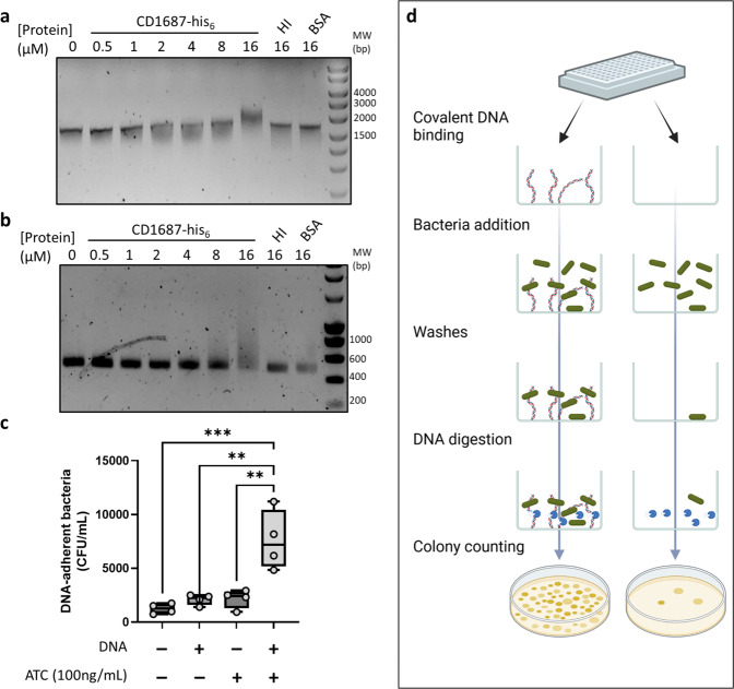
The ability of bacterial pathogens to establish recurrent and persistent infections is frequently associated with their ability to form biofilms. Clostridioides difficile infections have a high rate of recurrence and relapses and it is hypothesized that biofilms are involved in its pathogenicity and persistence. Biofilm formation by C. difficile is still poorly understood. It has been shown that specific molecules such as deoxycholate (DCA) or metronidazole induce biofilm formation, but the mechanisms involved remain elusive. In this study, we describe the role of the C. difficile lipoprotein CD1687 during DCA-induced biofilm formation. We showed that the expression of CD1687, which is part of an operon within the CD1685-CD1689 gene cluster, is controlled by multiple transcription starting sites and some are induced in response to DCA. Only CD1687 is required for biofilm formation and the overexpression of CD1687 is sufficient to induce biofilm formation. Using RNAseq analysis, we showed that CD1687 affects the expression of transporters and metabolic pathways and we identified several potential binding partners by pull-down assay, including transport-associated extracellular proteins. We then demonstrated that CD1687 is surface exposed in C. difficile, and that this localization is required for DCA-induced biofilm formation. Given this localization and the fact that C. difficile forms eDNA-rich biofilms, we confirmed that CD1687 binds DNA in a non-specific manner. We thus hypothesize that CD1687 is a component of the downstream response to DCA leading to biofilm formation by promoting interaction between the cells and the biofilm matrix by binding eDNA.


Conflict of interest statement

The authors declare no competing interests.

Figures

Fig. 1
Fig. 1. The CD1685-CD1689 cluster in C. difficile strain 630Δerm forms an operon with multiple transcription start sites.
a.RT-PCR performed with primers EA043 and EA027 (Supplementary Table 1) from various nucleic acid templates. cDNA was obtained using the EA027 primer with total RNA extracted from 48 h biofilms grown in BHISG supplemented with DCA (240 µM). b 5’RACE results from amplification of the poly-guanylated cDNA obtained, respectively, with the EA021 and EA018 primers (Supplementary Table 1), then the P1686 or P1687 primers along with the universal amplification primer (AAP) from the 5’RACE kit. The RNA was extracted from 48 h cell cultures grown under biofilm-inducing conditions (BHISG + 240 µM DCA) or non-biofilm-inducing conditions (BHISG). c Organization of the CD1685-CD1689 cluster, the location of the primers used for RT-PCR and the amplicons from the 5’RACE results using the P1686 or P1687 primers (amplicon sizes were predicted from the TSS identified by Soutourina et al. (2020) and Fuchs et al. (2021). TSS: Transcriptional Start Site; cDNA: complementary DNA; gDNA: genomic DNA. Blots in a and b derive from the same experiments and were not processed.
Fig. 2
Fig. 2. Overexpression of CD1687 induces biofilm formation in the absence of DCA.
Biofilms formation was assayed 24 h or 48 h after inoculation in BHISG +/− ATC (100 ng/mL) with the wild-type strain (630Δerm) containing either a control empty vector (pDIA6103) or the vector allowing the expression of CD1687 under the inducible Ptet promoter (pDIA6920). Each data point represents an independent biological replicate composed of 2 to 4 technical replicates. The boxplot used to represent quantitative data figure the median, minimum, maximum, and upper and lower quartiles. Asterisks indicate statistical significance with a one-way ANOVA test followed by a Tukey’s multiple comparison test (ns: not significant; ****p < 0.0001).
Fig. 3
Fig. 3. Differences in gene expression in the two transcriptomics experiments.
Venn diagram of the genes differentially regulated in the two transcriptomics experiments performed in this study (Supplementary Table 4).
Fig. 4
Fig. 4. CD1687 localizes at the cell surface of C. difficile and displays heterogenous distribution within the biofilm.
In situ epifluorescence microscopy analysis was performed on 48 h biofilms grown in BHISG + ATC (100 ng/mL) either in the presence or absence of DCA (240 µM) as indicated. The strains tested were the wild-type strain (630Δerm) carrying the control vector pDIA6103 and with the ∆1687 strain carrying the plasmid with an inducible CD1687 (pDIA6920) or the control plasmid (pDIA6103). DNA is stained with DAPI (blue) and CD1687 is labeled with specific anti-CD1687 rabbit antibodies detected with a TexasRed-conjugated goat anti-rabbit antibody (red). Pictures are representative of three biological replicates and were taken with a Nikon Eclipse Ti inverted microscope (Nikon, Japan). Scale bar: 10 µm.
Fig. 5
Fig. 5. DCA-induced biofilm formation is inhibited in the presence of anti-CD1687 antibodies.
a Biofilm formation of the 630Δerm strain was assayed 48 h in BHISG with DCA (240 µM) cultures in presence of different concentration of anti-CD1687 rabbit antibodies (0.05 mg/mL to 0.2 mg/mL). b Growth kinetics (OD600nm) of the WT (630Δerm) in BHISG medium with PBS or DCA supplemented with different concentrations of anti-CD1687 rabbit antibodies (0.05 mg/mL to 0.2 mg/mL). Ab: antibody; nsAb: non-specific antibody. c The alphafold2 predicted structure of CD1687 show a N-terminal signal peptide S (red) connected to the α beta domain (purple) by a linker peptide (green), with another similar β beta domain (yellow) in the C-terminal region. d 48 h biofilms form by various Δ1687 strain complemented with an empty vector (pDIA6103) or plasmids overexpressing the full-length CD1687 (pDIA6920) or truncated CD1687 lacking either one of the two domains removed (pDIA7242 and pDIA7243, Supplementary Table 1) grown in BHISG with ATC (100 ng/mL) and DCA (240 µM). Each data point represents an independent biological replicate composed of 2 to 4 technical replicates. The boxplots used to represent quantitative data figure the median, minimum, maximum, and upper and lower quartiles. Asterisks indicate statistical significance with a one-way ANOVA test followed by Dunnett’s multiple comparison test (a) (ns: not significant; **p < 0.01) or a Tukey’s multiple comparison test (d) (ns: not significant; ****p < 0.001).
Fig. 6
Fig. 6. CD1687 binds DNA and shifts DNA migration.
Electrophoretic Mobility shift assay (EMSA) was performed with a E. coli plasmid pUC9 or b C. difficile DNA (450 bp PCR-amplicon) mixed with various concentrations of CD1687 (up to 16 µM), with 16 µM of heat-inactivated (HI) CD1687 or BSA used as controls. c C. difficile CFUs measured from the adhesion assay. The Δ1687 pDIA6920 strain was used expressing or not CD1687 in response to ATc as described in (d). Schema of the adhesion assay. We compared the adhesion of bacteria either expressing CD1687 or not in wells that contain or not covalently bound DNA. This schema was made with biorender.com. The boxplot used to represent quantitative data figure the median, minimum, maximum, and upper and lower quartiles. Each data point represents an independent biological replicate. Asterisks indicate statistical significance with a one-way ANOVA test followed by Dunnett’s multiple comparison test (**p < 0.01; ***p < 0.001). Blots in a and b derive from the same experiments and were not processed.

References

    1. Rupnik M, Wilcox MH, Gerding DN. Clostridium difficile infection: new developments in epidemiology and pathogenesis. Nat. Rev. Microbiol. 2009;7:526–536. doi: 10.1038/nrmicro2164. - DOI - PubMed
    1. Giles, J. & Roberts, A. in Advances in Protein Chemistry and Structural Biology Vol. 129 (ed. Donev, R.) 215–245 (Academic Press, 2022). - PubMed
    1. Barbut F, et al. Epidemiology of recurrences or reinfections of Clostridium difficile-associated diarrhea. J. Clin. Microbiol. 2000;38:2386–2388. doi: 10.1128/JCM.38.6.2386-2388.2000. - DOI - PMC - PubMed
    1. Guery B, Galperine T, Barbut F. Clostridioides difficile: diagnosis and treatments. BMJ. 2019;366:l4609. doi: 10.1136/bmj.l4609. - DOI - PubMed
    1. Sachsenheimer FE, et al. Genomic and phenotypic diversity of Clostridium difficile during long-term sequential recurrences of infection. Int. J. Med. Microbiol. 2018;308:364–377. doi: 10.1016/j.ijmm.2018.02.002. - DOI - PubMed

Publication types