. 2023 Sep;19(9):1054-1062.
doi: 10.1038/s41589-023-01326-1. Epub 2023 May 11.

Signal peptide mimicry primes Sec61 for client-selective inhibition

Affiliations

Signal peptide mimicry primes Sec61 for client-selective inhibition

Shahid Rehan et al. Nat Chem Biol. 2023 Sep.

Abstract

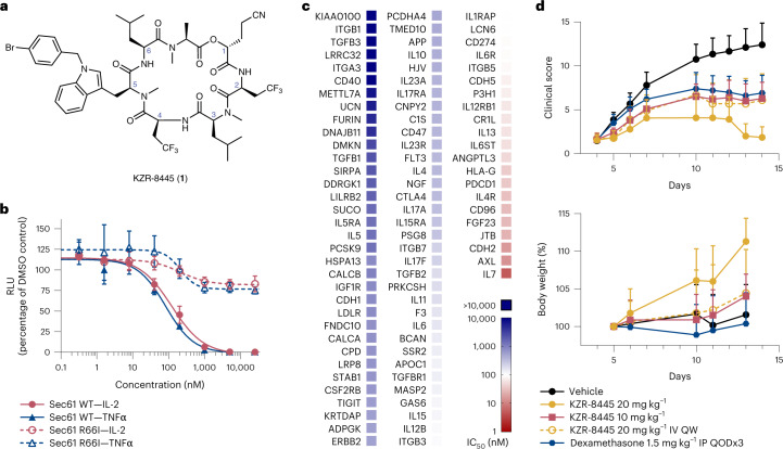
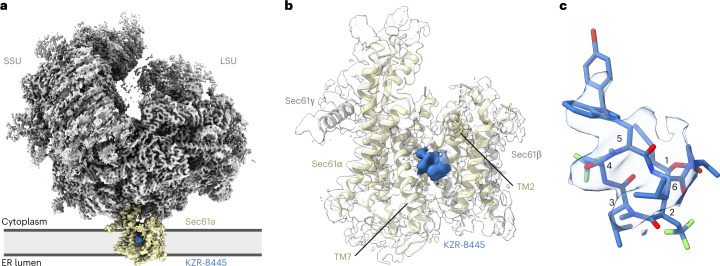
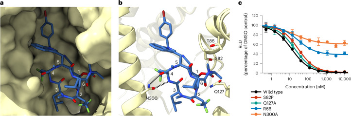
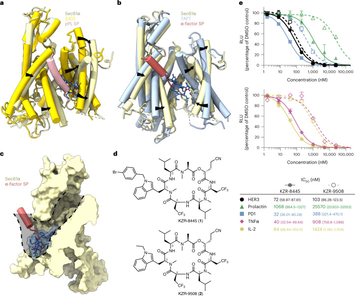
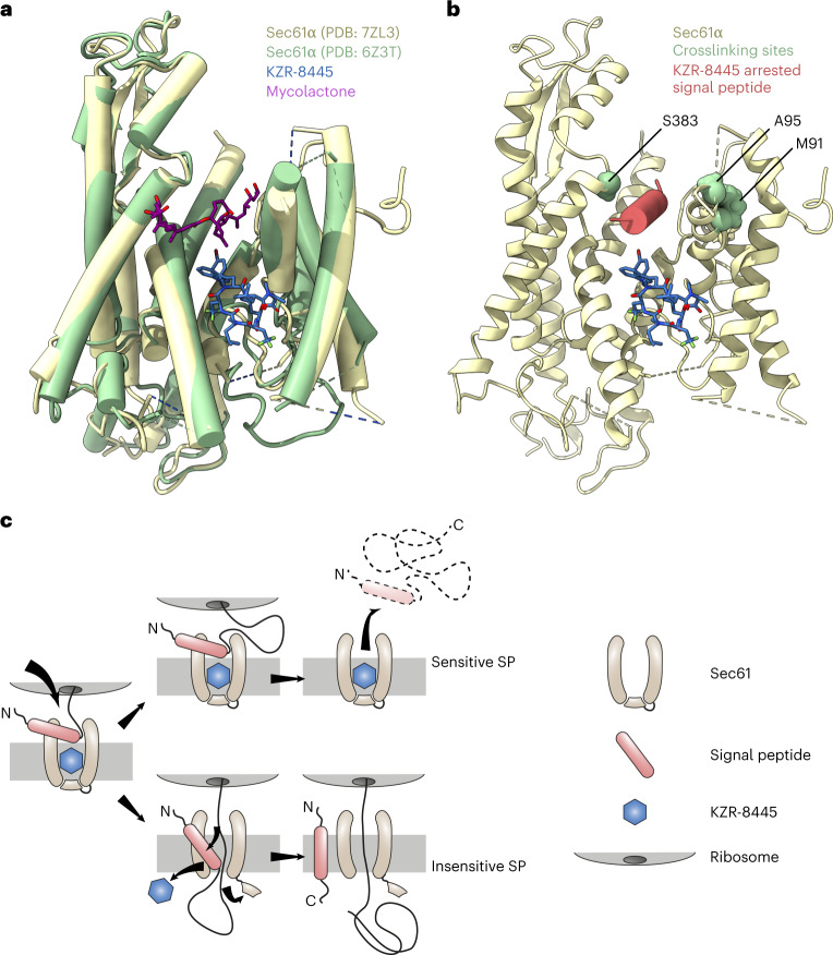
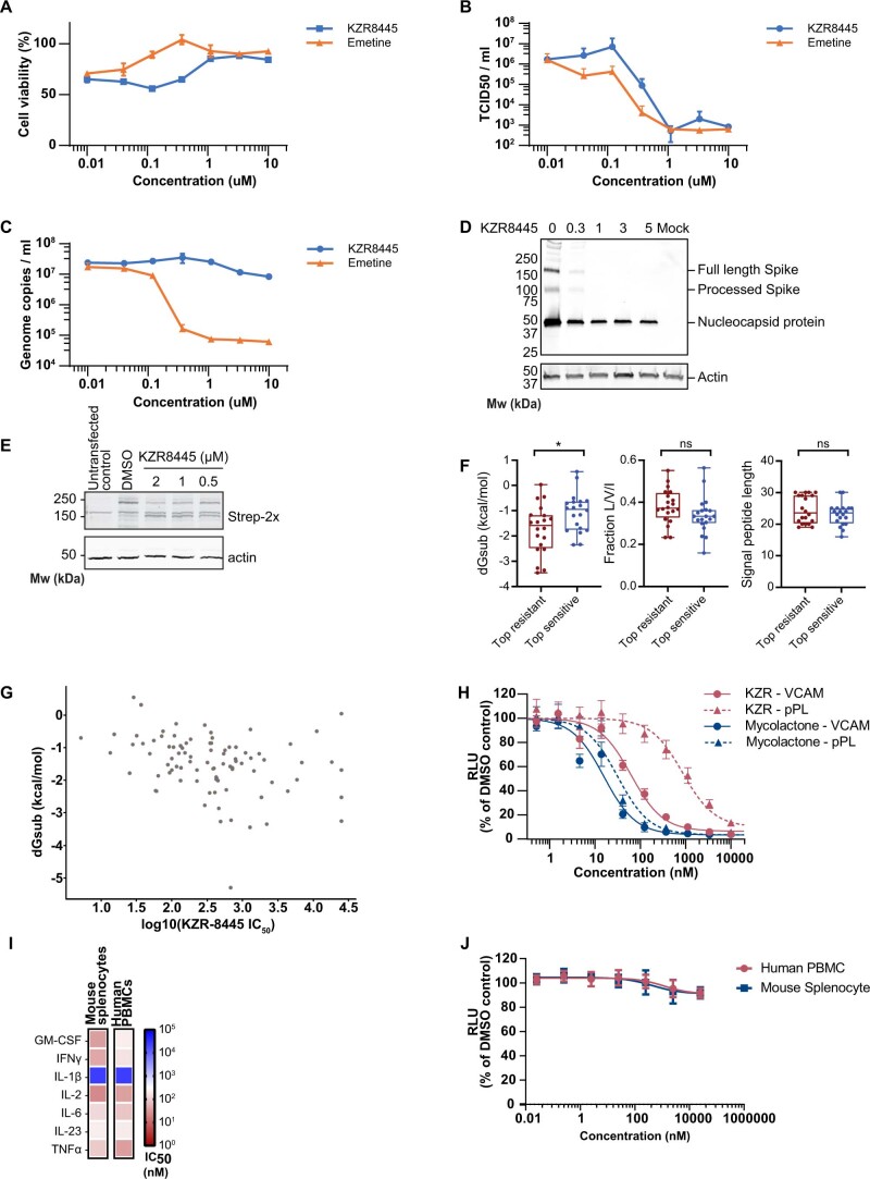
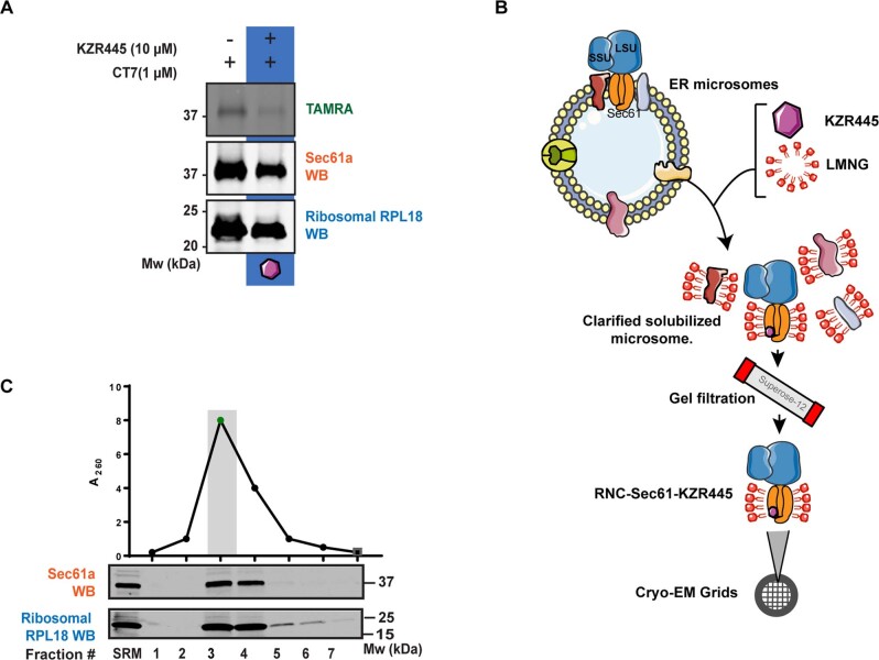
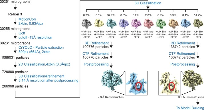
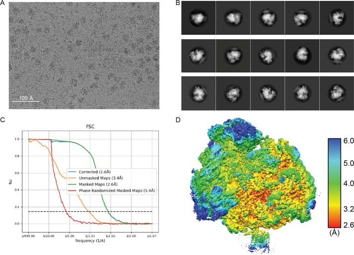
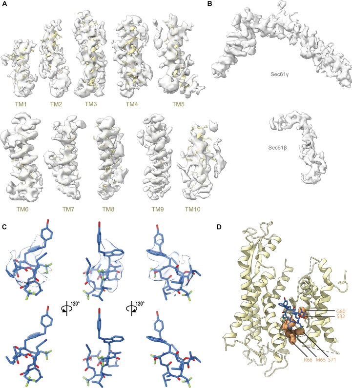
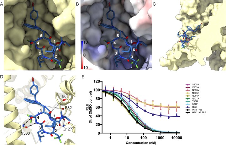
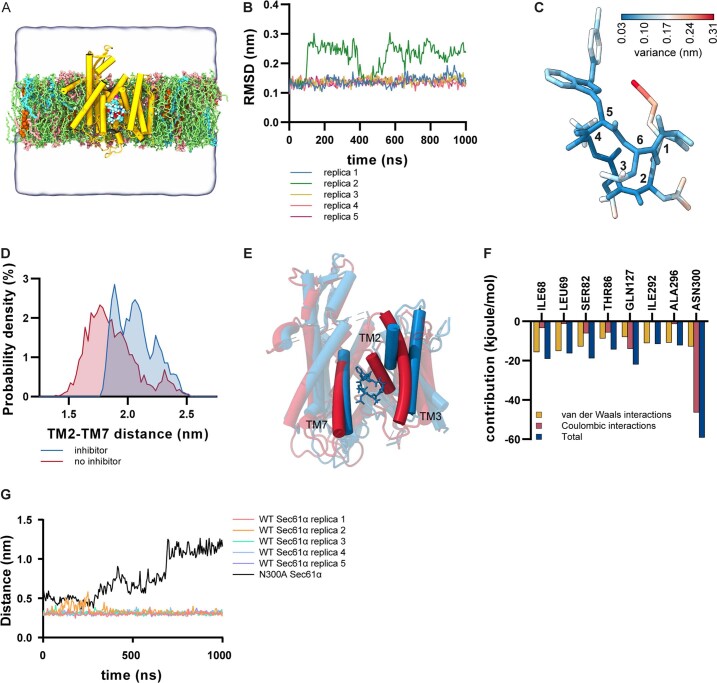
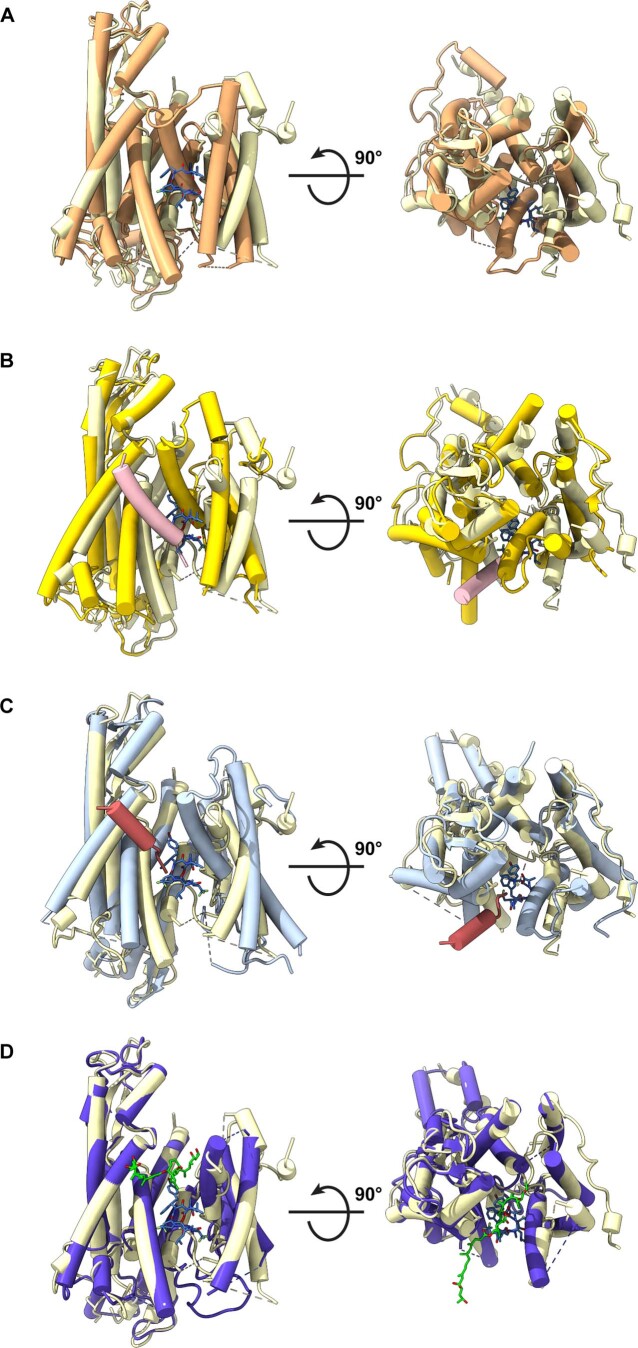
Preventing the biogenesis of disease-relevant proteins is an attractive therapeutic strategy, but attempts to target essential protein biogenesis factors have been hampered by excessive toxicity. Here we describe KZR-8445, a cyclic depsipeptide that targets the Sec61 translocon and selectively disrupts secretory and membrane protein biogenesis in a signal peptide-dependent manner. KZR-8445 potently inhibits the secretion of pro-inflammatory cytokines in primary immune cells and is highly efficacious in a mouse model of rheumatoid arthritis. A cryogenic electron microscopy structure reveals that KZR-8445 occupies the fully opened Se61 lateral gate and blocks access to the lumenal plug domain. KZR-8445 binding stabilizes the lateral gate helices in a manner that traps select signal peptides in the Sec61 channel and prevents their movement into the lipid bilayer. Our results establish a framework for the structure-guided discovery of novel therapeutics that selectively modulate Sec61-mediated protein biogenesis.


Conflict of interest statement

E.L., J.A., T.M. and C.J.K. are employees and shareholders of Kezar Life Sciences. D.M. is a shareholder of Kezar Life Sciences. J.T. is a founder of Global Blood Therapeutics, Kezar Life Sciences, Cedilla Therapeutics and Terremoto Biosciences, and is a scientific advisor to Entos. The remaining authors declare no competing interests.

Figures

Fig. 1
Fig. 1. KZR-8445 is a client-selective Sec61 inhibitor and is efficacious in a mouse arthritis model.
a, Chemical structure of KZR-8445. b, Cells stably expressing WT or R66I Sec61α were transfected with dox-inducible Gaussia luciferase (GLuc) reporter constructs fused to the C-terminus of IL-2 or TNF. After treatment with doxycycline and the indicated concentrations of KZR-8445 for 24 h, GLuc activity was quantified. Data are mean values ± s.d. from a single independent experiment. c, Cells were transfected with dox-inducible GLuc constructs fused to the indicated human signal peptides (SP-GLuc), treated with doxycycline and increasing concentrations of KZR-8445 for 24 h and assessed for GLuc activity. The heat map depicts IC50 values calculated from each SP-GLuc dose–response curve. d, Arthritis was induced in BALB/c mice with antibodies specific for type II collagen (mAb) on day 0 and endotoxin on day 3. On day 4, when the disease was present in all animals, mice were randomized and treated for 2 weeks with thrice weekly (QODx3) or weekly (QW) intravenous administration of vehicle or KZR-8445 or intraperitoneal administration of dexamethasone. Clinical scores (0–4 per paw; n = 10 per group) and body weights were followed until day 14. Data are presented as mean values ± s.d. mAb, monoclonal antibody. Source data
Fig. 2
Fig. 2. Structure of the mammalian Sec61 translocon with KZR-8445, a substrate-selective translocation inhibitor.
a, Cryo-EM map of the mammalian ribosome-bound heterotrimeric Sec61 translocon in complex with the cotransin analog KZR-8445. The map was low-pass filtered to 4 Å with density features corresponding to the ribosome (LSU and SSU), Sec61 and KZR-8445. b, Additional density assigned to KZR-8445 is located between TM2, TM3, TM7 and lumenal plug helices. KZR-8445 is bound to the center of the lateral gate, which adopts an open conformation. c, Fit of KZR-8445 to the observed density. LSU, ribosomal large subunit; SSU, ribosomal small subunit.
Fig. 3
Fig. 3. Detailed view of the KZR-8445 binding site.
a, Solvent-excluded surface view of the open Sec61α lateral gate bound to KZR-8445. b, Polar residues of the Sec61α cavity proximal to KZR-8445. c, KZR-8445 sensitivity of a VCAM-SP Gluc reporter construct in cells expressing the indicated Sec61α mutant. Data are mean values ± s.d. from n = 2 independent experiments. Source data
Fig. 4
Fig. 4. Structural insights lead to improved signal peptide selectivity.
a, Superimposition of Sec61–KZR-8445 complex with preprolactin signal peptide (salmon) engaged with Sec61 (PDB: 3JC2, bright yellow). b, Superimposition of Sec61–KZR-8445 complex with yeast α-factor signal peptide (red) engaged with Sec61 (PDB: 7AFT, blue). c, Sec61/KZR-8445 superimposed with a yeast α-factor signal peptide showing the solvent-excluded surface of the translocon. Indicated in gray is a putative route traversed by nascent signal peptides to reach the binding site occupied by the yeast α-factor signal peptide. d, Structures of KZR-8445 and KZR-9508, a cotransin with a truncated R-5 side chain. e, Cells were stably transfected with dox-inducible Gaussia luciferase (GLuc) reporter constructs fused to the C-terminus of the indicated signal peptides (top), or full-length IL-2 or TNFα (bottom). Following treatment with doxycycline and the indicated concentrations of KZR-8445 for 24 h, GLuc activity was quantified. Data are mean values ± s.d. from a single experiment. Source data
Fig. 5
Fig. 5. Proposed model for substrate-selective Sec61 inhibition.
a, Comparison of KZR-8445 (blue) bound to Sec61α (PDB: 7ZL3) and mycolactone (purple) bound to Sec61α (green, PDB: 6Z3T). b, KZR-8445-bound Sec61 (wheat) with residues previously shown to crosslink CT8-arrested TNFα highlighted in green. Placement of a putative KZR-8445-arrested signal peptide (red) was guided by the proximity of crosslinked residues. The bulky R-5 group of KZR-8445 projects toward the arrested signal peptide. c, Substrate-selective inhibitors, such as KZR-8445, arrest specific signal peptides in a nonproductive conformation in the cytosolic vestibule and in proximity to the KZR-8445 R-5 group, which is important for determining the range of inhibited Sec61 clients. Sensitive signal peptides are unable to progress in the insertion pathway and are displaced into the cytosol. Drug-resistant signal peptides are able to progress further along the insertion pathway. Intercalation between Sec61 lateral gate helices likely leads to inhibitor dissociation and allows translocation of the nascent polypeptide into the ER lumen.
Extended Data Fig. 1
Extended Data Fig. 1. Cotransin KZR-8445 inhibits SARS-CoV-2 viral replication and biogenesis of the Spike protein.
(a–c) Effect of KZR-8445 and emetine on cell viability (a), infectious virus particle production (b), and viral genome copy number (c) in Vero E6 cells infected with SARS-CoV-2 for 48 h. Data are shown as mean values ± SD (n = 3 replicates) from a single experiment. (d) Effect of KZR-8445 on Spike protein biogenesis in Vero E6 cells infected with SARS-CoV-2 for 48 h, assessed by immunoblotting. ‘Mock’: mock-infected cells. (e) Effect of KZR-8445 on SARS-Cov-2 Spike protein (2xStrep-tagged) transiently overexpressed in HEK293 cells. Data are representative of two independent experiments. (f) Comparison of signal peptide (SP) features from the top-20 most resistant versus top-20 most sensitive SP-GLuc IC50 values (related to Fig. 1c). dGsub was calculated for each SP using https://dgpred.cbr.su.se/index.php?p=TMpred (subsequence allowed; no length correction). P-value for dGsub (*) is 0.0282 (Wilcoxon rank-sum one-sided test; ns, not significant). (g) Scatterplot of SP-GLuc log(IC50) values (nM) (Fig. 1c) versus calculated dGsub. (h) Effect of KZR-8445 and mycolactone A/B on the secretion of eGluc2 fused to the signal peptide of human VCAM or bovine preprolactin (pPL) in transiently transfected HEK293T cells. Secreted luciferase data (RLU, relative light units) are shown as mean ± SD (n = 3 replicates), representative of two independent experiments. (i) Effect of KZR-8445 on cytokines secreted from human peripheral blood mononuclear cells (PBMCs) or mouse splenocytes stimulated with lipopolysaccharide or antibodies against CD3 and CD28 (see Methods). After 24 h, supernatants were collected for cytokine analysis by MSD U-PLEX electrochemiluminescent immunoassay and IC50 values calculated from dose-response curves. (j) Effect of KZR-8445 on cell viability of unstimulated human PBMCs and mouse splenocytes (CTG assay, 24 h treatment). Data are shown as mean values ± SD from 4 (PBMCs) or 2 (splenocytes) independent experiments. Source data
Extended Data Fig. 2
Extended Data Fig. 2. Isolation and biochemical characterization of the inhibitor-bound ribosome-Sec61 (RNC) complexes.
(a) Photocrosslinking experiment showing competitive binding of KZR-8445 with photo-cotransin (CT7) in the final purified sample. Highlighted sample (gray) was used for cryo-EM analysis. Gels are representative examples of n = 3 independent experiments. (b) Flowchart of purification of KZR-8445-bound RNC–Sec61 complex. Isolated sheep pancreatic rough ER microsomes were supplemented with 1 µM inhibitor and incubated on ice for 30 min followed by solubilization with 1% LMNG for 1 h. Solubilized material was clarified by centrifugation followed by gel filtration. (c) Gel filtration profile of clarified detergent-solubilized sample fractions with measured absorbance at A260. Western blot analysis demonstrates presence of Sec61 and ribosomes in the peak fractions confirming integrity of the purified complex. Gels are representative examples of n = 3 independent experiments. Source data
Extended Data Fig. 3
Cryo-EM data-processing workflow for ribosome-Sec61 translocon complexes.
Extended Data Fig. 4
Extended Data Fig. 4. Cryo-EM characterization of the ribosome/Sec61/KZR-8445 complex.
(a) A representative micrograph of ribosome-Sec61 complex on holey carbon grids coated. (b) Reference free 2D class averages showing different views of the ribosomes/Sec61 complex. (c) Fourier shell correlation (FSC) curves of the final reconstruction map. (d) Local resolution of the ribosome/Sec61 complex. Source data
Extended Data Fig. 5
Extended Data Fig. 5. Structure of the mammalian Sec61 translocon with a substrate-selective translocation inhibitor cotransin.
(a) Density of the fitted map within 3 Å of each transmembrane helix of Sec61α shown at 1.5 σ. (b) Density of the fitted map within 3 Å of Sec61γ (top) or Sec61β (bottom), shown at 1.5 σ. (c) Different views of density map about KZR-8445. (d) Locations of mutations conferring resistance to various Sec61 inhibitors.
Extended Data Fig. 6
Extended Data Fig. 6. KZR-8445 interfaces Sec61 and the lipid membrane in a multifaceted manner.
(a) Solvent excluded surface view of the KZR-8445 binding site within Sec61 viewed from the direction of the open lateral gate. (b) Same view, but surface colored based on the Coulombic potential surface. (c) Side view of the solvent excluded surface of the KZR-8445 binding site with intervening surfaces rendered transparent. Part of KZR-8445 is exposed to the lipid bilayer through the open lateral gate. (d) Polar residues of the Sec61α cavity proximal to KZR-8445. (e) KZR-8445 sensitivity of luciferase reporter constructs expressed in the background of Sec61 mutant cell lines in engineered human HEK293 cells. Data are mean values ± SD and are representative of n = 2 independent experiments. Source data
Extended Data Fig. 7
Extended Data Fig. 7. Molecular dynamics (MD) simulations of Sec61 bound to KZR-8445 in an intact membrane environment.
(a) Simulation system. Sec61 is shown in yellow and KZR-8445 based on chemical identity. Zwitterionic lipids (POPC, POPE, PSM) are shown in green, POPS in cyan, POPI in pink, and cholesterol in orange. Water is rendered as a transparent surface to highlight the simulation box dimensions, and ions are omitted for clarity. (b) Root mean squared deviation (RMSD) values for KZR-8445 as a function of simulation time, relative to its starting conformation. In one replica, the backbone around the nitrile group switches between two conformations, leading to large jumps in RMSD values. (c) Root mean squared fluctuation (RMSF) values mapped onto the KZR-8445 structure. The ring structure and the indole group reside stably in one conformation, whereas the trifluoromethyl groups are somewhat more mobile. The large RMSF values of the nitrile group mainly derive from a conformational change in the backbone observed in one simulation replica, leading to a different orientation of the nitrile group. The bromophenyl group rotates freely, leading to larger RMSF values. (d) The probability density of the TM2–TM7 distance that describes the degree of openness of the lateral gate. (e) Final structures (after 1 µs of MD simulation) of Sec61 with KZR-8445 present (blue) or without KZR-8445 (red). Both simulations were initiated from an open conformation. (f) Sec61 residues interacting with KZR-8445 with either van der Waals or Coulombic energy greater than 2 kcal/mol (8.4 kJ/mol). (g) Shortest distance between Sec61 N300 (or A300 in the mutant) and the peptide bond nitrogen between R-3 and R-4 in KZR-8445 as a function of simulation time. KZR-8445 remains stably bound in WT Sec61, but detaches from its binding site during the 1 µs MD simulation in the N300A mutant. Source data
Extended Data Fig. 8
Extended Data Fig. 8. Comparison of different structural states of mammalian Sec61 translocon.
(a–d) Comparisons of Sec61α (wheat) with bound KZR-8445 (royal blue) and (a) ‘idle’ mammalian Sec61α (PDB:3J7Q, orange), (b) mammalian Sec61 (PDB:3JC2, yellow) engaged with the preprolactin signal peptide (pink), (c) yeast Sec61 (PDB:7AFT, pale blue) engaged with the prepro-α factor signal peptide (red), or (d) mammalian Sec61α (PDB:6Z3T, green) with bound mycolactone (purple).

Comment in

References

    1. Zimmermann R, Eyrisch S, Ahmad M, Helms V. Protein translocation across the ER membrane. Biochim. Biophys. Acta. 2011;1808:912–924. - PubMed
    1. Rapoport TA, Li L, Park E. Structural and mechanistic insights into protein translocation. Annu. Rev. Cell Dev. Biol. 2017;33:369–390. - PubMed
    1. Manson, L. A. (ed.) Biomembranes Vol 2, pp. 193–195 (Springer, 1971).
    1. von Heijne G. Signal sequences. The limits of variation. J. Mol. Biol. 1985;184:99–105. - PubMed
    1. Elvekrog MM, Walter P. Dynamics of co-translational protein targeting. Curr. Opin. Chem. Biol. 2015;29:79–86. - PMC - PubMed

Publication types