. 2023 May 11;14(1):127.
doi: 10.1186/s13287-023-03356-x.

A pumpless monolayer microfluidic device based on mesenchymal stem cell-conditioned medium promotes neonatal mouse in vitro spermatogenesis

Affiliations

A pumpless monolayer microfluidic device based on mesenchymal stem cell-conditioned medium promotes neonatal mouse in vitro spermatogenesis

Selin Önen et al. Stem Cell Res Ther. .

Abstract

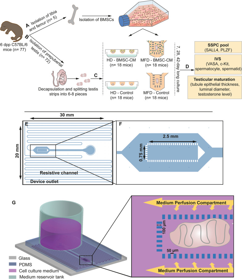
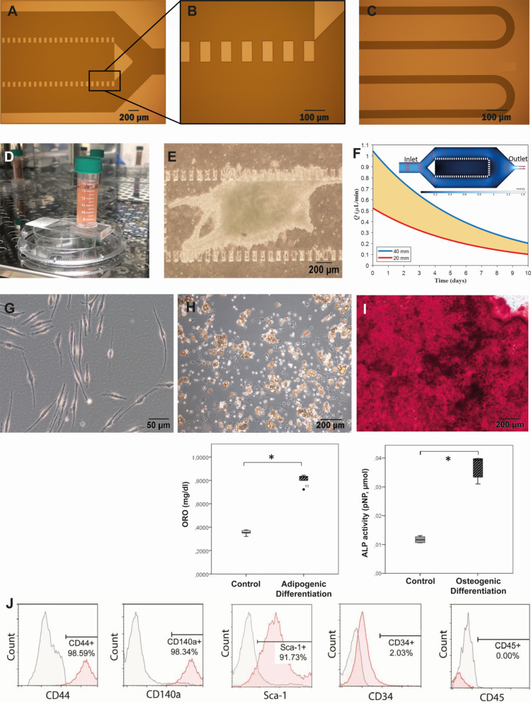
Background: Childhood cancer treatment-induced gonadotoxicity causes permanent infertility/sub-infertility in nearly half of males. The current clinical and experimental approaches are limited to cryopreservation of prepubertal testicular strips and in vitro spermatogenesis which are inadequate to achieve the expanded spermatogonial stem/progenitor cells and spermatogenesis in vitro. Recently, we reported the supportive effect of bone marrow-derived mesenchymal cell co-culture which is inadequate after 14 days of culture in static conditions in prepubertal mouse testis due to lack of microvascular flow and diffusion. Therefore, we generated a novel, pumpless, single polydimethylsiloxane-layered testis-on-chip platform providing a continuous and stabilized microfluidic flow and real-time cellular paracrine contribution of allogeneic bone marrow-derived mesenchymal stem cells.

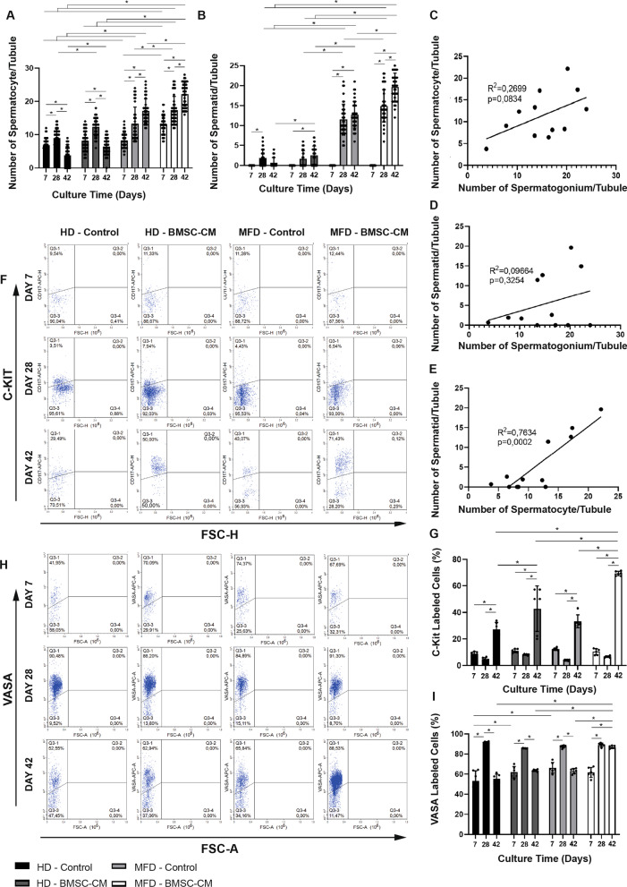
Methods: We aimed to evaluate the efficacy of this new setup in terms of self-renewal of stem/progenitor cells, spermatogenesis and structural and functional maturation of seminiferous tubules in vitro by measuring the number of undifferentiated and differentiating spermatogonia, spermatocytes, spermatids and tubular growth by histochemical, immunohistochemical, flow cytometric and chromatographic techniques.

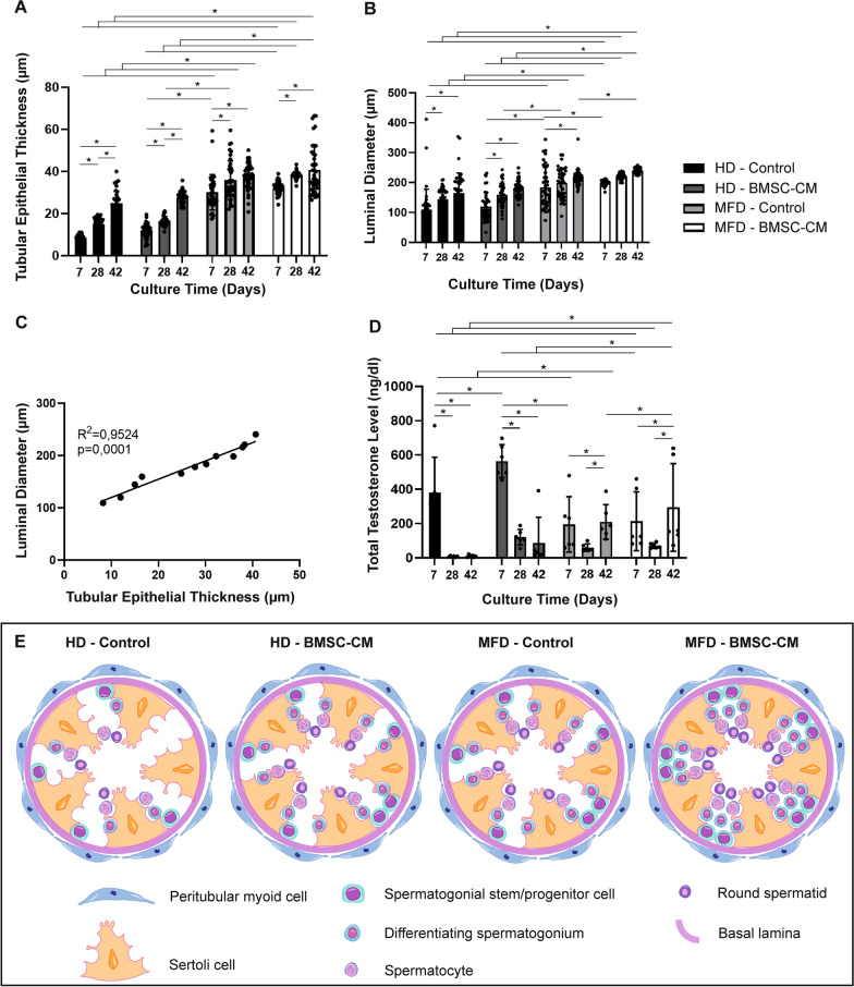
Results: Bone marrow-derived mesenchymal stem cell-based testis-on-chip platform supported the maintenance of SALL4(+) and PLZF(+) spermatogonial stem/progenitor cells, for 42 days. The new setup improved in vitro spermatogenesis in terms of c-Kit(+) differentiating spermatogonia, VASA(+) total germ cells, the meiotic cells including spermatocytes and spermatids and testicular maturation by increasing testosterone concentration and improved tubular growth for 42 days in comparison with hanging drop and non-mesenchymal stem cell control.

Conclusions: Future fertility preservation for male pediatric cancer survivors depends on the protection/expansion of spermatogonial stem/progenitor cell pool and induction of in vitro spermatogenesis. Our findings demonstrate that a novel bone marrow-derived mesenchymal stem cell-based microfluidic testis-on-chip device supporting the maintenance of stem cells and spermatogenesis in prepubertal mice in vitro. This new, cell therapy-based microfluidic platform may contribute to a safe, precision-based cell and tissue banking protocols for prepubertal fertility restoration in future.

Keywords: In vitro spermatogenesis; Male infertility; Mesenchymal stem cells; Microfluidics; Prepubertal boys.


Conflict of interest statement

The authors declare that they have no competing interests.

Figures

Fig. 1
Fig. 1
Workflow of the study and design of MFD. a Isolation of BMSCs. b Testicular tissue collection. c Generation of HD and MFD culture systems. d Evaluation of SSPC pool, IVS and testicular maturation. e Sketch of the mold with resistive channel, device outlet and f organ chamber. g 3D illustration of MFD with organ placement
Fig. 2
Fig. 2
Validation of new MFD and characterization of BMSCs. Micrographs of a organ chamber, b pillars and c resistive channels after photolithography and d picture of MFD with BMSC-CM insert. e Micrograph of seminiferous tubules within organ chamber during culture. f The graph presents the flow rate in resistive channels with different medium levels in the reservoir tank (MATLAB R2018a). Surface plot represents the velocity field inside the microsystem where the red arrows indicate the flow direction (COMSOL Multiphysics v5.6). g Micrograph of passage three spindle-shaped BMSCs adhering to culture plate. h Adipogenic and i osteogenic differentiation of BMSCs (ORO, 20x; ARS, 100x, respectively, *p < 0.01). j Characteristic surface antigenic expression of BMSCs by FCM
Fig. 3
Fig. 3
BMSC-contributed MFD provides maintenance and enlargement of SALL4(+) and PLZF(+) SSPC pool and PAS-stained spermatogonia in vitro. Immune labeling of a SALL4(+) and c PLZF(+) SSPCs, and e PAS-stained of spermatogonia in prepubertal mice testes from day 7 to 42 in BMSC-CM-applied and non-applied HD and MFD (SALL4 IHC and PAS–hematoxylin, 1000x). Note the presence of SSPCs indicated by arrow on SALL4-labeled sections. In time change in b number of SALL4(+) cells, d ratio of PLZF(+) cells to total testicular cells and f spermatogonium count in BMSC-CM-applied and non-applied groups are illustrated in bar graph with standard deviation and data distribution (*p < 0.05, n = 6 testes, 50 tubules for SALL4; n = 6 testes for PLZF). g Line graph illustrates positive correlation of SALL4 with PLZF labeling and spermatogonium number for SSPCs in control and BMSC groups (R2 = 0.0319, p = 0.5785; R2 = 0.9994, p = 0.001, respectively) (arrows: SSPCs, asterisks: seminiferous tubules)
Fig. 4
Fig. 4
BMSC-CM-contributed MFD promotes IVS. In time change in number of a spermatocytes and b spermatids per tubules in BMSC-CM-applied and non-applied groups are shown in bar graph with standard deviation and data distribution (*p < 0.05, n = 6 testes, 50 tubules). Line graphs illustrate positive correlation between the c number of spermatocyte and spermatogonium, d spermatid and spermatogonium and e spermatocyte and spermatid in BMSC-CM-applied and non-applied HD and MFD (R2 = 0.02699, p = 0.0894; R2 = 0.09664, p = 0.3254; R2 = 0.7634, p = 0.0002, respectively). Flow cytometric analysis of f c-Kit(+) and h VASA(+) cell ratio to total testicular cells. Bar graph with standard deviation and data distribution illustrates in time change in g c-Kit and i VASA-labeled cell ratio to total testicular cells in BMSC-CM-applied and non-applied HD and MFD (*p < 0.05, n = 6 testes)
Fig. 5
Fig. 5
BMSC-CM-contributed MFD supports testicular maturation by increasing tubule epithelial thickness, luminal diameter and testosterone level in vitro. Bar graphs illustrate time-dependent change by standard deviation and data distribution of a tubule epithelial thickness, b luminal diameter and c testosterone concentration (*p < 0.05, n = 6 testes, 50 tubules). d Line graph illustrates a positive correlation of tubule epithelial thickness with luminal diameter in control and BMSC groups (R2 = 0.9524, p = 0.0001). e Illustration of germ cell populations in tubules on day 42

References

    1. ACS. Key Statistics for Childhood Cancers; 2022. https://www.cancer.org/cancer/cancer-in-children/key-statistics.html.
    1. Pco ASRM. Fertility preservation in patients undergoing gonadotoxic therapy or gonadectomy: a committee opinion. Fertil Steril. 2019;112(6):1022–1033. doi: 10.1016/j.fertnstert.2019.09.013. - DOI - PubMed
    1. ASRM ECo Fertility preservation and reproduction in patients facing gonadotoxic therapies: an Ethics Committee opinion. Fertil Steril. 2018;110(3):380–386. doi: 10.1016/j.fertnstert.2018.05.034. - DOI - PubMed
    1. Wasilewski-Masker K, Seidel KD, Leisenring W, Mertens AC, Shnorhavorian M, Ritenour CW, et al. Male infertility in long-term survivors of pediatric cancer: a report from the childhood cancer survivor study. J Cancer Surv. 2014;8(3):437–447. doi: 10.1007/s11764-014-0354-6. - DOI - PMC - PubMed
    1. Siegel RL, Miller KD, Fuchs HE, Jemal A. Cancer statistics, 2022. CA Cancer J Clin. 2022;72(1):7–33. doi: 10.3322/caac.21708. - DOI - PubMed

Publication types

Substances

LinkOut - more resources