. 2021 Jun 11;6(1):20.
doi: 10.1186/s40850-021-00084-8.

A comparative analysis of the nervous system of cheilostome bryozoans

Affiliations

A comparative analysis of the nervous system of cheilostome bryozoans

Jakob Prömer et al. BMC Zool. .

Abstract

Background: Bryozoans are sessile aquatic suspension feeders in mainly marine, but also freshwater habitats. Most species belong to the marine and calcified Cheilostomata. Since this taxon remains mostly unstudied regarding its neuroanatomy, the focus of this study is on the characterization and ground pattern reconstruction of the autozooidal nervous system based on six representatives.

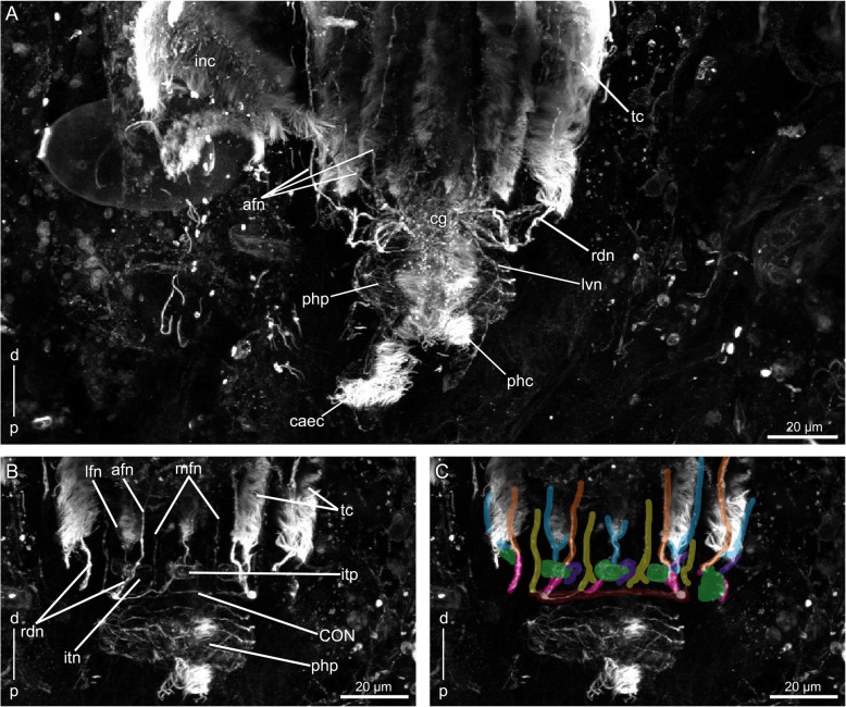
Results: A common neuronal innervation pattern is present in the investigated species: a cerebral ganglion is located at the base of the lophophore, from where neurite bundles embrace the mouth opening to form a circumoral nerve ring. Four neurite bundles project from the cerebral ganglion to innervate peripheral areas, such as the body wall and parietal muscles via the tentacle sheath. Five neurite bundles comprise the main innervation of the visceral tract. Four neurite bundles innervate each tentacle via the circumoral nerve ring. Mediofrontal tentacle neurite bundles emerge directly from the nerve ring. Two laterofrontal- and one abfrontal tentacle neurite bundles emanate from radial neurite bundles, which originate from the cerebral ganglion and circumoral nerve ring in between two adjacent tentacles. The radial neurite bundles terminate in intertentacular pits and give rise to one abfrontal neurite bundle at the oral side and two abfrontal neurite bundles at the anal side. Similar patterns are described in ctenostome bryozoans.

Conclusions: The present results thus represent the gymnolaemate situation. Innervation of the tentacle sheath and visceral tract by fewer neurite bundles and tentacular innervation by four to six tentacle neurite bundles support cyclostomes as sister taxon to gymnolaemates. Phylactolaemates feature fewer distinct neurite bundles in visceral- and tentacle sheath innervation, which always split in nervous plexus, and their tentacles have six neurite bundles. Thus, this study supports phylactolaemates as sistergroup to myolaemates.

Keywords: Alpha tubulin staining; Gymnolaemata; Lophotrochozoa; Myolaemata; Neuroanatomy.


Conflict of interest statement

The authors declare that they have no competing interests.

Figures

Fig. 1
Fig. 1
Colony morphology of few cheilostome bryozoan species. A Electra posidoniae encrusting colony on Posidonia oceanica. B Bugula neritina erect dichotomous branching colony with biserial branches. C Fenestrulina joannae encrusting colony on P. oceanica. D Myriapora truncata thick massive radial branch of irregularly branching colony. Abbreviations: anc – ancestrula, o – operculum, oc – ovicell, z – zooid
Fig. 2
Fig. 2
Schematic overview on the morphology of cheilostome bryozoans based on Electra posidoniae from the frontal side. The cystid (gray) is calcified. The polypide is composed of a lophophore (blue), digestive tract (green), musculature (pink) and the nervous system (cerebral ganglion in yellow). Abbreviations: a – anus, ca – cardia, cae – caecum, d – distal, db – duplicature bands, eso – esophagus, int – intestine, o – operculum, opo – operculum occlusor, p – proximal, pdi – parietodiaphragmatic muscle, ph – pharynx, pm – parietal muscle, py – pylorus, rm. – retractor muscle, sph – sphincter muscle, ts – tentacle sheath
Fig. 3
Fig. 3
Electra posidoniae. Acetylated alpha-tubulin-lir in orange in all images. A Overview of few zooids. B Overview from the frontal side, showing alpha-tubulin-lir and f-actin (gray). Volume rendering. C Overview from the frontal side, showing alpha-tubulin-lir structures. Volume rendering. D Detail of the lophophoral base. Volume rendering. E Detail of the apertural area. Volume rendering. Abbreviations: abr – annular branch of the trifid neurite bundle, afn – abfrontal tentacle neurite bundle, caec – caecum cilia, cl – caecum ligament, cg – cerebral ganglion, CON – circumoral nerve ring, ctsn – compound tentacle sheath nerve, d – distal, izp – interzooidal pores, dtsn – direct tentacle sheath nerve, lb. – lophophoral base, lvn – laterovisceral neurite bundle, mvn – mediovisceral neurite bundle, o – operculum, opn – opercular nerve, opo – operculum occlusor, p – proximal, pbs – parietal branching site, pdi – parietodiaphragmatic muscle, pdin – parietodiaphragmatic nerve, php – pharyngeal nerve plexus, pm – parietal muscle, pmn – parietal muscle nerve, rm. – retractor muscle, sph – sphincter muscle, spn – sphincter nerve, tc – tentacle cilia, tfd – trifid neurite bundle, tm – tentacle muscle, white arrows indicate the position of duplicature bands
Fig. 4
Fig. 4
Fenestrulina joannae. A Overview of a zooid. B Overview from the frontal side, showing alpha-tubulin-lir in orange and f-actin in gray. Volume rendering. C Overview from the frontal side, showing alpha-tubulin-lir structures in orange. Volume rendering. D Detail of the lophophoral base. Alpha-tubulin-lir in orange. Volume rendering. E Detail of apertural area. Alpha-tubulin-lir in gray. Maximum intensity projection. Abbreviations: afn – abfrontal tentacle neurite bundle, cg – cerebral ganglion, CON – circumoral nerve ring, ctsn – compound tentacle sheath nerve, d – distal, dtsn – direct tentacle sheath nerve, int – intestine, lb. – lophophoral base, lvn – laterovisceral neurite bundle, mvn – mediovisceral neurite bundle, opn – opercular nerve, opo – operculum occlusor, p – proximal, pbs – parietal branching site, pdi – parietodiaphragmatic muscle, pdin – parietodiaphragmatic nerve, phm – pharyngeal musculature, php – pharyngeal nerve plexus, pm – parietal muscle, pmn – parietal muscle nerve, rm. – retractor muscle, sph – sphincter muscle, spn – sphincter nerve, tc – tentacle cilia, tfd – trifid neurite bundle, tm – tentacle muscle
Fig. 5
Fig. 5
Myriapora truncata. A Overview of a few zooids. B Overview from the basal side, showing alpha-tubulin-lir in orange and f-actin in gray. Volume rendering. C Overview from the basal side, showing alpha-tubulin-lir structures in orange. Volume rendering. D Detail of the lophophoral base. Lateral view, alpha-tubulin-lir in orange. Volume rendering. E Detail of apertural area. Alpha-tubulin-lir in gray Maximum intensity projection. Abbreviations: abr – annular branch of trifid neurite bundle, afn – abfrontal tentacle neurite bundle, caec – caecum cilia, cg – cerebral ganglion, CON – circumoral nerve ring, ctsn – compound tentacle sheath nerve, d – distal, dtsn – direct tentacle sheath nerve, lb. – lophophoral base, mvn – mediovisceral neurite bundle, o – operculum, opn – opercular nerve, opo – operculum occlusor muscle, p – proximal, pbs – parietal branching site, pdi – parietodiaphragmatic muscle, pdin – parietodiaphragmatic nerve, phc – pharyngeal cilia, phm – pharyngeal musculature, pm – parietal muscle, pmn – parietal muscle nerve, rm. – retractor muscle, sph – sphincter muscle, spn – sphincter nerve, tc – tentacle cilia, tfd – trifid neurite bundle, tm – tentacle muscle
Fig. 6
Fig. 6
Bugula neritina. Acetylated alpha-tubulin-lir in orange in all images. A Overview of few zooids. B Overview from the frontal side, showing alpha-tubulin-lir and f-actin (gray). Volume rendering. C Overview from the frontal side, showing alpha-tubulin-lir structures. Volume rendering. D Detail of the lophophoral base in lateral view. Volume rendering. E Detail of apertural area in lateral view. Volume rendering. Abbreviations: abr – annular branch of trifid neurite bundle, afn – abfrontal tentacle neurite bundle, cac – cardia cilia, cg – cerebral ganglion, CON – circumoral nerve ring, ctsn – compound tentacle sheath nerve, d – distal, dtsn – direct tentacle sheath nerve, lb. – lophophoral base, lvn – laterovisceral neurite bundle, mlvn – mediolaterovisceral neurite bundle, mvn – mediovisceral neurite bundle, p – proximal, pbs – parietal branching site, pdi – parietodiaphragmatic muscle, pdin – parietodiaphragmatic nerve, phc – pharyngeal cilia, phm – pharyngeal musculature, php – pharyngeal nerve plexus, pm – parietal muscle, pmn – parietal muscle nerve, rdn – radial nerve, rm. – retractor muscle, sph – sphincter muscle, spn – sphincter nerve, tc – tentacle cilia, tfd – trifid neurite bundle, tm – tentacle muscle, white arrows indicate the position of duplicature bands
Fig. 7
Fig. 7
Collarina balzaci. A Overview of a zooid. B Overview from the frontal side, showing alpha-tubulin-lir in orange and f-actin in gray. C Overview from the frontal side, alpha-tubulin-lir in orange. D Detail of the lophophoral base, alpha-tubulin-lir in gray. Maximum intensity projection. E Detail of apertural area, alpha-tubulin-lir in orange. Volume rendering. Abbreviations: afn – abfrontal tentacle neurite bundle, cg – cerebral ganglion, CON – circumoral nerve ring, ctsn – compound tentacle sheath nerve, d – distal, dtsn – direct tentacle sheath nerve, lb. – lophophoral base, lvn – laterovisceral neurite bundle, mvn – mediovisceral neurite bundle, opn – opercular nerve, opo – operculum occlusor, p – proximal, pbs – parietal branching site, pdi – parietodiaphragmatic muscle, pdin – parietodiaphragmatic nerve, pm – parietal muscle, pmn – parietal muscle nerve, rm. – retractor muscle, sph – sphincter muscle, spn – sphincter nerve, tc – tentacle cilia, tfd – trifid neurite bundle, tm – tentacle muscle, white arrows indicate the position of duplicature bands
Fig. 8
Fig. 8
Chorizopora brongniartii. Acetylated alpha-tubulin-lir in orange in all images. A Overview of a zooid. F-actin in gray. B Overview from the frontal side, showing alpha-tubulin-lir and f-actin structures. Volume rendering. C Overview from the frontal side, showing alpha-tubulin-lir structures. Volume rendering. D Detail of the lophophoral base from the anal side. Volume rendering. E Detail of apertural area. Volume rendering. Abbreviations: afn – abfrontal tentacle neurite bundle, caec – caecum cilia, cg – cerebral ganglion, ctsn – compound tentacle sheath nerve, d – distal, dtsn – direct tentacle sheath nerve, int – intestine, lb. – lophophoral base, mvn – mediovisceral neurite bundle, opn – opercular nerve, opo – operculum occlusor, p – proximal, pbs – parietal branching site, pdi – parietodiaphragmatic muscle, pdin – parietodiaphragmatic nerve, phc – pharyngeal cilia, pm – parietal muscle, pmn – parietal muscle nerve, rm. – retractor muscle, sph – sphincter muscle, spn – sphincter nerve, tc – tentacle cilia, tfd – trifid neurite bundle, tm – tentacle muscle
Fig. 9
Fig. 9
Ground pattern of the autozooidal innervation in cheilostomes. A Lateral view of the autozooidal innervation pattern without lophophore. B View from the distal side showing the innervation pattern of the lophophore C Cross section of a tentacle. Abbreviations: abr – annular branch of trifid nerve, afc – abfrontal cilia, afn – abfrontal tentacle neurite bundle, cg – cerebral ganglion, CON – circumoral nerve ring, ctsn – compound tentacle sheath nerve, dtsn – direct tentacle sheath nerve, fc – frontal cilia, itn – intertentacular neurite, itp – intertentacular pit, lc – lateral cilia, lfc – laterofrontal cilia, lfn – laterofrontal tentacle neurite bundle, lvn – laterovisceral neurite bundle, mfn – mediofrontal tentacle neurite bundle, mlvn – mediolaterovisceral neurite bundle, mvn – mediovisceral neurite bundle, pbs – parietal branching site, pdin – parietodiaphragmatic nerve, php – pharyngeal nerve plexus, pmn – parietal muscle nerve, opn – opercular nerve, rdn – radial nerve, spn – sphincter nerve, tco – tentacle coelom, tfd – trifid neurite bundle
Fig. 10
Fig. 10
Innervation schemes shown from the frontal side based on data from this study. A Electra posidoniae. B Myriapora truncata. C Fenestrulina joannae. D Collarina balzaci. E Chorizopora brongniartii. F Bugula neritina. Abbreviations: abr – annular branch of trifid nerve, cg – cerebral ganglion, CON – circumoral nerve ring, ctsn – compound tentacle sheath nerve, dtsn – direct tentacle sheath nerve, lvn – laterovisceral neurite bundle, mlvn – mediolaterovisceral neurite bundle, mvn – mediovisceral neurite bundle, opn – opercular nerve, pbs – parietal branching site, pdin – parietodiaphragmatic nerve, php – pharyngeal nerve plexus, pmn – parietal muscle nerve, spn – sphincter nerve, tfd – trifid neurite bundle
Fig. 11
Fig. 11
Acetylated alpha-tubulin-lir (gray) in the lophophore of Electra posidoniae. A Detail of the lophophoral base and of the digestive tract. View from anal side of lophophoral base. B Detail of tentacular innervation at the lophophoral base. C Same as in B but labeled with a color code. Abbreviations and color code: afn – abfrontal tentacle neurite bundle (orange), caec – caecal cilia, cg – cerebral ganglion (red), CON – circumoral nerve ring (brown), d – distal, itn – intertentacular neurite (purple), itp – intertentacular pit (green), lfn – laterofrontal tentacle neurite bundle (blue), lvn – laterovisceral neurite bundle, mfn – mediofrontal tentacle neurite bundle (yellow), mlvn – mediolaterovisceral neurite bundle, mvn – mediovisceral neurite bundle, p – proximal, php – pharyngeal nerve plexus, rdn – radial nerve (pink), tc – tentacle cilia
Fig. 12
Fig. 12
Acetylated alpha-tubulin-lir (gray) in the lophophore of Fenestrulina joannae. A Detail of the lophophoral base and the visceral tract. B Detail of tentacular innervation at the lophophoral base. C Same as in B but labeled with a color code. Abbreviations and color code: afn – abfrontal tentacle neurite bundle (orange), caec – cardia cilia, cg – cerebral ganglion (red), CON – circumoral nerve ring (brown), d - distal, itn – intertentacular neurite (purple), inc – intestinal cilia, itp – intertentacular pit (green), lfn – laterofrontal tentacle neurite bundle (blue), lvn – laterovisceral neurite bundle, mfn – mediofrontal tentacle neurite bundle (yellow), p – proximal, phc – pharyngeal cilia, php – pharyngeal nerve plexus, rdn – radial nerve (pink), tc – tentacle cilia
Fig. 13
Fig. 13
Acetylated alpha-tubulin-lir (gray) in the lophophore of Myriapora truncata. A Detail of the lophophoral base. B Detail of tentacular innervation at the lophophoral base. C Same as in B but labeled with a color code. Abbreviations and color code: abr – annular branch of trifid nerve, afn – abfrontal tentacle neurite bundle (orange), cg – cerebral ganglion (red), CON – circumoral nerve ring (brown), d – distal, dtsn – direct tentacle sheath nerve, itn – intertentacular neurite (purple), itp – intertentacular pit (green), lfn – laterofrontal tentacle neurite bundle (blue), lvn – laterovisceral nerve, mfn – mediofrontal tentacle nerve (yellow), mlvn – mediolaterovisceral neurite bundle, mvn – mediovisceral neurite bundle, p – proximal, php – pharyngeal nerve plexus, rdn – radial nerve (pink), tc – tentacle cilia, tfd – trifid neurite bundle
Fig. 14
Fig. 14
Acetylated alpha-tubulin-lir in the lophophore of Bugula neritina. A Detail of the lophophoral base. Acetylated alpha-tubulin-lir in orange. B Detail of the annular branch of trifid nerve. Acetylated alpha-tubulin-lir in gray. C Detail of tentacular innervation at the lophophoral base. Acetylated alpha-tubulin-lir in gray D Same as in C but labeled with a color code. Abbreviations and color code: abr – annular branch of trifid nerve, afn – abfrontal tentacle neurite bundle (orange), cae – caecum, cg – cerebral ganglion (red), CON – circumoral nerve ring (brown), d – distal, dtsn – direct tentacle sheath nerve, itn – intertentacular neurite (purple), itp – intertentacular pit (green), lfn – laterofrontal tentacle neurite bundle (blue), lvn – laterovisceral neurite bundle, mfn – mediofrontal tentacle neurite bundle (yellow), mlvn – mediolaterovisceral neurite bundle, mvn – mediovisceral neurite bundle, p – proximal, php – pharyngeal nerve plexus, rdn – radial nerve (pink), tc – tentacle cilia
Fig. 15
Fig. 15
Acetylated alpha-tubulin-lir (gray) in the lophophore of Collarina balzaci. A Detail of the lophophoral base. B Detail of tentacular innervation at the lophophoral base. C Same as in B but labeled with a color code. Abbreviations and color code: afn – abfrontal tentacle neurite bundle (orange), cg – cerebral ganglion (red), CON – circumoral nerve ring (brown), ctsn – compound tentacle sheath nerve, d – distal, dtsn – direct tentacle sheath nerve, itn – intertentacular neurite (purple), itp – intertentacular pit (green), lfn – laterofrontal tentacle neurite bundle (blue), lvn – laterovisceral neurite bundle, mfn – mediofrontal tentacle neurite bundle (yellow), p – proximal, phc – pharyngeal cilia, php – pharyngeal nerve plexus, rdn – radial nerve (pink), tc – tentacle cilia, tfd – trifid neurite bundle
Fig. 16
Fig. 16
Acetylated alpha-tubulin-lir (gray) in the lophophore of Chorizopora brongniartii. A Detail of the lophophoral base. B Detail of tentacular innervation at the lophophoral base. C Same as in B but labeled with a color code. Abbreviations and color code: afn – abfrontal tentacle neurite bundle (orange), cg – cerebral ganglion (red), CON – circumoral nerve ring (brown), d – distal, itn – intertentacular neurite (purple), itp – intertentacular pit (green), lfn – laterofrontal tentacle neurite bundle bundle (blue), lvn – laterovisceral neurite bundle, mfn – mediofrontal tentacle nerve (yellow), mvn – mediovisceral neurite bundle, p – proximal, rdn – radial nerve (pink), tc – tentacle cilia, tfd – trifid neurite bundle
Fig. 17
Fig. 17
Comparison of the acetylated alpha-tubulin-lir (gray) in the lophophoral base of six cheilostome species. A & C are from the anal side showing the cerebral ganglion, B slightly laterally of the latter two showing a thick circum-oral nerve ring, and D-F are from the oral side where the nerve ring is only thin. A Electra posidoniae. B Bugula neritina. C Myriapora truncata. D Chorizopora brongniartii. E Fenestrulina joannae. F Collarina balzaci. Abbreviations and color code: abfrontal tentacle neurite bundle (orange), cerebral ganglion (red), circumoral nerve ring (brown), d – distal, intertentacular neurite (purple), intertentacular pit (green), laterofrontal tentacle neurite bundle (blue), mediofrontal tentacle neurite bundle (yellow), p – proximal, radial nerve (pink)

References

    1. Bock P, Gordon DP. Phylum Bryozoa Ehrenberg, 1831. Zootaxa. 2013;3703(1):67–74. doi: 10.11646/zootaxa.3703.1.14. - DOI - PubMed
    1. Nesnidal MP, Helmkampf M, Bruchhaus I, Ebersberger I, Hausdorf B, Lophophorata monophyletic - after all . In: Deep Metazoan Phylogeny: The Backbone of the Tree of Life: New Insights from Analyses of Molecules, Morphology, and Theory of Data Analysis. Wägele JW, Bartolomaeus T, editors. Berlin, Boston: Walter de Gruyter; 2014. pp. 127–142.
    1. Marlétaz F, Peijnenburg KTCA, Goto T, Satoh N, Rokhsar DS. A New Spiralian Phylogeny Places the Enigmatic Arrow Worms among Gnathiferans. Curr Biol. 2019;29(2):312–318.e313. doi: 10.1016/j.cub.2018.11.042. - DOI - PubMed
    1. Schwaha T. Morphology of bryozoans. In: Schwaha T, editor. Handbook of Zoology: Bryozoa. Berlin: DeGruyter; 2020.
    1. Schwaha T, Ostrovsky AN, Wanninger A. Key novelties in the evolution of aquatic colonial phylum Bryozoa: evidence from soft body morphology. Biol Rev. 2020;95(3):696–729. doi: 10.1111/brv.12583. - DOI - PMC - PubMed

LinkOut - more resources