. 2023 May 11;15(1):93.
doi: 10.1186/s13195-023-01172-2.

Phenotype and imaging features associated with APP duplications

Affiliations

Phenotype and imaging features associated with APP duplications

Lou Grangeon et al. Alzheimers Res Ther. .

Abstract

Background: APP duplication is a rare genetic cause of Alzheimer disease and cerebral amyloid angiopathy (CAA). We aimed to evaluate the phenotypes of APP duplications carriers.

Methods: Clinical, radiological, and neuropathological features of 43 APP duplication carriers from 24 French families were retrospectively analyzed, and MRI features and cerebrospinal fluid (CSF) biomarkers were compared to 40 APP-negative CAA controls.

Results: Major neurocognitive disorders were found in 90.2% symptomatic APP duplication carriers, with prominent behavioral impairment in 9.7%. Symptomatic intracerebral hemorrhages were reported in 29.2% and seizures in 51.2%. CSF Aβ42 levels were abnormal in 18/19 patients and 14/19 patients fulfilled MRI radiological criteria for CAA, while only 5 displayed no hemorrhagic features. We found no correlation between CAA radiological signs and duplication size. Compared to CAA controls, APP duplication carriers showed less disseminated cortical superficial siderosis (0% vs 37.5%, p = 0.004 adjusted for the delay between symptoms onset and MRI). Deep microbleeds were found in two APP duplication carriers. In addition to neurofibrillary tangles and senile plaques, CAA was diffuse and severe with thickening of leptomeningeal vessels in all 9 autopsies. Lewy bodies were found in substantia nigra, locus coeruleus, and cortical structures of 2/9 patients, and one presented vascular amyloid deposits in basal ganglia.

Discussion: Phenotypes associated with APP duplications were heterogeneous with different clinical presentations including dementia, hemorrhage, and seizure and different radiological presentations, even within families. No apparent correlation with duplication size was found. Amyloid burden was severe and widely extended to cerebral vessels as suggested by hemorrhagic features on MRI and neuropathological data, making APP duplication an interesting model of CAA.

Keywords: APP duplication; Alzheimer disease; Autosomal dominant; Cerebral MRI; Cerebral amyloid angiopathy.


Conflict of interest statement

The authors declare that they have no competing interests.

Figures

Fig. 1
Fig. 1
Reduced pedigrees of 24 APP duplication families
Fig. 2
Fig. 2
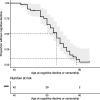
Kaplan-Meier curve showing cognitive decline over time. In the cox-proportional hazard model, the two individuals without complaint were censored. At 52 of age, 48.1% [29,2–71.6%] of patients suffer from cognitive decline. At 59 of age, 91.6% [72,1–97.5%] of patients suffer from cognitive decline
Fig. 3
Fig. 3
Size of APP locus duplications (n = 16) for each carrier with MRI available and associated genes involved. Rectangles show the duplicated regions in each family. For the family BES-262, MRIs of three related patients were available, and therefore, the number of ICH or CMBs varied between individuals. Otherwise, only one MRI was available per family. First column shows the position on chromosome 21 (GRCh37); second column shows name of genes corresponding to the position or the exon number within the APP gene; following columns represent the genomic duplication carried by each family (with family number and size of duplication in Mb). Each duplication contains the complete grey line corresponding to the APP gene. CAA, cerebral amyloid angiopathy diagnosis on MRI according to Boston revised criteria (except age criterion); ICH, intracerebral hemorrhage; CMB, cerebral microbleed; CSS, cortical superficial siderosis. Intra-familial heterogeneity was observed in the BES_262 family, in which 3 MRIs of symptomatic patients were available. The number of CMBs ranged from 42 to 420 (MRI performed at 54 and 56 years of age, respectively), and one patient from this family showed CAA-related inflammation at 52 years of age [17]

References

    1. Greenberg SM, Bacskai BJ, Hernandez-Guillamon M, Pruzin J, Sperling R, van Veluw SJ. Cerebral amyloid angiopathy and Alzheimer disease—one peptide, two pathways. Nat Rev Neurol. 2020;16(1):30-42. - PMC - PubMed
    1. Cacace R, Sleegers K, Van Broeckhoven C. Molecular genetics of early-onset Alzheimer’s disease revisited. Alzheimers Dement. 2016;12(6):733–748. doi: 10.1016/j.jalz.2016.01.012. - DOI - PubMed
    1. Carcaillon-Bentata L, Quintin C, Boussac-Zarebska M, Elbaz A. Prevalence and incidence of young onset dementia and associations with comorbidities: a study of data from the French national health data system. PLoS Med. 2021;18(9):e1003801. doi: 10.1371/journal.pmed.1003801. - DOI - PMC - PubMed
    1. Hendriks S, Peetoom K, Bakker C, van der Flier WM, Papma JM, Koopmans R, et al. Global prevalence of young-onset dementia: a systematic review and meta-analysis. JAMA Neurol. 2021;78(9):1080. doi: 10.1001/jamaneurol.2021.2161. - DOI - PMC - PubMed
    1. Lanoiselee HM, Nicolas G, Wallon D, Rovelet-Lecrux A, Lacour M, Rousseau S, et al. APP, PSEN1, and PSEN2 mutations in early-onset Alzheimer disease: a genetic screening study of familial and sporadic cases. PLoS Med. 2017;14(3):e1002270. doi: 10.1371/journal.pmed.1002270. - DOI - PMC - PubMed

MeSH terms