

Salivary proteins offer insights into keratinocyte death during aphthous stomatitis. A case-crossover study
- PMID: 37170213
- PMCID: PMC10176878
- DOI: 10.1186/s12903-023-02955-7
Salivary proteins offer insights into keratinocyte death during aphthous stomatitis. A case-crossover study
Abstract
Background: The death of oral keratinocytes is a crucial step in the emergence of recurrent aphthous stomatitis (RAS, also known as aphthae or aphthous ulcers). Since there are no experimental models available to research aphthous ulcers, little is understood about this process. We hypothesize that saliva can be a data bank of information that offers insights on epithelial damage.
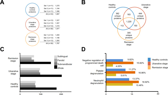
Methods: In this case-crossover study, we assessed the salivary proteome of patients with RAS (n = 36) in the presence and absence of ulcers using discovery proteomics and bioinformatics. Additionally, we contrasted these patterns with those of healthy individuals (n = 31) who had no prior aphthous ulceration.
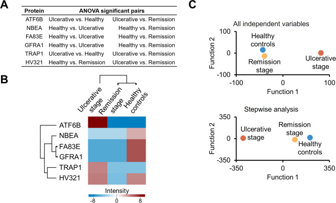
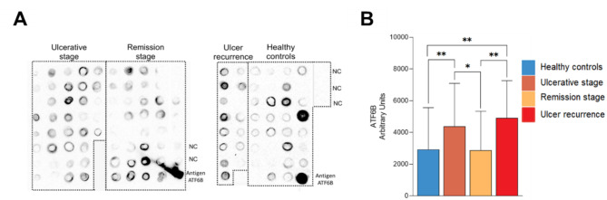
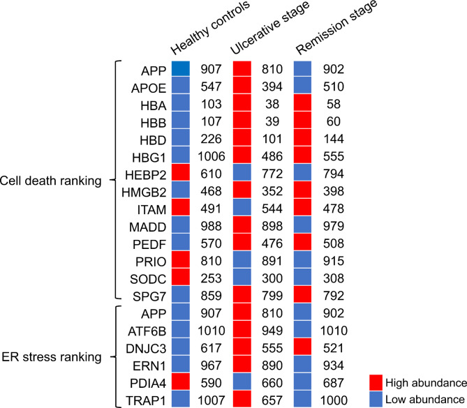
Results: Salivary proteome showed that during the ulcerative phase, controlled cell death was downregulated. Due to its ability to distinguish between individuals with and without ulcers, the ATF6B protein raises the possibility that endoplasmic reticulum (ER) stress is responsible for the damage that leads to the death of oral keratinocytes. The high abundance of TRAP1 and ERN1 matches with this biological discovery. The type of death is immunogenic, according to the functional data found in a cell death database.
Conclusion: We identified a cellular process that can lead to the death of oral keratinocytes in the etiopathogenesis process of RAS. Future studies should be conducted to identify what is responsible for the increase in ER stress signaling that would lead to an anti-cell death response.
Keywords: Endoplasmic reticulum stress; Proteomics; Recurrent aphthous stomatitis; Regulated cell death.
© 2023. The Author(s).
Conflict of interest statement
The authors declare that they have no competing interests.
Figures
Similar articles
-
Salivary proteome of aphthous stomatitis reveals the participation of vitamin metabolism, nutrients, and bacteria.Sci Rep. 2021 Aug 2;11(1):15646. doi: 10.1038/s41598-021-95228-8. Sci Rep. 2021. PMID: 34341431 Free PMC article.
-
Salivary mucin MUC7 oligosaccharides in patients with recurrent aphthous stomatitis.Clin Oral Investig. 2015 Nov;19(8):2147-52. doi: 10.1007/s00784-015-1495-3. Epub 2015 Jun 9. Clin Oral Investig. 2015. PMID: 26051835 Clinical Trial.
-
Decreased levels of salivary prostaglandin E2 and epidermal growth factor in recurrent aphthous stomatitis.Arch Oral Biol. 1995 Dec;40(12):1093-8. doi: 10.1016/0003-9969(95)00095-x. Arch Oral Biol. 1995. PMID: 8850647
-
Recurrent aphthous stomatitis: genetic aspects of etiology.Postepy Dermatol Alergol. 2013 Apr;30(2):96-102. doi: 10.5114/pdia.2013.34158. Epub 2013 Apr 12. Postepy Dermatol Alergol. 2013. PMID: 24278055 Free PMC article. Review.
-
Recurrent aphthous stomatitis: clinical characteristics and associated systemic disorders.Semin Cutan Med Surg. 1997 Dec;16(4):278-83. doi: 10.1016/s1085-5629(97)80017-x. Semin Cutan Med Surg. 1997. PMID: 9421219 Review.
Cited by
-
Aphthous stomatitis - computational biology suggests external biotic stimulus and immunogenic cell death involved.BMC Oral Health. 2024 Sep 29;24(1):1154. doi: 10.1186/s12903-024-04917-z. BMC Oral Health. 2024. PMID: 39343890 Free PMC article.
-
Causal associations between estradiol and mouth ulcers: A Mendelian randomization study.Medicine (Baltimore). 2024 Apr 26;103(17):e37989. doi: 10.1097/MD.0000000000037989. Medicine (Baltimore). 2024. PMID: 38669373 Free PMC article.
References
Publication types
MeSH terms
Substances
LinkOut - more resources
Full Text Sources
Medical
Miscellaneous
