. 2023 May 11;14(1):126.
doi: 10.1186/s13287-023-03361-0.

HSPB7 oppositely regulates human mesenchymal stromal cell-derived osteogenesis and adipogenesis

Affiliations

HSPB7 oppositely regulates human mesenchymal stromal cell-derived osteogenesis and adipogenesis

Shuang Zhang et al. Stem Cell Res Ther. .

Abstract

Background: Recent evidence suggests that accumulation of marrow adipose tissue induced by aberrant lineage allocation of bone marrow-derived mesenchymal stromal cells (BMSCs) contributes to the pathophysiologic processes of osteoporosis. Although master regulators of lineage commitment have been well documented, molecular switches between osteogenesis and adipogenesis are largely unknown.

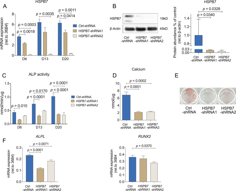
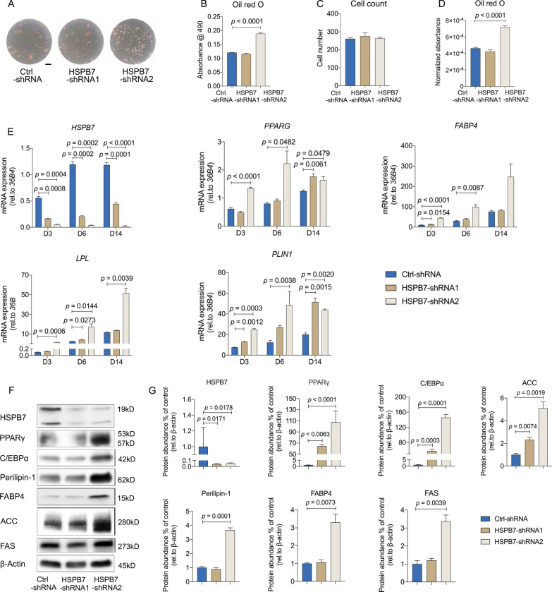
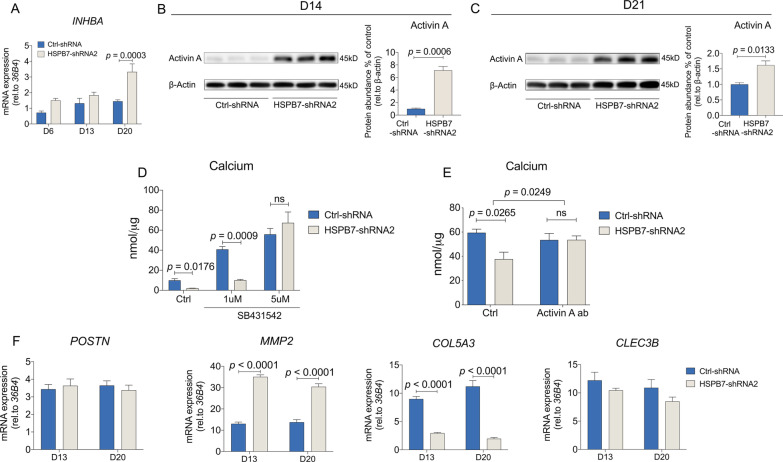
Methods: HSPB7 gene expression during osteogenic and adipogenic differentiation of BMSCs was evaluated by qPCR and Western blot analyses. Lentiviral-mediated knockdown or overexpression of HSPB7 and its deletion constructs were used to assess its function. The organization of cytoskeleton was examined by immunofluorescent staining. ALP activity, calcium assay, Alizarin Red S staining and Oil Red O staining were performed in vitro during osteoblast or adipocyte differentiation. SB431542 and Activin A antibody were used to identify the mechanism of Activin A in the regulation of osteogenic differentiation in BMSCs.

Results: In this study, we identified HSPB7 capable of oppositely regulating osteogenic and adipogenic differentiation of BMSCs. HSPB7 silencing promoted adipogenesis while reducing osteogenic differentiation and mineralization. Conversely, overexpression of HSPB7 strongly enhanced osteogenesis, but no effect was observed on adipogenic differentiation. Deletion of the N-terminal or C-terminal domain of HSPB7 led to decreased osteoblastic potency and mineralization. Mechanistically, our data showed that Activin A is a downstream target participating in HSPB7 knockdown-mediated osteogenic inhibition.

Conclusions: Our findings suggest that HSPB7 plays a positive role in driving osteoblastic differentiation, and with the capability in maintaining the osteo-adipogenesis balance. It holds great promise as a potential therapeutic target in the treatment of bone metabolic diseases.

Keywords: Activin A; Adipogenic differentiation; Bone marrow-derived mesenchymal stromal cells; HSPB7; Lineage commitment; Osteogenic differentiation.


Conflict of interest statement

The authors declare no competing interests.

Figures

Fig. 1
Fig. 1
HSPB7 expression is upregulated during osteogenic differentiation and downregulated during early adipogenic differentiation. A Cellular localization of HSPB7 in BMSCs with or without induction at day 3. B Relative mRNA expression levels of HSPB7 in BMSCs were assessed by qRT-PCR over 3 weeks. BMSC cultured in osteogenic condition was indicated with blue bars, and adipogenic condition was indicated with pink bars. Data are presented as means ± SEM and analyzed by one-way ANOVA (n = 2 per group) followed by post-hoc testing. Scale bars: 200 μm
Fig. 2
Fig. 2
HSPB7 silencing in BMSCs inhibits osteogenic differentiation and mineralization. AB HSPB7 silencing was assessed by quantification of HSPB7 mRNA expression by qRT-PCR at different time points (A) and Western blot (B) on day 3 under osteogenic induction. Full-length blots are presented in Additional file 7: Fig. S6. C ALP activity was evaluated at different time points under osteogenic induction. DE Osteoblast mineralization was assessed by calcium deposition assay (D) and Alizarin Red S staining (E) after three weeks osteogenic induction. F osteogenesis-related genes were evaluated on day 6. Data are presented as means ± SEM and analyzed by one-way ANOVA (B, D and F, n = 3–4 per group) or two-way ANOVA (A, n = 4 per group) followed by post-hoc testing
Fig. 3
Fig. 3
HSPB7 silencing in BMSCs affects cytoskeleton organization. A Representative images of immunostainings against F-actin (phalloidin-rhodamine), α-tubulin (Alexa Fluor 488) and Nuclei (DAPI) at day 14 following osteogenic induction. Arrows indicate spindle-shaped fibroblast-like cells. Scale bars: 200 μm
Fig. 4
Fig. 4
HSPB7 silencing in BMSCs enhances adipogenesis. AD Representative images (A) and quantitative data (B) of Oil Red O staining performed after 14 days with adipogenic induction. Total cell number was determined by DAPI staining (C). Quantitative data of Oil Red O staining were adjusted by cell number (D). E The expression of adipogenic markers was evaluated by qRT-PCR at indicated time points. FG Representative images (F) and quantitative data (G) of adipogenic genes were assessed on day 7 following adipogenic induction by Western blot. Full-length blots are presented in Additional file 7: Fig. S6. Abbreviations: Peroxisome proliferator-activated receptor gamma (PPARγ), Fatty acid binding protein 4 (FABP4), CCAAT/enhancer binding protein alpha (C/EBPα) and Perilipin-1 (PLIN1) Fatty acid synthase (FASN) and Acetyl-CoA carboxylase (ACC). Data are presented as means ± SEM and analyzed by one-way ANOVA (BD and G, n = 3–4 per group) or two-way ANOVA (E, n = 4 per group) followed by post-hoc testing. Scale bars: 200 μm
Fig. 5
Fig. 5
HSPB7 overexpression in BMSCs enhances osteogenic differentiation and mineralization, but not adipogenesis. A HSPB7 overexpression was assessed by quantification of HSPB7 mRNA expression at different time points. B Representative images and quantitative expression of HSPB7 were assessed by Western blot using HSPB7 antibody on day 3 following osteogenic induction. Full-length blots are presented in Additional file 7: Fig. S6. C ALP activity was evaluated at multiple time points with osteogenic media. DE Osteoblast mineralization was assessed by calcium deposition assay (D) and Alizarin Red S staining (E) after 3 weeks osteogenic induction. F Osteogenesis-related gene were evaluated at different time points with osteogenic induction. G Representative images of immunostainings against F-actin (phalloidin-rhodamine), α-tubulin (Alexa Fluor 488) and Nuclei (DAPI) on day 14 following osteogenic induction. HI Representative images (H) and quantitative data (I) of Oil Red O staining performed after 14 days with adipogenic induction. Total cell number was determined by DAPI staining. Quantitative data of Oil Red O staining was adjusted by cell number. Data are presented as means ± SEM and analyzed by two-tailed Student’s t-test ANOVA (B, D, F and I, n = 3–4 per group) or two-way ANOVA (A and C, n = 4 per group) followed by post-hoc testing. Scale bars: 200 μm
Fig. 6
Fig. 6
The ability of HSPB7 to enhance osteogenic differentiation depends on N- and C-terminal domains. A A schematic representation of full-length HSPB7 and its truncated mutants. His tag is located at the C terminal in each construct B 3D structures of FL HSPB7 and its truncated mutants generated by SWISS-MODEL (https://swissmodel.expasy.org). These models were predicated with reference to the template of 4jut.1.A and were selected on the basis of GMQE and QMEANDisCo global scores. C BMSCs expressing the indicated HSPB7 constructs were immunoblotted with anti-histidine antibody in non-differentiating conditions. Control samples were transduced with dsRED. Full-length blots are presented in Additional file 7: Fig. S6. D ALP activity was evaluated in BMSCs expressing dedicated constructs under osteogenic induction at day 13. EF Osteoblast mineralization was assessed by calcium deposition assay (E) and Alizarin Red S staining (E) after three weeks osteogenic induction in BMSCs. Abbreviations: ΔN, N-terminus deletion; ΔC, C-terminus deletion; SRS, serine-rich sequence stretch; FL, full-length. Data are presented as means ± SEM and analyzed by one-way ANOVA (n = 4 per group) followed by post-hoc testing
Fig. 7
Fig. 7
Blocking Activin A overcomes HSPB7 silencing-mediated osteogenic inhibition. A INHBA mRNA expression was assessed by qRT-PCR following HSPB7 silencing at different time points following osteogenic induction. BC INHBA mRNA expression was assessed by Western blot following HSPB7 silencing at day 14 (B) and day 21 (C) following osteogenic induction. Full-length blots are presented in Additional file 7: Fig. S6. D Osteoblast mineralization was assessed by calcium deposition assay after three weeks osteogenic induction. BMSCs were induced to osteogenic differentiation following HSPB7 silencing in the presence of SB431542 (D) or neutralizing Activin A antibodies (E). F ECM genes were evaluated by qRT-PCR following HSPB7 silencing at day 13 and day 20. Data are presented as means ± SEM and analyzed by two-tailed Student’s t-test (BC, n = 3 per group) or two-way ANOVA (A, DF, n = 4 per group) followed by post-hoc testing
Fig. 8
Fig. 8
A schematic diagram of HSPB7-regulated osteogenesis and adipogenesis. HSPB7 regulates the osteogenic differentiation through Activin A

References

    1. Seeman E, Delmas PD. Bone quality—the material and structural basis of bone strength and fragility. N Engl J Med. 2006;354:2250–2261. doi: 10.1056/NEJMra053077. - DOI - PubMed
    1. Teitelbaum SL, Ross FP. Genetic regulation of osteoclast development and function. Nat Rev Genet. 2003;4:638–649. doi: 10.1038/nrg1122. - DOI - PubMed
    1. Veldhuis-Vlug AG, Rosen CJ. Clinical implications of bone marrow adiposity. J Intern Med. 2018;283:121–139. doi: 10.1111/joim.12718. - DOI - PMC - PubMed
    1. Meunier P, Aaron J, Edouard C, Vignon G. Osteoporosis and the replacement of cell populations of the marrow by adipose tissue. A quantitative study of 84 iliac bone biopsies. Clin Orthop Relat Res. 1971;80:147–154. doi: 10.1097/00003086-197110000-00021. - DOI - PubMed
    1. Caplan AI. Mesenchymal stem cells. J Orthop Res. 1991;9:641–650. doi: 10.1002/jor.1100090504. - DOI - PubMed

Publication types

Substances

LinkOut - more resources