. 2022 Nov 18;7(1):55.
doi: 10.1186/s40850-022-00157-2.

Morphological and morphometric studies on the axial skeleton of the sitatunga (Tragelaphus spekii gratus)

Affiliations

Morphological and morphometric studies on the axial skeleton of the sitatunga (Tragelaphus spekii gratus)

Kenechukwu Tobechukwu Onwuama et al. BMC Zool. .

Abstract

Background: Anatomical features of the skeleton of wild animals contribute largely to their adaptation. A dearth of information on the skeletal anatomy of the sitatunga (Tragelaphus spekii gratus) necessitated this study. Two adult sitatunga carcasses weighing 54 kg and 57 kg were obtained after post-mortem examination. Bone preparation was achieved through cold water maceration protocol.

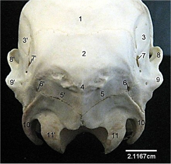
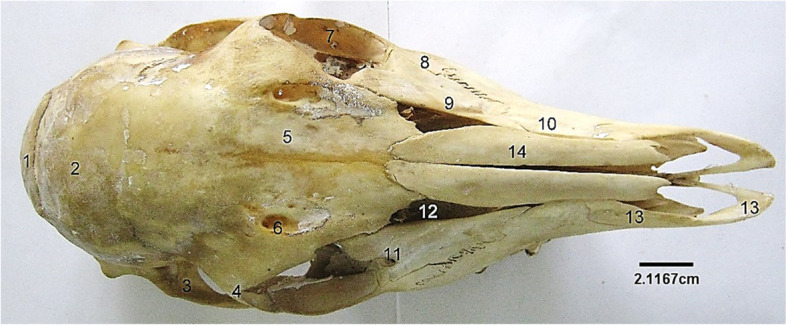
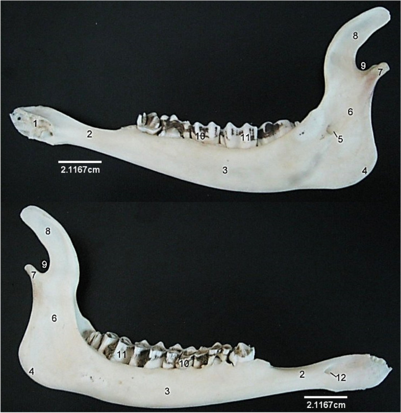
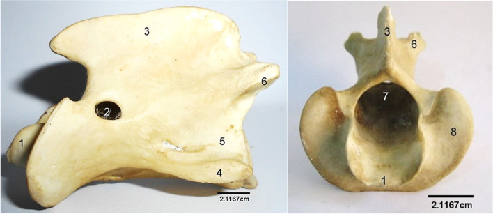
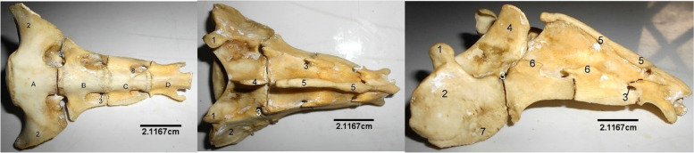
Result: The tympanic bulla was elongated and massive, resulting in the rudimentary appearance of the styloid and muscular processes of the temporal bone. The lacrimal bone had a somewhat triangular presentation with the lacrimal foramen on the caudal border of the facial surface while its dorsal border formed the lateral margin of the frontal sinus. There was no observable lacrimal fossa on this facial surface of the lacrimal bone. The facial tubercle was absent. The vertebral column formula was C7 T13 L6 S4 C10-14, and the atlas dorsal median tubercle was smooth, devoid of ridges. The spinous process of the axis extended the entire arch length to hang little above the odontoid process. The thoracic spinous processes were oriented dorso-caudally from T1 to T11; spinous process of T12 was vertical, while that of T13 was oriented dorso-cranially. The length of the transverse process of L1 and L6 were the same, and smaller than the length of those of L2-L5. There was incomplete fusion of sacral spinous processes. Three dorsal and ventral sacral foramina were identified laterally on each side of the vertebrae. The ribs were 26 in number (13 pairs). The sternum was comprised of 5 sternabrae separated by intersternal cartilage. The average number of bones of the axial skeleton was 75. Morphometric in formation included the length of skull, mandible and ribs; body length of vertebrae and spinous process length and height of the vertebrae.

Conclusion: This study recorded anatomical features and biometric information on axial skeletal bones of the Sitatunga (Tragelaphus spekii gratus) thereby providing baseline data for future biomedical, archaeological and comparative skeletal anatomical studies.

Keywords: Bone; Ribs; Sitatunga; Skull; Vertebrae.


Conflict of interest statement

The authors declare that they no competing interests.

Figures

Fig. 1
Fig. 1
Sitatunga skull (Caudal view). 1, 3, Parietal; 2, Occipital; 4, Occipital protuberance; 5, Squamous occipital; 6, 7, Temporal canal; 8, 9, Squamous temporal; 10, Paramastoid process; 11, Occipital condyle
Fig. 2
Fig. 2
Sitatunga skull (Ventral view). 1, Foramen magnum; 2, Occipital condyle; 3, Condyloid fossa; 4, Paracondyloid process; 5, Jugular foramen; 6, Mastoid process; 7, External acoustic meatus; 8, Temporal; 8’, Zygomatic process of temporal; 9, Basilar part of occipital; 10, Basisphenoid; 11, Muscular process in front of large tympanic bulla; 12, Foramen orbitorotundum; 13, Wing of sphenoid; 14, Oval foramen; 15, Pterygoid; 16, Presphenoid; 17, Malar; 17’, Zygomatic process of malar; 18, Vomer; 19, Vertical part of palatine; 20, Maxillary tuberosity; 21, Horizontal plate of palatine; 22, Palatine foramen; 23, Molars; 24, Palatine process of maxilla; 25, Premolars; 26, Palatine process of premaxilla
Fig. 3
Fig. 3
Sitatunga skull (dorsal view). 1, Occipital, 2, Parietal; 3, Temporal; 4, Orbital process of frontal; 5, Frontal; 6, Supraorbital foramen; 7, Zygomatic process of malar; 8, Malar or Zygomatic; 9, Lacrimal; 10, Maxilla; 11, Lacrimal foramen; 12, Triangular sinus; 13, Premaxilla; 14, Nasal
Fig. 4
Fig. 4
Sitatunga skull (lateral view). 1, Occipital; 2, Paramastoid process; 3, Mastoid process; 5, External acoustic meatus; 6, Squamous part of temporal; 6’, Zygomatic process of temporal; 7, Tympanic bulla; 7’, Muscular process; 8, Parietal; 9, Frontal; 10, Malar or Zygomatic10’, Zygomatic process of malar; 11, Hamulus of pterygoid; 12, Supraorbital foramen; 12’, Orbit; 13, Lacrimal foramen; 14, Lacrimal; 16, Nasal; 17, Maxilla; 18, Premaxilla; 19, Molars; 20, Premolars
Fig. 5
Fig. 5
Sitatunga skull (cranial view). 1, Lacrimal; 2, 3, Maxilla; 4, Premaxilla; 5, Sinus; 6, Perpendicular plate; 7, Ethmoturbinate; top 8, Dorsal turbinate; below 8, Vomer; 10, Palatine process of premaxilla; 11, Palatine fissure
Fig. 6
Fig. 6
Sitatunga mandible (Medial (A) and lateral (B) views). 1, Mandibular symphysis; 2, Diastema; 3, Horizontal ramus; 4, Mandibular angle; 5, Mandibular foramen; 6, Vertical ramus; 7, Mandibular condyle; 8, Coronoid process; 9, Mandibular notch; 10, Premolars; 11, Molars, 12, Mental foramen
Fig. 7
Fig. 7
Sitatunga Atlas (Dorsal and ventral views). 1, Dorsal tubercle; 2, Wing; 3, Alar foramen; 4, Intervertebral foramen; 5, Ventral tubercle; 6, Atlanta fossa; 7, Cranial articular surface; 8, Caudal articular surface
Fig. 8
Fig. 8
Sitatunga Axis (Lateral and cranial views). 1, Odontoid process; 2, Lateral vertebral foramen; 3, Spinous process; 4, Transverse process; 5, Body; 6, Caudal articular process; 7, Vertebral foramen; 8, Cranial articular surface
Fig. 9
Fig. 9
Sitatunga cervical vertebrae (Lateral view). 1, dorsal tubercle; 2, Spinous process; 3, Cranial articular process; 4, Caudal articular process; 5, Transverse process; 6, Ventral process; 7, Intervertebral foramina
Fig. 10
Fig. 10
Sitatunga thoracic vertebrae (lateral view). A-M, 1st to 13th thoracic vertebrae; 1, Spinous process; 2, Mammillary process; 3, Transverse process; 4, Intervertebral foramen; 5, Body; 6, Cranial articular process; 7, Caudal articular process
Fig. 11
Fig. 11
Sitatunga lumbar vertebrae (Lateral view). A-F, 1st to 6th lumbar vertebrae; 1, Spinous process; 2, Cranial articular process; 3, Caudal articular process; 4, Transverse process; 5, Body
Fig. 12
Fig. 12
Sitatunga Sacrum (Ventral, dorsal and lateral views). 1, Cranial articular process; 2, Wing; 3, Dorsal sacral foramina; 4, Spinous process; 5, Dorsal sacral crest; 6, Interrupted lateral sacral crest; 7, Auricular surface; 8, Ventral Sacral foramina. A-D, Sacral vertebra
Fig. 13
Fig. 13
Sitatunga Caudal vertebrae (dorsal view). 1, Spinous process; 2, Transverse process
Fig. 14
Fig. 14
Sitatunga Ribs (lateral view). 1, Head; 2, Neck; 3, Tubercle; 4, Groove; 5, Shaft; 6, Distal extremity
Fig. 15
Fig. 15
Sitatunga Sternum (Dorsal view). 1, First sternabra; 2, Second sternabra; 3, Third sternabra; 4, Fourth sternabra; 5, Fifth sternabra

References

    1. Meng F, Liu Y, Hu K, Zhao Y, Kong L, Zhou S. A comparative study of the skeletal morphology of the temporo-mandibular joint of children and adults. J Postgrad Med. 2008;54:191–194. doi: 10.4103/0022-3859.40960. - DOI - PubMed
    1. Onwuama KT, Salami SO, Kigir ES, Jaji ZA. Gross anatomical studies on the Hind limb of the sitatunga (Tragelaphus spekii) Anatomy Journal of Africa. 2021;10(1):1974–1979.
    1. Camille HW, Mark SB. Population density of sitatunga in riverine wetland habitats. Global Ecology and Conservation. 2020;24:2351–9894. doi: 10.1016/j.gecco.2020.e01212. - DOI
    1. Rose P, Robert, R. Evaluating the activity patterns and enclosure usage of a little studied zoo species, the sitatunga (Tragelaphus spekii). J Zoo Acquarium Res. 2013;1(1):14–9.
    1. McPherson JM, Sammy J, Sheppard DJ, Mason JJ, Brichieri-Colombi TA, Moehrenschlager A. Integrating traditional knowledge when it appears to conflict with conservation: lessons from the discovery and protection of sitatunga in Ghana. Ecol Soc. 2016;21(1):24. doi: 10.5751/ES-08089-210124. - DOI

LinkOut - more resources